Een abonnement op JoVE is vereist om deze inhoud te bekijken. Log in of start vandaag met uw gratis proefperiode.

Methodenartikel

Metastatic Cancer Cell Colony Isolation from Chicken CAM: A Procedure to Isolate Cancer Cells from Shell-less Egg Cultures

3.1K weergaven

⸱

30 april 2023

In dit artikel

Samenvatting

Source: Stoletov, K. et al. Discovery of Metastatic Regulators Using a Rapid and Quantitative Intravital Chick Chorioallantoic Membrane Model. J. Vis. Exp. (2021)

In this video, we describe the isolation of metastatic cancer cell colonies from the chicken chorioallantoic membrane (CAM) using shell-less egg culture. This method helps screen for factors that may be responsible for cancer metastasis.
Protocol

All procedures involving animal models have been reviewed by the local institutional animal care committee and the JoVE veterinary review board.

1. Isolation of Metastatic Colonies

  1. At day 5 post injection, remove embryos from the incubator and inspect the embryo CAMs for metastatic colony distribution. Identify the embryos with uniform colony distribution in which compact (or overly invasive) colonies are present.
    NOTE: The process of culturing chicken embryos is not sterile, therefore, all the screening steps must be performed under highly clean conditions to avoid future tissue culture ....

Toegang beperkt. Log in of start een proefperiode om deze inhoud te bekijken.

Resultaten

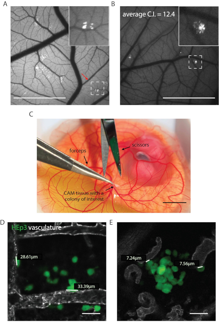
Chick embryo CAM assay with HEp3 cells showing vascularization and colony measurement, microscopy images.
Figure 1. Representative results for different steps of the screening protocol. (A) Metastatic colonies formed by heterogenous (library transduced cancer cell line HEp3 cells), 5 days post injection. Red arrow shows com.......

Toegang beperkt. Log in of start een proefperiode om deze inhoud te bekijken.

Openbaarmakingen

No conflicts of interest declared.

Materialen

Lijst van materialen gebruikt in dit artikel
NaamBedrijfCatalogusnummerOpmerkingen
15 mL conical centrifuge tubes.CorningCLS430791-500EA
18 gauge x 1 1/2 BD precision<br/> needleBDBD305196we use 1.2mm x 40mm, it is possible<br/> to use shorter needles if preferred
Benchtop centrifuge.many sources are availableAny TC compatible centrifuge that<br/> can be used to spin down the cells is<br/> suitable
CollagenaseSigmaC0130-100MG
Cotton swabsmany sources are availablemust be sterilized before use
Culture media appropriate for the cell<br/> lines usedmany sources are availableWe grow HT1080, HEp3 and b16 cell<br/> lines in DMEM, 10% FBS media
Egg incubatormany sources are availableAn exact model that is necessary<br/> depends on the scale of the screen.<br/> Available sources are MGF Company<br/> Inc., Savannah, GA, or Lyon Electric<br/> Company Inc., Chula Vista, CA
Eppendorf tubes , 1.5mlSigmaT4816-250EA
Fertilized White Leghorn eggsany local supplyer
Fine forcepsmany sources are availablemust be sterilized before use
Image analysis softwareWe use Nikon Elements
PBS (1x)many sources are available
Plastic weighting dishesSimportCA11006-614dimensions are 78x78x25mm; many<br/> other sources are available
Small surgical scissorsmany sources are availablemust be sterilized before use
Stereo fluorescent microscopeWe use Zeiss Lumar v12

Meer artikelen verkennen

Isolatie van kankercellencollagenasebehandelingfluorescentiemicroscopieweefselexcisiecelkweekmetastatische koloniënselectieve mediacentrifugatie