Method Article

Amphotericin-B Mediated Perforated Patch-Clamp Analysis: An Electrophysiological Technique to Study Ionic Currents in Urinary Bladder Cells

April 30th, 2023

In This Article

Abstract

Loading...
$$\rightleftharpoonup{xx}$$ $$\longleftharp{xx}$$, $$\longrightharp{xx}$$,

Source: Malysz, J. et al. Preparation and Utilization of Freshly Isolated Human Detrusor Smooth Muscle Cells for Characterization of 9-Phenanthrol-Sensitive Cation Currents. J. Vis. Exp. (2020)

This video demonstrates the characterization of cationic currents from transient receptor potential melastatin type 4 (TRPM4) ion channels in detrusor smooth muscle cells using amphotericin-B mediated perforated patch-clamp technique. The technique helps evaluate the biophysical and pharmacological properties of ion channels.

Protocol

Loading...
$$\rightleftharpoonup{xx}$$ $$\longleftharp{xx}$$, $$\longrightharp{xx}$$,

1. Recording Voltage-step Induced Cation Currents from DSM Cells Using Amphotericin-B Perforated Whole-cell Voltage Patch-Clamp Technique

  1. Pipette 0.25–1 mL of cell suspension onto a glass-bottom chamber sitting on a stage of an inverted microscope and allow the cells to adhere to the glass bottom.
  2. After incubation for at least 45 min, remove digestion solution (DS), from the bath and replace with the E solution (Table 1) by superfusion where the gravity-aided-solution flow via inlet tubing slowly replaces DS with the new solution while outlet tubing connected to a vacuum waste vessel removes the chamber solution and prevents overflow.
    NOTE: E solution contains tetraethylammonium (TEA+) and cesium (Cs+) ions to inhibit K+ currents.
  3. Prepare a working stock solution of amphotericin-B in dimethyl sulfoxide (DMSO) (1 mg per 10 µL of DMSO). To fully dissolve amphotericin powder, sonicate (at least 15 min) and vortex the solution well.
    NOTE: This step usually takes less than 10 min. Dissolving 3–4 mg of amphotericin-B in 30–40 µL of DSMO in a 1.5 mL microcentrifuge tube works well. Higher quantities of amphotericin-B require more DSMO solvent typically resulting in a longer interval for mixing and incomplete solubilization of the amphotericin-B solid particles present in the tube.
  4. Dissolve stock solution of amphotericin-B in the pipette solution (solution P, Table 1) to obtain a final concentration of 200–500 µg/mL. This step requires extensive sonication and vortexing at a high-speed setting (8–10/10) for ~30 to 60 min per step to ensure optimal mixing and prevention of amphotericin-B precipitate formation in the pipette solution.
    NOTE: Amphotericin-B will precipitate over time and is light-sensitive. The working pipette solution containing Amphotericin-B is checked for solubility, hand-mixed prior to pipette filling, and kept in the dark.
  5. Pull multiple patch electrodes, fire-polish electrode tips, and (if needed) coat the tips with dental wax.
  6. Fill the tip of a patch electrode with the pipette solution (solution P, Table 1) without amphotericin-B by briefly dipping the electrode in the solution.
  7. Backfill the electrode with the same pipette solution containing amphotericin-B.
  8. Mount the electrode onto a holder connected to a patch-clamp amplifier headstage.
  9. Using a micromanipulator, place the electrode just below the surface of the extracellular solution so that the tip of the electrode is just submerged.
  10. In the voltage-clamp mode, set the holding potential to 0 mV and adjust the current to 0 pA with the pipette offset dial on the commercial amplifier (Table of Materials).
  11. Determine the electrode resistance using the Membrane Test window/function of the commercial acquisition software (Table of Materials). To activate click Tools>Membrane Test>Play or a shortcut icon in the software. The determined electrode resistance should be in the range of 2 to 5 MΩ.
    NOTE: Membrane Test function provided in the commercial acquisition software or Seal Test option on the amplifier can be used to monitor electrode resistance by applying voltage steps repetitively.
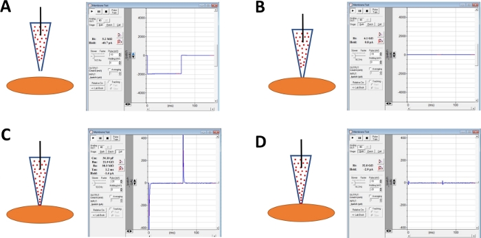
  12. Continue monitoring electrode resistance while advancing the electrode toward a chosen DSM cell with a micromanipulator (Figure 1A).
    NOTE: To be considered a viable DSM cell, the cell must show spindle-shaped elongated morphology, a well-defined halo around the cell, crisp edges, and semi-contractile (serpentine) appearance.
  13. When touching the cell surface with the electrode—indicated by a rapid increase in the electrode resistance measured with the Membrane Test function—form a giga-seal by applying gentle rapid negative pressure to the electrode holder via tubing. This results in negative pressure created at the tip of the electrode that pulls cell membrane into electrode aiding in the formation of a giga-seal or a very tight contact between the electrode and plasma membrane (Figure 1B).
  14. Once the giga-seal forms, compensate pipette capacitance by adjusting fast and slow dials on the commercial amplifier and monitor giga-seal stability (leak current) using the Membrane Test function.
  15. Allow time, typically 30–60 min, for amphotericin-B to diffuse down the pipette and be inserted into the plasma membrane forming pores primarily selective to monovalent cations. During this step, continue monitoring the giga-seal with the Membrane Test function. As cell perforation increases so does the amplitude of the capacitance transients (compare Figure 1B versus Figure 1C displaying no and effective cell perforation, respectively) measured with the Membrane Test function.
  16. When the patch perforation is optimal (judged by stable series resistance typically below 50 MΩ), cancel out the capacitance transients by adjusting the dials for cell capacitance and series resistance on the amplifier. Series resistance compensation can also be performed at this time (Figure 1D).
  17. Once stable voltage-step induced cation currents evoked by the specified protocol are observed, apply a compound or physiological condition to test by superfusion and record the responses for the control-, test-condition, and washout (if possible) with the commercial acquisition software.
    1. Record currents with a routine voltage-step protocol that involves holding DSM cells at -64 or -74 mV and stepping the voltage in 10 mV increments for 400 or 500 ms from -94 to +96 or +106 mV and returning to the holding potential.
      NOTE: The membrane potential values are adjusted for a liquid junction potential of 14 mV (using P and E solutions, Table 1). The liquid junction potential is obtained in the commercial acquisition software (Table of Materials) by clicking Tools>Junction Potentials and entering the concentrations of solution ion components. A ramp protocol can also be used to obtain current recordings.
    2. Run the voltage protocol in continuous ~1 min interval during an experiment recording currents for pre-addition control, test condition, and washout
Solution TypeComposition (in mM)
DS (Dissection/ Digestion Solution)80 Na-glutamate, 55 NaCl, 6 KCl, 10 HEPES, 2 MgCl2, and 11 glucose, pH adjusted to 7.4 (with 10 M NaOH)
DS-P (Papain-containing DS)DS containing 1–2 mg/ml papain, 1 mg/ml dithiothreitol and 1 mg/ml bovine serum albumin
DS-C (Collagenase-containing DS)DS solution containing 1–2 mg/ml collagenase type II, 1 mg/ml bovine serum albumin, 0 or 1 mg/ml trypsin inhibitor and 100-200 μM Ca2+
P (Pipette)110 CsOH, 110 aspartic acid, 10 NaCl, 1 MgCl2, 10 HEPES, 0.05 EGTA, and 30 CsCl,pH adjusted to 7.2 with CsOH, and supplemented with amphotericin-B (300-500 μg/ml)
E (Extracellular)10 tetraethylammonium chloride (TEA), 6 CsCl, 124 NaCl, 1 MgCl2, 2 CaCl2, 10 HEPES, and 10 glucose, pH adjusted to 7.3–7.4 with NaOH or CsOH, and 0.002–3 (2–3 mM) nifedipine

Table 1: Compositions of dissection/digestion solution (DS), and pipette and extracellular solutions used in perforated patch-clamp experiments.

Access restricted. Please log in or start a trial to view this content.

Results

Loading...
$$\rightleftharpoonup{xx}$$ $$\longleftharp{xx}$$, $$\longrightharp{xx}$$,

Atomic force microscopy process with force-distance curves; diagram displaying measurement results.
Figure 1: Schematic illustration of steps involved in giga-seal formation and amphotericin-B perforation of human DSM cells. Illustrated are spatial positions of an amphotericin-B containing pipette and a DSM cell along with associated r...

Access restricted. Please log in or start a trial to view this content.

Disclosures

Loading...
$$\rightleftharpoonup{xx}$$ $$\longleftharp{xx}$$, $$\longrightharp{xx}$$,
No conflicts of interest declared.

Materials

List of materials used in this article
NameCompanyCatalog NumberComments
5 ml polystyrene round-bottom tubeFalcon352054Tubes for DS containing enzymes used in digestion steps
Amphotericin-BFisherBP928-250Used for patch/cell perforation
Amphotericin-BEuropean Pharmacopoeia Reference Standards5Used for patch/cell perforation
Amphotericin-BSigma-AldrichA9528-100MGUsed for patch/cell perforation
Analog vortex mixerVWR58816-121
Aspartic acidSigma-AldrichA9006Intracellular pipette solution
Bovine serum albuminSigma-AldrichA7906DS
CaCl2Sigma-AldrichC1016Extracellular solution and DS
Capillary GlassSutterBF150-110-7.5Capillary for preparation of pulled patch electrodes
Cesium hydroxide hydrateSigma-AldrichC8518Intracellular pipette solution
Clampex ver. 10 software includes data acqusition (Clampex) and analysis (Clampfit) programsAxon Instruments/ Molecular DevicespCLAMP-10Commerical software and part of patch-clamp rig setup
CsClSigma-Aldrich203025Extracellular and intracellular solutions
Dental waxMiltex Dental Wax Technologies, Inc.18058351
Dimethyl sulfoxide (DMSO)Sigma-AldrichD2650Solvent
EGTASigma-AldrichE3889Ca2+ chelator, used in intracellular pipette solution
Flaming/Brown micropipette pullerSutterP-97Required to pull electrodes with very fine tips
Floating foam tube rack/holderVWR Scientific82017-634Used for holding tubes with enzymes for temperature control
GlucoseSigmaG8270
Glutamic acid (Na salt)Sigma-AldrichG1626DS
HEPESSigma-AldrichH3375pH Buffer
KClFisher ScientificBP366-1Extracellular solution
Low Noise Data Acquisition SystemAxon Instruments/ Molecular DevicesDigidata 1440APart of patch-clamp rig setup
Magnetic stirrerVWR01-442-684
MgCl2 (hexahydrate)Sigma-AldrichM2670Extracellular and intracellular solutions
MicroForgeNarishigeMF-830Used for fire-polishing electrodes
NaClSigma-AldrichS7653Extracellular and intracellular solutions
NaOHSigma-AldrichS8045
NifedipineSigma-AldrichN7634L-type voltage-gated Ca2+ channel blocker
Nikon inverted microscope, TS100 with T1-SM stage with 5x, 10x, 20x, and 40x objectivesNikonDiscontinuedPart of Patch-clamp rig setup
Non-metalic syringe needle, MicroFilWPIMF-34G-5Filling of intracellular pipette solution
Pasteur pipetteFisherBrand13-678-20ATips are broken off and fire-polished and used for titration of enzymatically treated tissues to release single DSM cells from pieces
Patch-clamp amplifierAxon Instruments/ Molecular DevicesAxon Axopatch 200BPart of patch-clamp rig setup
PC computerDELLCustom configurationPart of patch-clamp rig setup
pH MeterAspera InstrumentsPH700
Polyethylene tubingIntramedic427-436Tubing for superfusion of extracellular bath connected to glass-bottom recording chamber
Tetraethylammonium chlorideSigma-AldrichT2265Ion channel blocker of Kv and BK channels added to the extracellular bath solution
Weighting scaleMettler ToledoXS64
ZeissAxiovert 40C inverted microscope with 10x and 40x objectivesCarl-ZeissDiscontinuedPart of patch-clamp rig setup

Reprints and Permissions

Request permission to reuse the text or figures of this JoVE article

Request Permission

Tags

Amphotericin B Perforated Patch ClampIonic Currents MeasurementDetrusor Smooth Muscle CellsUrinary Bladder CellsTRPM4 Ion ChannelsExtracellular Solution PreparationGiga Seal FormationMembrane Perforation TechniqueElectrophysiological Current RecordingIon Channel Characterization

Related Articles