Een abonnement op JoVE is vereist om deze inhoud te bekijken. Log in of start vandaag met uw gratis proefperiode.

Methodenartikel

Laser-assisted Selective Ablation of Porcine Corneal Endothelial Cells: A Technique to Induce Focused Porcine Corneal Endothelial Injury for Ex Vivo Studies

2.8K weergaven

⸱

8 juli 2025

In dit artikel

Samenvatting

Source: Holzhey, A. et al., Development of a Noninvasive, Laser-Assisted Experimental Model of Corneal Endothelial Cell Loss. J. Vis. Exp. (2020)

This video demonstrates a non-invasive laser-assisted procedure to selectively damage the corneal endothelial cells in the porcine eye. The developed ex vivo model of endothelial cell damage can be used to study diseases of the corneal endothelium.
Protocol

All procedures involving animal models have been reviewed by the local institutional animal care committee and the JoVE veterinary review board.

1. Preparation of organ culture and laser treatment

  1. Obtain freshly enucleated porcine eyes from the local abattoir. Keep them cool (4 °C) in Dulbecco's modified Eagle medium (DMEM) with high glucose, supplemented with L-glutamine, sodium pyruvate, penicillin/streptomycin (1%), and porcine serum (10%), henceforth referred to in this article as full medium.
  2. Remove extracellular tissues with scissors and soak the eyes in 5% povidone-iodine ophthalmic solution for 5 min before placing them in sterilized phosphate-buffered saline (PBS) until use.
  3. Screen eyes for major anterior segment pathologies, such as corneal scarring, edema, and other opacities with a spectral-domain optical coherence tomography device (Table of Materials).
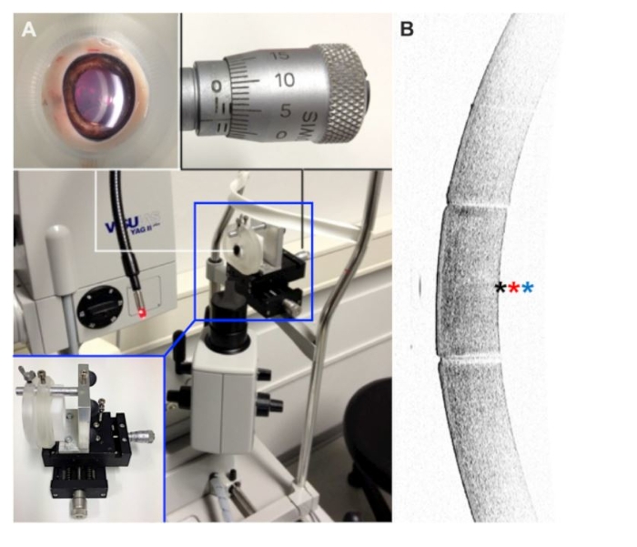
  4. Position the eyes in front of a slit-lamp unit equipped with a Nd:YAG laser (Table of Materials), which has a wavelength of 1,064 nm and a focal spot diameter of 10 µm in air.
    NOTE: For optimal positioning a 3D-printed holding apparatus is used, which was designed to hold the eye firm, without putting too much pressure on it (Figure 1).
  5. Use a magnification of 12x and deflect the illumination to visualize the individual corneal layers.
  6. Set the pulse energy (e.g., 1.6 mJ) and focus point (e.g., 0.16 mm) for selective ablation of endothelial cells.
  7. Place a clear cornea paracentesis close to the limbus and inject viscoelastic (Table of Materials) to stabilize the anterior chamber.
  8. Excise the laser-treated central cornea using an 8 mm trephine.
  9. Place the excised cornea in a well of a 12-well plate with the endothelial site facing upwards and incubate the specimen in 3 mL of full medium at 37 °C for up to 3 days.
    NOTE: Potential cytoprotective agents can be added to the medium during this step.

Toegang beperkt. Log in of start een proefperiode om deze inhoud te bekijken.

Resultaten

YAG laser setup, microscopy image of optic interface; optical measurement and analysis.
Figure 1: Experimental setup. (A) Eyes were fixed in a partially 3D-printed holding apparatus, which allowed precise alignment with respect to the laser beam. (B) Before laser treatment, the tissue was evaluated with an anterior seg...

Toegang beperkt. Log in of start een proefperiode om deze inhoud te bekijken.

Openbaarmakingen

No conflicts of interest declared.

Materialen

Lijst van materialen gebruikt in dit artikel
NaamBedrijfCatalogusnummerOpmerkingen
Barron vacuum trephine KatenaK20-2058
CryostatLeicaCM 3050S
Dulbecco's Modified Eagle's Medium - high glucosePAAE-15009
Eye holderSelfN/A
Inverted MicroscopeLeicaDMI 6000 B
KH2PO4Merck529568
Na2HPO4Merck1065860500
Nd:YAG laserZeiss MeditecvisuLAS YAG II plus
OCT Tissue TekSakura Finetechnical4583
Penicillin-StreptomycinSigma-AldrichP4333
Phosphate Buffered Saline (PBS)Gibco10010056
Porcine serumSigma-Aldrich12736C
Spectral-domain optical coherence tomographHeidelberg EngineeringSpectralis
Tissue culture plate 12-wellSarstedt833921
ViscoelasticOmniVisionMethocel

Tags

Nd:YAG-laserspectrale domein optische coherentietomografiespleetlampunittrephine-excisievisco-elastische injectiecorneaale transparantie