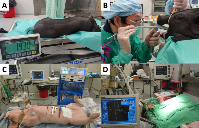
All procedures involving animal models have been reviewed by the local institutional animal care committee and the JoVE veterinary review board.
1. Tracheal intubation (Figure 1B)
- Prepare the equipment and materials required for electromyography (EMG) tube intubation: a size #6 EMG endotracheal tube, a face mask for assisted ventilation, two slings to hold the mouth open, one gauze strip to pull the tongue, a blunt tip suction catheter, a veterinary laryngoscope with 20cm straight blades, an elastic bougie, a 20-mL syringe, a stethoscope, and adhesive tape.
- Position the anesthetized piglet in a prone position on the operating table. Align the head and body to ensure clear visualization of the upper airway.
- Direct the assistant to apply traction of the upper and lower jaw to maintain an adequate mouth opening and to avoid rotation or overextension of the head. Cover the tongue with gauze and pull the tongue out to optimize the visual field.
- Hold the laryngoscope and place it directly in the oral cavity to depress the tongue.
- Directly visualize the epiglottis and use the laryngoscope to press the epiglottis downward toward the tongue base.
- When the vocal cords are clearly identified, gently advance the elastic bougie into the trachea. Slight rotation of the elastic bougie may be required to overcome resistance. Next, advance the EMG tube at the mouth angle to a depth of 24 cm.
- Inflate the EMG tube cuff to a volume no larger than 3 mL. If ventilation by manual bagging reveals no obvious air leakage, in situ deflation of the EMG tube is feasible.
- When the EMG tube is placed at the proper depth, confirm the free passage of fresh gas by manual bagging. Further confirm the proper tracheal intubation by end-tidal carbon dioxide (etCO2) monitoring (capnography) and chest auscultation for early identification of inadvertent esophageal or endobronchial intubation.
NOTE: Capnography showed both the etCO2 waveform and the digital value in mmHg. When esophageal intubation occurred, etCO2 was absent or near zero after 6 breaths. When the EMG tube was in the correct place, the typical etCO2 waveform and adequate value (usually >30 mmHg) was noted. Furthermore, the breathing sound of a bilateral lung filled is clear and symmetric as determined by chest auscultation.
- Use medical tape to fix the EMG tube at the mouth angle. Since the tube usually requires adjustment during intraoperative neural monitoring (IONM) experiments, do not fasten the tube to the snout.
- Connect the EMG tube to the ventilator. Continuous capnography is mandatory for monitoring the etCO2 value and curve throughout the experiment.
2. Anesthesia maintenance (Figure 1C)
- After the EMG tube is fixed, position the piglet on its back with the neck extended (Figure 1C). Maintain general anesthesia with 1-3% sevoflurane in oxygen at 2 L/min.
- Ventilate the lungs in volume-control mode at a tidal volume of 8-12 mL/kg, and set the respiratory rate to 12-14 breaths/min.
- Begin physiologic monitoring, including capnography, electrocardiography (ECG)and monitoring of oxygenation (SaO2).