Methodenartikel

Isolating Short Posterior Ciliary Arteries: A Protocol to Excise Intact Short Posterior Ciliary Microvessels from Porcine Eye

8 juli 2025

In dit artikel

Samenvatting

Loading...
$$\rightleftharpoonup{xx}$$ $$\longleftharp{xx}$$, $$\longrightharp{xx}$$,

Source: Perumal, N. et al., Sample Preparation for Mass-spectrometry-based Proteomics Analysis of Ocular Microvessels. J. Vis. Exp. (2019).

This video describes the protocol to isolate the short posterior ciliary arteries from the porcine eye. The isolated short posterior arteries are a suitable source for elucidating the molecular insights and protein profiles of the ocular blood vessels.

Protocol

Loading...
$$\rightleftharpoonup{xx}$$ $$\longleftharp{xx}$$, $$\longrightharp{xx}$$,

All procedures involving animal models have been reviewed by the local institutional animal care committee and the JoVE veterinary review board.

1. Isolation of Short Posterior Ciliary Arteries

Porcine eyes together with optic nerve and extraocular tissues were obtained immediately post-mortem. Enucleated eyes were transported to the laboratory in ice-cold phosphate buffered saline (PBS) and used immediately.

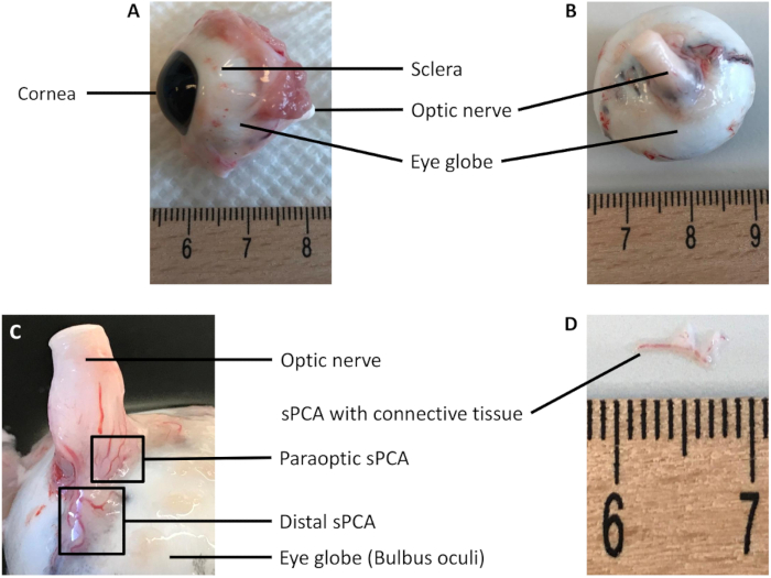
NOTE: The porcine eye is basically divided into the anterior (Figure 1A) and posterior sections (Figure 1B).

  1. Place the eye globe in a dissection chamber containing ice-cold Krebs-Henseleit (K-H) buffer. Carefully cut away surrounding muscle and tissues with a pair of sharp Mayo scissors.
    NOTE: To prepare Krebs-Henseleit (K-H) buffer, weigh the following chemicals (27.68 g of NaCl, 8.4 g of NaHCO3,1.4 g of KCl, 1.18 g of MgSO4, and 0.64 g of KH2PO4) into one dry and clean 50 mL conical centrifuge tube and, 1.47 g of CaCl2 and 8.0 g of glucose into another tube. The powder mixture of these chemicals should be stored in a cool and dry place. The CaCl2 and glucose powder mixture is best prepared fresh to prevent absorption of moisture and clumping.
  2. Make an incision with a scalpel and cut the globe along the equatorial plane with a pair of sharp Mayo scissors until the eye is separated into the anterior and posterior halves. Remove as much vitreous body as possible from the posterior half of the eye using a pair of standard pattern forceps.
    NOTE: Separation of the eye globe into two halves facilitates the isolation of short posterior ciliary arteries (sPCA) with ease without having a moving eye in the dissection dish. However, this step is not mandatory. The vessels can also be isolated without opening the eye globe.
  3. Use dissection pins to carefully pin down the posterior half of the eye with the retina side down and the optic nerve facing up towards the experimenter.
  4. Gently cut away the connective tissues surrounding the optic nerve to expose the underlying retrobulbar vasculature with a pair of Student Vannas spring scissors.
  5. The sPCA can be seen as short branches of vessels (between 5-8 branches) that penetrate the sclera and circumferentially surround the optic nerve head (Figure 1C). Isolate the paraoptic and distal sPCA together with the surrounding connective tissues with a pair of type 5 precision tweezers and Vannas capsulotomy scissors (Figure 1D).
    NOTE: Ensure that the K-H buffer remains ice-cold throughout the isolation procedure. It is highly recommended to change the buffer every 20-30 min, depending on the ambient temperature.

2. Sample Preparation

  1. Gently remove connective tissues from the arterial segments using extra fine-tipped type 5 precision tweezers and Vannas capsulotomy scissors under a stereomicroscope and rinse the isolated arteries in ice-cold PBS to remove contaminants and blood residues.
    NOTE: If samples are not subjected to the next steps immediately, snap-freeze them in liquid nitrogen and store in -80 °C until further use.
Toegang beperkt. Log in of start een proefperiode om deze inhoud te bekijken.

Resultaten

Loading...
$$\rightleftharpoonup{xx}$$ $$\longleftharp{xx}$$, $$\longrightharp{xx}$$,

Eye dissection labeled diagram; sclera, optic nerve, cornea; anatomy study.
Figure 1: Representative photographs of the porcine eyes.

(A) Lateral view of the eye globe shows the cornea, which is located in the anterior part of the eye, the sclera, surrounding muscle and optic nerve. (B) Pos...

Toegang beperkt. Log in of start een proefperiode om deze inhoud te bekijken.

Openbaarmakingen

Loading...
$$\rightleftharpoonup{xx}$$ $$\longleftharp{xx}$$, $$\longrightharp{xx}$$,
No conflicts of interest declared.

Materialen

Lijst van materialen gebruikt in dit artikel
NaamBedrijfCatalogusnummerOpmerkingen
Calcium chloride dihydrate (CaCl<sub>2</sub>)Carl Roth&nbsp;5239.12.5 mM
Dulbecco's phosphate-buffered saline (PBS)Thermo Fisher Scientific&nbsp;14190169
Sodium chloride (NaCl)&nbsp;Carl Roth9265.2118.3 mM
Sodium hydrogen carbonate (NaHCO<sub>3</sub>)Carl Roth&nbsp;965.325 mM
&alpha;-(D)-(+)- Glucose monohydrate&nbsp;Carl Roth&nbsp;6780.111 mM
Disposable scalpels #21&nbsp;pfm Medical&nbsp;200130021
Dissection pins&nbsp;Carl Roth&nbsp;PK47.1
Extra Fine Bonn Scissors&nbsp;Fine Science Tools&nbsp;14084-08
Falcon conical centrifuge tubes (50 mL)Fisher Scientific&nbsp;14-432-22
Mayo scissors, Tough cut&nbsp;Fine Science Tools&nbsp;14130-17
Precision tweezers&nbsp;Fine Science Tools&nbsp;11251-10&nbsp;Type 5
Precision tweezers, straight with extra fine tipsCarl Roth&nbsp;LH53.1&nbsp;Type 5
Standard pattern forceps&nbsp;Fine Science Tools&nbsp;11000-12
Student Vannas spring scissors&nbsp;Fine Science Tools&nbsp;91501-09
Vannas capsulotomy scissors&nbsp;Geuder&nbsp;19760&nbsp;Straight, 77 mm

Herprints en machtigingen

Toestemming aanvragen om de tekst of afbeeldingen van dit JoVE-artikel te hergebruiken

Toestemming aanvragen

Trefwoorden

Isolatie van het varkensoogexcisie van oculaire microvatenvisualisatie van de retrobulbaire vasculatuurtechniek voor circumferenti le incisieverwijdering van de glasachtig lichaamlaagdissectie van bindweefselisolatie met precisie pincettenijskoude Krebs bufferwassen van arteri n met PBS

Gerelateerde artikelen