1. Lentivirus production
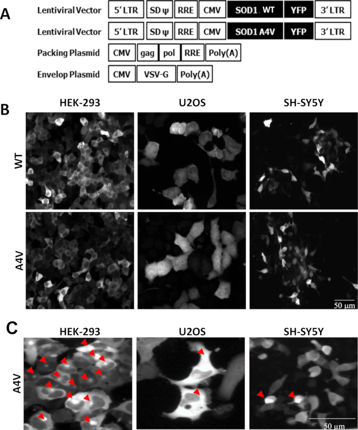
NOTE: The production and manipulation of lentiviral vectors was carried out according to the National Institutes of Health (NIH) guidelines for research involving recombinant DNA. The plasmid encoding the wild-type and A4V mutant SOD1 tagged with enhanced YFP (SOD1WT-YFP and SOD1A4V-YFP) are used. Both gene fusion products were amplified using the PCR primer pair 5′-ATCGTCTAGACACCATGGCGACGAAGGTCGTGTGC-3′ and 5′-TAGCGG CCGCTACTTGTACAGCTCGTCCATGCC-3′and inserted into the pTRIP-delta U3 CMV plasmid using XhoI and BsrGI restriction sites. Avoidance of more than 20 passages and maintenance of cells at less 60% confluency helps to ensure good transfection efficiencies.
- On Day 1, split HEK-293T cells at 30 - 40% confluence in a 10-cm diameter tissue culture plate (3 x 10⁶ cells/dish) in 10 mL of cell culture medium (high-glucose DMEM with 10% FBS and 4 mM L-glutamine) for virus production.
- Keep the 10-cm diameter tissue culture plate in a CO₂ incubator (37 °C, 5% CO₂) overnight.
- On Day 2, prepare a DNA transfection solution containing 10 µg of lentiviral vectors (pTRIP-delta U3 CMV - SOD1WT-YFP or -SOD1A4V-YFP) together with the lentiviral packaging plasmids (5 µg of pVSVg; 10 µg of pCMV-dR8.71) (Figure 1A). Add 125 µL of 1 M CaCl₂ and adjust the volume to 500 µL using distilled water. Gently add 500 µL of 0.05 M HEPES into the mixture and incubate for 10 min at room temperature.
- Replace HEK-293T cells medium with 10 mL pre-warmed fresh culture medium antibiotic-free mixed with 1 mL DNA transfection solution and incubate overnight at 37 °C, 5% CO₂.
- On Day 3, replace the cell supernatant with fresh culture medium.
- On Day 4, harvest the supernatant in a 15 mL tube and centrifuge for 5 min at 500 x g to remove the dead cells and debris. Further purify the supernatant by passing it through a 0.45 µm filter using a large 60 mL syringe. Immediately dispense into single-use aliquots (300 µL), and store at -80 °C. To maintain maximum product activity, avoid a freeze-thaw cycle.
2. Lentiviral transduction
- Split the cell line to be infected at 60% confluence (HEK-293 cells and SH-SY5Y; 5 x 10⁵ cells per well and U2OS; 1 x 10⁵ cells per well) in a 6-well tissue culture plate in 2 mL of DMEM media supplemented with 10% FBS.
- Keep the 6-well tissue culture plate in a 37 °C incubator at 5% CO₂ overnight.
- Remove the frozen lentivirus from the -80 °C freezer and thaw an aliquot on ice before each use; do not refreeze.
- While the virus is thawing, warm the cell culture medium containing the serum compatible with the cell line of interest. Once the virus is fully thawed, prepare a range of dilutions (1:3, 1:10, 1:30, and 1:100) in DMEM in a fresh 1.5 mL microfuge tube.
- Bring up the volume in the tubes to 1 mL with reduced serum media.
- Add 2 µL of Polybrene (at a stock of 4 µg/µL) to 1 mL of virus/media. Mix well by pipetting and add 1 mL of mixture to the cells. Incubate the cells with the virus for 24 h.
- Remove the virus media and replace with normal DMEM media supplemented with 10% FBS. Maintain the cells at 37 °C, 5% CO₂.
- Monitor the growth of the cells and change the culture media every two days. At confluence, expand the 6-well dish into a 10-cm diameter tissue culture plate.
- Once the cells have been sufficiently expanded, seed 1.5 x 10⁴ cells per well in 50 µL of DMEM media on 384-well assay plates to check the expression level of the YFP tagged protein by microscopy. If the YFP tagged protein cell expression shows a signal-to-noise ratio ≥3, prepare cell stock for the corresponding cell line.
3. Time dependent effect of proteasome inhibitor on protein aggregation
NOTE: Perform image acquisition using an automated microscope (see Materials).
- Dispense 0.25 µL of DMSO or proteasome inhibitor dissolved in 100% DMSO (ALLN, 2 mM) on 384-well flat bottom plates.
- Manually seed 1.5 x 10⁴ cells per well in 50 µL of DMEM media on the same assay plate. The proteasome inhibitor (ALLN) final concentration should be 10 µM.
- Set up the microscope system environmental control unit to 37 °C and 5% CO₂. A screenshot of the experimental setup is shown in Figure 2.
- Operate the microscope in wide field fluorescence mode, using the software with the following settings: LWD 20X objective, non-confocal mode, 4 fields/well, with a capture interval of an hour. At the end of each run, captured images are automatically uploaded to the server.
4. Time lapse imaging for YFP expression in living cells, and image analysis (single channel)
NOTE: The following steps describe application of the software (e.g., Columbus).
- Select the appropriate algorithm to segment the primary objects (cytosol and aggregates). If required, adjust background threshold and contrast parameters (Figure 3).
- Count cells using 'find cells' with an individual threshold of 0.1 for the YFP intensity channel. Determine aggregates using a 'find spots' algorithm with a relative YFP spot intensity >0.2 and a splitting coefficient of 1.0.
5. Dose response effect of proteasome inhibitors on protein aggregation on living cells stained for their nuclei (two channels)
NOTE: Perform image acquisition using an automated microscope. Determine the concentration range of proteasome inhibitors (ALLN, Epoxomicin, and MG132) based on the expected IC50 value to ensure an optimal curve fit.
- Prepare serial dilutions of compounds on a 384-well storage polypropylene plate by the standard 1:3 dilution series. Make sure to mix the compound dilutions well to ensure that the compound concentrations are accurate.
- Dispense 0.25 µL of proteasome inhibitors on empty flat-bottom black 384-well assay plates. We use a liquid handler equipped with a 384-capillary head capable of transferring volumes.
- Seed 1.5 x 10¹⁰⁴ cells per well in 50 µL of DMEM Media on assay plates. Incubate the assay plates at 37 °C and 5% CO₂ for 24 h.
- Add 10 µL of DMEM Media (pre-warmed at 37 °C) with staining solution (Hoechst 33342 at a stock of 10 mg/mL) and incubate at room temperature for 10 min.
- Launch the microscope operating software. A screenshot of the experimental setup is shown in Figure 4. Select the "Configuration" tab and select 20X objective and the correct plate type. Ensure "collar" is set to the correct value on the objective allowing proper focus with different plate types.
- Select the "Microscope" tab. Define exposure 1 as YFP (488 laser) and exposure 2 as Hoechst (405 laser). Activate the filter on both exposures and assign exposure 1 to camera 1 and exposure 2 to camera 2. Set exposure times to ~800 ms for exposure 1 and ~40 ms for exposure 2.
- Select exposure 1. Set focus height to 0 µm. Select "Focus". Once focused, expose camera 1. Adjust the focus height to optimize the exposure plane and click on "Take height". Change the exposure times and laser power to give a maximum pixel intensity of ~3,000. Save exposure parameters. Repeat for exposure 2.
- Select "Experiment Definition" tab. Create a layout and sublayout. Drag and drop the relevant layout, exposure, reference image, skewcrop file, and sublayout. Save the experiment.
- Select "Automatic Experiment" tab and acquire images. At the end of each run, captured images are automatically uploaded to the server.
6. Image analysis of two channel images
- Select the software algorithm to segment the primary objects (nuclei, cytosol, and aggregates) (Figure 5).
- Select the method that segments nuclei accurately by visual inspection of the segmented objects. Hoechst-stained objects in channel 1 (Ch1) will be used to determine the number of cells. The YFP staining from the mutant SOD1-A4V in channel 2 (Ch2) will be used to determine the amount of aggregates.
- Count cells using the 'find nuclei' algorithm as Hoechst-staining regions >20 µm², with a split factor of 7.0, an individual threshold of 0.40, and a contrast >0.10.
- Create the data table and determine EC50 for each of the compounds using the 'Non-linear regression' equation in the graphing software.