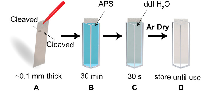
1. Functionalization of Mica Surface for Static AFM Imaging of Nucleosomes
- Prepare a 50 mM 1-(3-Aminopropyl) silatrane APS stock solution in deionized water as described. Store 1 mL aliquots of this solution at 4 °C until use.
NOTE: The aliquots can be stored for more than a year at 4 °C.
- Prepare a working APS 1:300 solution for mica modification by dissolving 50 µL of the 50 mM APS stock in 15 mL dd H2O.
NOTE: This working solution can be stored at room temperature for several days.
- Cut 1 x 3 cm strips of mica from high-quality mica sheets (see Table of Materials for mica used here).
- Check that the piece fits when placed diagonally in a cuvette. Use the tip of sharp tweezers, a razor blade, or scotch tape, to cleave layers of the mica until both sides are freshly cleaved and the piece is as thin as ~0.1 mm (Figure 1A). Immediately place the mica piece into the APS-filled cuvette and incubate for 30 min (Figure 1B).
- Transfer the mica piece to a cuvette filled with dd H2O and soak for 30 s (Figure 1C). Completely dry both sides of the APS-mica strip under an argon flow.
NOTE: A non-woven cellulose and polyester cleanroom wipe (recommended wipe detailed in materials) can be used to aid in wicking water from the edge of the mica when drying.
- Use the dry mica strip now for the sample preparation. Otherwise, store the piece in a clean, dry cuvette (Figure 1D).
NOTE: Additional storage in a vacuum for 1-2 h is recommended when the environment is humid. The protocol can be paused here.
2. Preparation of Nucleosome Samples on APS-Mica for Static AFM Imaging
- Apply double-faced adhesive tape to several magnetic pucks and place them to the side.
- Cut the APS-mica substrate to the desired size (1 x 1 cm squares for the MM AFM instrument used here). Place these pieces in a clean petri dish and keep them covered.
- Prepare three dilutions of the assembled nucleosomes (final nucleosome concentrations of 0.5, 1.0, and 2.0 nM) using a 0.22 µm filtered buffer containing 10 mM HEPES pH 7.5 and 4 mM MgCl2.
NOTE: To limit the loss of nucleosomes at the low final concentration, the dilutions should be done one at a time, immediately prior to deposition on the APS-mica.
- Deposit 5-10 µL of the diluted nucleosome sample at the center of the APS-mica piece, and let it incubate for two minutes. Gently rinse the sample with 2-3 mL of dd H2O to remove all buffer components. After each ~0.5 mL of dd H2O is used, gently shake the mica to remove the excess rinse water.
NOTE: A disposable syringe is recommended for this rinsing step.
- Dry the deposited sample under a light flow of clean argon gas.
NOTE: The sample is now ready to be imaged or can be stored in a vacuum cabinet or desiccator filled with argon. Samples prepared and stored as described have been imaged one year following preparation with no quality loss. The protocol can be paused here.
3. Static AFM Imaging of Nucleosomes
- Mount an AFM tip on the tip holder. Use a tip that has a spring constant of ~40 N/m and a resonance frequency between 300 and 340 kHz (see the Table of Materials for the cantilevers used here).
- Mount the sample prepared in section 3 on the AFM stage being careful not to contact the sample surface.
- Position the laser over the cantilever until the sum is at the maximum and adjust the vertical and lateral deflection values to near zero.
- Tune the AFM probe to find its resonance frequency and adjust the drive amplitude and set the image size to 100 x 100 nm. Click the engage button to begin the approach.
- Once approached, gradually optimize the Amplitude Setpoint until the surface of the sample is clearly seen. Increase the scan size to 1 x 1 µm and the resolution to 512 x 512 pixels. Click the capture button followed by the engage button to begin image acquisition.
NOTE: The images in Figure 2 show the smooth background that can be expected when imaging these samples.