All procedures involving animal models have been reviewed by the local institutional animal care committee and the JoVE veterinary review board.
1. Preparation of Media, Enzyme Solutions, and Animals
- Prepare 1% iodine alcohol by dissolving 1 g of iodine in 100 ml ethanol.
- Prepare 2x 500 ml Dulbecco´s phosphate buffered saline (PBS) without Ca2+ and Mg2+ supplemented each with 7.5 ml D-glucose (100 g/L) and 5 ml 100x Penicillin/Streptomycin stock solution (15 mg/ml D-glucose, 50 U/ml Penicillin and 50 µg/ml Streptomycin final).
- Supplement 1x 500 ml Roswell Park Memorial Institute (RPMI) 1640 medium with 5 ml 100x Penicillin/Streptomycin stock solution (50 U/ml Penicillin and 50 µg/ml Streptomycin final).
- Prepare a Trypsin-DNase I solution (2.5 mg/ml Trypsin and 10 µg/ml DNase I) by adding 25 mg Trypsin and 0.1 ml DNase I (1 mg/ml DNase I) to 9.9 ml PBS.
- Dissolve 50 mg Trypsin inhibitor in 5 ml PBS for Trypsin inhibitor A solution (10 mg/ml). Dissolve 25 mg Trypsin inhibitor in 10 ml PBS for Trypsin inhibitor B solution (2.5 mg/ml).
- For a Collagenase-Hyaluronidase-Deoxyribonuclease I (DNase I) solution dissolve 10 mg Collagenase A (1 mg/ml final), 10 mg Hyaluronidase (1 mg/ml final) and 0.1 ml DNase I (1 mg/ml DNase I) (10 µg/ml final) in 10 ml PBS.
- Prepare a Hyaluronidase-DNase I solution by adding 10 mg Hyaluronidase (1 mg/ml final) and 0.1 ml DNase I solution (1 mg/ml) (10 µg/ml final) to 9.9 ml PBS.
- Order Wistar rats on time so that they are 19 days old on the day of Sertoli cell isolation.
Note: The described protocol is designed for 10 rats but can be modified for a minimum of 5 and a maximum of 20 rats. The volumes of all solutions have to be adjusted accordingly.
In the following protocol a "pipette" stands for a serological pipette.
2. Preparation of Seminiferous Tubules
- Anesthetize 10 male Wistar rats one by one in a desiccator using CO2 and a flow rate that displaces at least 20% of the chamber volume per minute. Wait until breathing stops, test anesthesia by squeezing a foot pad, and if there is no reaction euthanize each rat by cervical dislocation. Drain the blood by sectioning the jugular vein and carotid artery (vagina carotica) under flowing water. Disinfect the abdomen by wiping it with 70% ethanol.
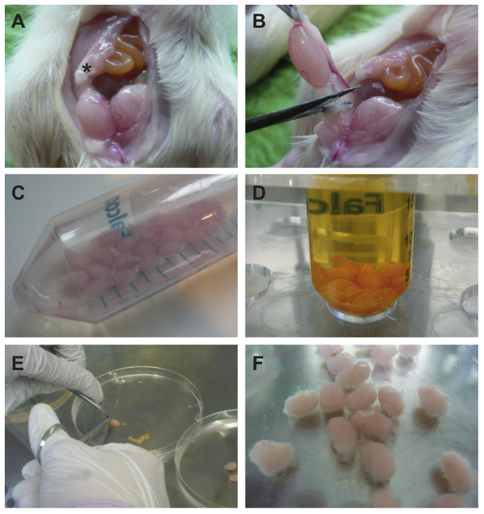
- Using forceps lift the skin from the abdominal muscles and cut out an oval-shaped skin lobe that is extending from the pubic symphysis to the sternum (Figure 1A) and flap it upwards onto the chest. Next, grab the abdominal muscles and make a medial incision from the pubic symphysis to the sternum.
- Squeeze the abdomen with the thumbs from the pelvis upwards in order to push the testes out of the lower pelvis. Pick the epididymal fat pad (Figure 1B) with forceps for pulling up the testis further. Cut the spermatic cord (Figure 1B), leaving the tunica albuginea intact, and collect all testes in 20 ml PBS in a 50 ml conical tube (Figure 1C).
- After collecting all testes, disinfect them by adding an equal volume (20 ml) of 1% (w/v) iodine in ethanol. Invert the tube twice and decant the supernatant immediately. Quickly wash the testes twice with 25 ml PBS each (Figure 1D).
- Transfer the testes to a Petri dish containing 15 ml PBS (Figure 1E). Grasp each testis firmly by forceps at one end, make a small incision into the tunica albuginea at the opposite end, and squeeze out the tubules using closed scissor blades as a scraping tool.
Note: The tubules should still form a compact testicle-shaped bundle with only a few tubules extending into the solution in order to enable homogeneous enzymatic digestion (Figure 1F).
- Transfer the de-capsulated testes to a 100 ml screw-cap bottle containing 10 ml Trypsin-DNase I solution. Perform the digestion in a shaking water bath at 32 °C (120 oscillations/min). After 4 min evaluate the tubules with the naked eye for dispersion of tubules. Once tubules start to disperse into the solution, stop the digestion immediately. If necessary, continue incubation for up to 2 min.
- Stop trypsin digestion by adding 5 ml of trypsin inhibitor solution A. Thoroughly mix the solution by pipetting up and down 3-4 times using a 10 ml pipette and transfer it to a 50 ml conical tube.
- After 5 min of incubation observe the tubules settle by unit gravity, carefully remove the supernatant by using the 10 ml pipette from step 2.6, and add 10 ml of trypsin inhibitor B solution in order to fully stop trypsin digestion. Resuspend the tubules by pipetting up and down 2-3 times.
- In order to remove contaminating interstitial cells wash the tubules 9 times with 25 ml PBS each. Resuspend tubules in each wash by using a 25 ml pipette and allow them to settle by unit gravity for 10 min. Always use the same 25 ml pipette for pipetting PBS, resuspension, and removal of the supernatant.
3. Removal/Isolation of Peritubular Cells (PTCs)
- Transfer all tubules to a 100 ml screw-cap bottle and add the Collagenase-Hyaluronidase-DNase I solution (Figure 2A). Perform digestion for 10 min in a shaking water bath at 32 °C (120 oscillations/min).
- Pipet a drop of the suspension on a glass slide and quickly check the tubules under an inverted light microscope for routine cell culture at 100x magnification. If digestion is not advanced enough continue incubation for an additional 2 min (do not count the time needed for the microscopical check).
Note: During this step all tubules get shortened in length and tubule edges should become rough (Figure 2B and C). Rough edges are indicative of the release of peritubular cells.
- Transfer the suspension with digested tubules to a 50 ml conical tube using the 25 ml pipette from step 2.8. Wash the 100 ml bottle with 10 ml PBS and add it to the tubules in the conical tube. Allow the digested tubules to settle by unit gravity for 10 min, and carefully aspirate the supernatant, containing the PTCs, using the 25 ml pipette again, and transfer it to a new conical tube. For aspiration of the last few milliliters use a 5 ml pipette in order to prevent any carry-over of tubules.
4. Isolation of Sertoli Cells (SCs)
- Resuspend the settled tubules from step 3.3 thoroughly in 25 ml of PBS by using a 25 ml pipette and allow them to settle by unit gravity for 12 min. Use the same 25 ml pipette for pipetting PBS, resuspension of tubules, and removal of supernatants. Repeat the wash 3 times (4 washes in total).
- After the last wash transfer the digested tubules to another 100 ml screw-cap bottle containing the Hyaluronidase-DNase I solution. Perform the digestion in a shaking water bath at 32 °C (120 oscillations/min).
- After 5 min check the tubules again by light microscopic inspection at 100X magnification as described in step 3.2.
Note: Short tubules, tubular aggregates, and released cells should be visible (Figure 2D).
- Allow the tubular aggregates to settle for 10 min, and aspirate the supernatant using a 25 ml pipette. Wash aggregates 4 times using 25 ml PBS and a sedimentation time of 10 min for each washing step (Figure 2E).
- Add 20 ml of RPMI 1640 medium and pass the suspension 10 times through an 18 G needle mounted on a 20 ml syringe. Avoid air bubbles.
Note: Pulling the suspension in and out through the needle is considered as one pass. The disruption and homogenization of cells by hydrodynamic shearing is a standard technique in the preparation of cells that tend to aggregate. The passing of cells through a hypodermic needle of small inner diameter is unlikely to have an impact on their functional properties. Figure 3C and D shows the normal ultrastructure of purified Sertoli cells on day 4 of culture.
- Pipet a drop of the suspension on a glass slide and quickly check the tubules under an inverted light microscope for routine cell culture at 10X magnification if the aggregates are all dispersed (Figure 2F). Filter the cell suspension through a 70 µm cell strainer in order to obtain a pure single-cell suspension in the filtrate.
- Centrifuge the filtrate of step 4.4 at 200 x g for 10 min at RT without using the break. Carefully aspirate the supernatant using a 25 ml pipette, and resuspend the Sertoli cells in 40 ml RPMI 1640 medium (serum-free).
- Mix 100 µl cell suspension with 100 µl of Trypan Blue Stain and count cells with a Neubauer Improved chamber. Adjust the cell concentration to 3 x 10⁶ cells/ml according to the counting result. Seed 1 ml per well on a 6-well plate (=day 1). Note: Sertoli cells in suspension settle quickly so the tube should be briefly shaken every time before a cell aliquot is taken with the pipette. Until day 4 they firmly attach to the substratum.