Een abonnement op JoVE is vereist om deze inhoud te bekijken. Log in of start vandaag met uw gratis proefperiode.

Methodenartikel

Mesenteric Lymph Duct Cannulation in a Rat Model to Harvest Lymph Fluid

2.7K weergaven

8 juli 2025

In dit artikel

Samenvatting

Source: Trevaskis, N. L., et al. The Mesenteric Lymph Duct Cannulated Rat Model: Application to the Assessment of Intestinal Lymphatic Drug Transport. J. Vis. Exp. (2015).

This video demonstrates a technique to cannulate the mesenteric lymph duct in rats. This method enables direct access to the entire volume of components present in the lymph fluid.

Protocol

All procedures involving animal models have been reviewed by the local institutional animal care committee and the JoVE veterinary review board.

1. Cannulation of the Mesenteric Lymph Duct

  1. Place the animal with its right side facing toward the operator. Perform the surgery with or without the assistance of a surgical microscope as required.
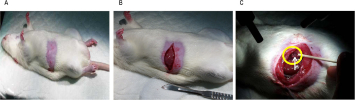
  2. Open the top layer of the abdominal muscle wall with a straight 4 cm incision extending from the midline (xiphoid process) to the right flank approximately 2 cm below the ribcage (costal margin) using a sterile scalpel blade (see Figure 1B).
  3. Open the remaining layers of the abdominal muscle wall from 4 - 5 mm lateral to the midline to the right flank with a small pair of surgical scissors (see Figure 1B).
  4. Retract the small intestine under the left abdominal muscle wall and keep it in place using 2 - 3 pieces of sterile gauze saturated with normal saline.
  5. Bridge the rat over a 10 ml plastic syringe placed horizontally under the rat's back at the level of the right kidney to ease visualization of the mesenteric lymph duct.
  6. Locate the superior mesenteric lymph duct: a vessel approximately 0.5 - 1 mm in diameter that is perpendicular to the right kidney and immediately rostral and parallel to the mesenteric artery (a pulsating dark red blood vessel; see Figure 1C).
    NOTE: In non-fasted or oil-pre-dosed rats the vessel is white, opaque, and easier to visualize than in fasted rats in which it is quite translucent.
  7. Inspect the area immediately caudal to the large superior mesenteric lymph duct and the mesenteric artery to determine if a second, smaller accessory lymph duct is present. If present, block the flow of lymph through the duct by severing the duct and occluding with superglue, or tying a suture around the duct, to ensure that the entire volume of lymph flowing from the intestine is collected from the superior mesenteric lymph duct.
  8. Pass a pair of straight forceps through the peri-renal fat bed at the lower margin of the right kidney, and through the connective tissue layers immediately below the vena cava, in a direction parallel with the superior mesenteric lymph duct.
  9. Using the tip of the forceps take hold of the lymph cannula and pull it through the peri-renal fat bed with one end immediately adjacent to the mesenteric lymph duct and the other exteriorized from the animal at the level of the right kidney.
  10. Isolate the mesenteric lymph duct from overlying layers of connective and fat tissue by blunt dissection. Take care not to pierce or damage the lymph duct.
  11. After isolating the lymph duct, make a small hole in the lymph duct with either micro-scissors, the sharp tip of the jeweler's forceps, or a 25 G needle. Take care not to completely sever the vessel.
  12. Ensure that the lymph cannula is completely filled with an anti-coagulant solution (using a syringe and 25 G needle) with no air gaps. Insert the lymph cannula approximately 2 - 4 mm inside the mesenteric lymph duct via the small hole with the aid of a small pair of forceps.
  13. Observe the lymph cannula for a couple of minutes. Observe a gradual flow of intestinal lymph from the free-collecting end of the cannula if the cannulation is successful. If the cannulation is not successful, repeat steps 1.8 - 1.12.
  14. If the cannulation is successful, secure the cannula by placing a small drop of veterinary adhesive over the entrance hole into the lymph duct. Take care not to occlude the vessel via the application of excess veterinary adhesive.
  15. Whilst waiting for the veterinary adhesive to set, observe the flow of lymph for several minutes to ensure that the cannulation procedure was successful. Once certain of the success of the cannulation, remove the gauze pieces that were placed in the abdominal cavity carefully and place the small intestine in its original position.
  16. Pre-weigh lymph collection tubes, label tubes for lymph and blood sample collection and add anti-coagulant solution. Add sufficient anti-coagulant solution to achieve a final concentration of 1 mg/ml EDTA in sterile water or 10 - 20 IU/ml heparin in the collected lymph or blood.
  17. Place a lymph collection tube containing an anti-coagulant (step 1.16) at the end of the cannula to collect the free-flowing lymph.
  18. Next, cannulate the duodenum for infusion of rehydration solutions and/or formulations.

Toegang beperkt. Log in of start een proefperiode om deze inhoud te bekijken.

Resultaten

Rodent surgical procedure, wound healing experiment; step-by-step incision and biopsy diagram.
Figure 1. Photographs of (A) the abdomen shaved in preparation to cannulate the lymph duct, (B) the abdominal muscle wall opened with a straight 4 cm incision extending from the midline to the right flank to access the lymph ...

Toegang beperkt. Log in of start een proefperiode om deze inhoud te bekijken.

Openbaarmakingen

No conflicts of interest declared.

Materialen

Lijst van materialen gebruikt in dit artikel
NaamBedrijfCatalogusnummerOpmerkingen
Sterile salineBaxter healthcareAHB 1307Any brand can be used. Example here is Baxter 100 ml saline bags, box of 50
70 % ethanol in waterAnyAny brand can be used
Chlorhexidine gluconate solution (Microshield 4)Livingstone InternationalJJ60243LAny brand can be used. http:// www.livingstone.com.au/? PG=search_result&CAT=6&search =JJ60243L
Betadine solutionLivingstone InternationalBU0510Any brand can be used. http:// www.livingstone.com.au/? PG=search_result&CAT=6&search =BU0510
Ilium Ketamil (Ketamine 100 mg/ ml)PROVET VICTORIAKETA I 1http://www.provet.vom.au
Heparin (35000I.U. in 35 mLSigmaPharmaceuticals337220http://www.sigmaco.com.au
Polyethylene (PE) cannula o.d. 0.96 mm x i.d. 0.58 mmMicrotube extensionsPE8050Any brand can be used. Example here is PE tubing 0.96 x0.58 mm, 30 m
Polyethylene (PE) cannula o.d. 0.8
mm x i.d. 0.5 mm
Microtube extensionsPE9658Any brand can be used. Example here is PE tubing 0.8 x0.5 mm, 30 m
MarkersAnyAny brand can be used
Veterinary adhesiveAnyAny brand can be used
23 gauge needlesLivingstone InternationalDN23GX0.75LVAny brand can be used. Example here is Livingstone Disposable Needle, Sterile, 23GX0.75inch, 100/BOX. http:// www.livingstone.com.au/? PG=search_result&CAT= 6&search=DN23GX0.75LV
25 gauge needlesLivingstone InternationalDN25GX1.0LVAny brand can be used. Example here is Livingstone Disposable Needle, Sterile, 25GX1.0inch, 100/BOX. http:// www.livingstone.com.au/? PG=search_result&CAT=6&search= DN25GX1.0LV
1 ml syringeLivingstone InternationalT3SS01TAAny brand can be used. Example here is Terumo syringe 1 ml Slip Tuberculin 100/Box. http:// www.livingstone.com.au/? PG=search_result&CAT=6&search =T3SS01TA
10 ml syringeLivingstone InternationalT3SS10SAAny brand can be used. Example here is Terumo syringe 10 ml Slip 100/Box. http:// www.livingstone.com.au/? PG=search_result&CAT=6&search =T3SS10SA
Gauze swabsLivingstone InternationalGSC075Any brand can be used and cut to required size. Example here is gauze swabs cotton filled 7.5x7.5 cm, 8 ply. http:// www.livingstone.com.au/? PG=search_result&CAT=6&search =GSC075
Heating padRatekWT1Any brand that keeps temperature at 37C can be used. Example here is Ratek warming tray.
Surgical microscopeZeiss495005-0014-000Any brand can be used. Example here is Zeiss Stereomicroscope Stemi 2000-C with Stand S Double Spot and KL 300 LED. https:// www.micro-shop.zeiss.com/? l=en&p=us&f=e&i=10143
1 x Small surgical scissorsFine Science Tools (FST)14060-09Any brand can be used. Example here is FST Fine Scissors, 9 cm with 21 mm cutting edge, sharp, straight. http://www.finescience.ca/ Special-Pages/Products.aspx? ProductId=40&CategoryId=17
2 x Forceps with serrated curved tipFine Science Tools (FST)11001-13Any brand can be used. Example here is FST 13 cm standard pattern forceps with curved 2.8x1.4 mm tip. http://www.finescience.ca/ Special-Pages/Products.aspx? ProductId=405&CategoryId=32

Trefwoorden

Oogsten van lymfevloeistofcanulatie bij ratmodelchirurgische canulatietechniekantistollingsoplossingperirenaal vetbedarteria mesentericainbrengen van lymfecanuleveterinair plakmiddeltransport van intestinale lymfe