Een abonnement op JoVE is vereist om deze inhoud te bekijken. Log in of start vandaag met uw gratis proefperiode.

Methodenartikel

Fluorescein Staining in an Autoimmune Dry Eye Rat Model to Examine Corneal Damage

1.8K weergaven

⸱

8 juli 2025

In dit artikel

Samenvatting

Source: Hou Ahiua, et al. A Chronic Autoimmune Dry Eye Rat Model with Increase in Effector Memory T Cells in Eyeball Tissue. J.Vis. Exp. (2017)

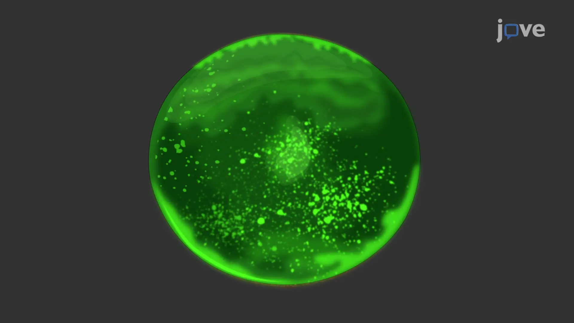
This video demonstrates the staining of the damaged cornea using fluorescein dye to evaluate the extent of autoimmune dry eye disease in a rat model. The green fluorescence spots of the stained collagen fibers indicate corneal epithelial damaged cells.

Protocol

All procedures involving animal models have been reviewed by the local institutional animal care committee and the JoVE veterinary review board.

1. Assessment of Dry Eye Features

Measure corneal damage with fluorescein staining.

  1. Add 2 µL of 0.2% fluorescein to the rat cornea. Passively open and close the rat eyelids 3 times with a gloved finger to spread the fluorescein dye on the surface of the eye.
  2. After 1 min, draw 1 mL of saline with a 3 mL syringe (without any needle attachment), position the syringe about 2-3 mm anterior to the cornea, and gently propel saline onto the eye.
  3. Acquire images under the cobalt blue filter (i.e., ~400 nm) of an ocular-imaging microscope with the background lights switched off.
    NOTE: Images were subsequently analyzed by a grading system modified from previous publications. Images were then analyzed by subjectively grading the number, area, and intensity of the green spots from 0 to 2, where 0 indicates their absence, 1 indicates punctate staining of less than 50 spots, and 2 indicates punctate staining of more than 50 spots.
Toegang beperkt. Log in of start een proefperiode om deze inhoud te bekijken.

Openbaarmakingen

No conflicts of interest declared.

Materialen

Lijst van materialen gebruikt in dit artikel
NaamBedrijfCatalogusnummerOpmerkingen
Reagent
Fluorescein sodium solutionBausch & Lomb U.K LimitedNA
Equipment
Stereo microscope with ring light illuminator and cameraCarl ZeissNA

Meer artikelen verkennen

Fluoresceïne-kleurstofOculaire BeeldvormingKobaltblauw FilterGroene FluorescentieCollageenkleuringEpitheliale Beschadiging