Method Article

Detection of Vitronectin Binding to Bacterial Surfaces using Flow Cytometry

July 8th, 2025

In This Article

Abstract

Loading...
$$\rightleftharpoonup{xx}$$ $$\longleftharp{xx}$$, $$\longrightharp{xx}$$,

Source: Singh, B. et al., Assays for Studying the Role of Vitronectin in Bacterial Adhesion and Serum Resistance. J. Vis. Exp. (2018)

This video demonstrates the detection of vitronectin's interaction with bacterial surface proteins. The flow-cytometry analysis measures the fluorescence signal of fluorescently labeled antibodies bound to vitronectin, confirming its presence at the bacterial surface. This technique provides insights into host-pathogen interactions and potential bacterial infection therapies.
Protocol

Loading...
$$\rightleftharpoonup{xx}$$ $$\longleftharp{xx}$$, $$\longrightharp{xx}$$,

1. Analysis of Vitronectin, Vn as a Bacterial Surface Protein-Ligand

  1. Detection of Vn-binding at the bacterial surface using flow cytometry
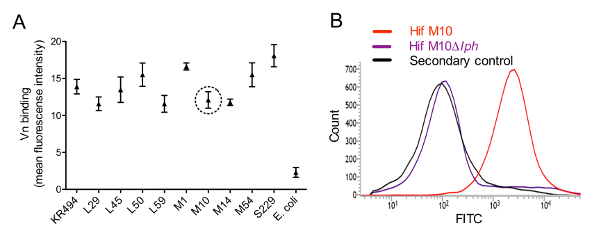
    NOTE: In flow cytometry, we used side scatter and forward scatter to gate positive events. To examine the interactions with Vn, Haemophilus influenzae type f, Hif clinical isolates (n=10) were selected together with E. coli BL21 (DE3) as a negative control (Figure 1A).
    1. Culture Hif clinical isolates in brain-heart infusion (BHI) medium supplemented with 10 µg/mL nicotinamide adenine dinucleotide (NAD) and hemin at 37 °C with shaking at 200 rpm. Use Luria-Bertani medium to culture E. coli. Harvest Hif at mid-log phase (Optical density, OD600 = 0.3), and resuspend the bacteria in phosphate-buffered saline (PBS), pH 7.2, containing 1% (w/v) bovine serum albumin (BSA) (blocking buffer; PBS-BSA). Adjust the suspension to 109 colony-forming units, CFU/mL.
    2. Transfer the aliquots containing 5 x 10⁶ CFU to 5 mL polystyrene round-bottom tubes (12 x 75 mm2) and add 1 mL of blocking buffer. Centrifuge the suspension at 3,500 × g at room temperature (RT) for 5 min to pellet the bacteria, then carefully aspirate to remove the supernatant without disturbing the pellet.
    3. Resuspend the bacterial pellets with 50 μL of blocking buffer containing 250 nM Vn and incubate the samples for 1 h at RT without shaking. After incubation, pellet the bacteria by centrifugation at 3,500 x g for 5 min, then wash the pellets three times using PBS and similar centrifugation steps.
    4. To the bacterial pellet, add 50 μL of primary sheep anti-human Vn polyclonal antibodies (pAbs) at 1:100 dilution in PBS-BSA. Incubate the suspension for 1 h at RT, then wash the bacteria three times with PBS to remove unbound antibodies (as described in step 1.1.3).
    5. Next, add 50 μL of PBS-BSA containing fluorescein isothiocyanate (FITC)-conjugated donkey anti-sheep pAbs (1:100 dilution) and incubate at RT for 1 h in the dark.
    6. Prepare a blank control by incubating bacteria with blocking buffer only and pAbs without Vn.
    7. Wash bacteria three times with 1 mL of blocking buffer and pellet the suspension by centrifugation, as described in section 1.1.2. Finally, resuspend the bacterial pellet with 300 µL of PBS and analyze it by flow cytometry.

Access restricted. Please log in or start a trial to view this content.

Results

Loading...
$$\rightleftharpoonup{xx}$$ $$\longleftharp{xx}$$, $$\longrightharp{xx}$$,

Fluorescence intensity graphs for Vn binding analysis and FITC flow cytometry comparison with control.
Figure 1: Haemophilus influenzae serotype f binds Vn via surface-expressed PH. (A) Flow cytometry results showing binding of Vn to cells of Hif clinical isolates. Each clinical isolate (5 x 10⁶ CFU...

Access restricted. Please log in or start a trial to view this content.

Disclosures

Loading...
$$\rightleftharpoonup{xx}$$ $$\longleftharp{xx}$$, $$\longrightharp{xx}$$,
No conflicts of interest declared.

Materials

List of materials used in this article
NameCompanyCatalog NumberComments
5 mL polystyrene round-bottom tube BD Falcon60819-13812 × 75 mm style
Bovine Serum Albumins (BSA)Sigma-AldrichA2058Suitable for cell culture
Flow cytometer BD Biosciences651154Cell analysis grade for research applications
Vitronectin (Vn) from human plasmaSigma-AldrichV8379-50UGCell culture grade
E. coli host (E. Coli BL21)Novagen69450-3Protein expression host
FITC-conjugated donkey anti-sheep antibodies AbD SerotecSTAR88FPolyclonal
Sheep anti-human Vn antibodiesAbD SerotecAHP396Polyclonal

Reprints and Permissions

Request permission to reuse the text or figures of this JoVE article

Request Permission

Tags

Vitronectin BindingFlow CytometryBacterial SurfaceAnti vitronectin AntibodiesFluorescence SignalWild type BacteriaMutant StrainProtein HSecondary AntibodiesCentrifugation Wash

Related Articles