1. Preparation
- Cell Plating
Note: Human cervical epithelial cells, HeLa and HeLa expressing Histone 2B-GFP (H2B-GFP), were used in this protocol, but this assay can be adapted to other mammalian cells.
- Detach adherent cells from a 75 cm2 cell culture flask by washing the cells with 2 mL of Trypsin-EDTA 0.25%. Replace the used trypsin with 2 mL of fresh trypsin-EDTA 0.25%.
- Incubate the cells at 37 ˚C for 5 min until the cells have rounded and detached from the flask.
- Resuspend the cells in 8 mL of growth medium (DMEM containing 10% heat-inactivated fetal bovine serum, 100 U/mL penicillin, and 100 µg/mL streptomycin).
- Determine the cell concentration using a hemocytometer and 10 µL of cell suspension.
- Dilute the cells in a growth medium to a concentration of 2.5 x 10⁵ cells/mL.
- Pour the cell suspension into a sterile pipette basin and thoroughly mix the suspension using a 10 mL serological pipette.
- Using a 12-multichannel micropipette and 200 µL tips, distribute HeLa cells (2.5 x 10⁴ cells/100 µL/well) in triplicate (or quadruplicate) in a 96-well flat, clear bottom, black polystyrene tissue culture-treated plate.
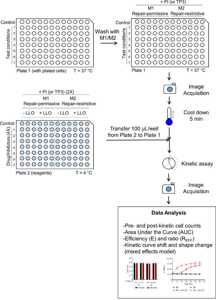
Note: A plating arrangement is presented as an example in Figure 1.
- Culture the cells for 24 h in a humidified cell culture incubator at 37 ˚C and 5% CO2.
- Stock Solution Preparation
- Prepare 1 L of a 10x stock of buffer M (used to prepare M1 and M2) by adding 95 g of Hanks Balanced Salt Solution, 0.476 g of MgCl2 (5 mM), and 23.83 g of HEPES (100 mM) to 900 mL of water. Adjust the pH to 7.4 and raise the volume to 1 L. Filter sterilize.
- Prepare 50 mL of a 50x (1.25 M) stock of glucose by adding 11.26 g of D-(+)-Glucose to a total of 50 mL of water. Filter sterilize the solution.
- Prepare 50 mL of a 100x (120 mM) stock of calcium by adding 0.666 g of CaCl2 to a total of 50 mL of water. Filter sterilize the solution.
- Prepare 50 mL of a 10x (50 mM) stock of ethylene glycol-bis(2-aminoethylether)-N,N,N',N', tetraacetic acid (EGTA) by adding 0.951 g of EGTA to 40 mL of water. Increase the pH to 8 using NaOH to dissolve the EGTA, then raise the volume to 50 mL. Filter sterilize the solution.
- For a single 96-well plate, prepare 50 mL of Medium 1 (M1, contains Ca2+), 50 mL of Medium 2 (M2, Ca2+-free), and 15 mL of Medium 2 supplemented with EGTA, accordingly:
- For M1, add 5 mL of 10x Buffer M, 0.5 mL of 100x CaCl2, and 1 mL of 50x glucose to 43.5 mL of water.
- For M2, add 5 mL of 10x Buffer M and 1 mL of 50x glucose to 44 mL of water.
- For M2/EGTA, add 1.5 mL of 10x Buffer M and 1.5 mL of 10x EGTA to 12 mL of water.
Note: All solutions containing propidium iodide (PI) should be prepared directly prior to adding to the cells.
- Plate Reader/Imaging Cytometer Settings
Note: Use a multi-mode plate reader equipped with two detection units: a spectrofluorometer and an imaging cytometer. Limit the fluorescence exposure to avoid photobleaching the fluorophores.
- Pre-warm the plate reader to 37 °C before performing the assay.
- Set up the parameters for the kinetic assay accordingly within the Settings mode:
- Choose Monochromator, FL (fluorescence), and Kinetic for the optical configuration, read modes, and read type, respectively.
- Under Wavelength Settings, select a 9 and 15 nm excitation and emission bandpass, respectively. For assays using propidium iodide (PI), set the excitation and emission wavelengths to 535 and 617 nm, respectively.
- Under Plate Type, select 96 Wells for the plate format and a pre-set plate configuration corresponding to a black-wall clear bottom plate.
- Under Read Area, highlight the wells that will be analyzed throughout the kinetic.
- Under PMT and Optics, preset the flashes per read to 6 and check the box for Read from Bottom.
- Under Timing, insert 00:30:00 in the Total Run Time box for a 30-minute kinetic assay, and insert 00:05:00 for the Interval.
Note: For each time point and one wavelength, the reading time of a full 96-well plate is 30 s.
- Confirm the specified settings in the Settings Information to the right and select OK. Press Read to initiate the kinetic run.
- Set up the imaging parameters accordingly within the Settings mode:
- Choose Minimax, Imaging, and Endpoint for the optical configuration, read modes, and read type, respectively.
- Under Wavelengths, select transmitted light, and either or both the fluorescence boxes corresponding to excitation and emission wavelengths of 456/541 nm (GFP) and 625/713 nm (PI).
- Use the same options for the Plate Type and Read Area as defined in steps 1.3.2.3 and 1.3.2.4.
- Under Well Area Setting, select the number of sites within a well to be imaged.
Note: 12 sites correspond to a full-well image.
- Under the Image Acquisition Settings, select the exposure times for transmitted light, 541 (GFP), and 713 (PI). For GFP, image the entire well with an exposure time of 20 ms/image. For transmitted light (TL) and PI fluorescence, acquire a single image of the center of each well with exposure times of 8 and 20 ms, respectively.
- Confirm the specified settings in the Settings Information to the right and select OK. The acquisition time for imaging the entire surface of each well (12 images/well) of a 96-well plate and for one wavelength is ~15 min. Press Read to initiate imaging.
Note: The acquisition time of a single image/well of a 96-well plate requires ~2.5 min/plate for one wavelength. The parameters described above correspond to the specific equipment in our laboratory. Spectrofluorometric measurements: A xenon flash lamp displaying 1.0 nm increment excitation wavelengths (250-850 nm) with an adjustable 9 or 15 nm bandpass, a photomultiplier tube detector with a > 6 log dynamic range, and an adjustable 15 or 25 nm emission bandpass. Imaging cytometer: An illumination light source capable of white light, 460 nm, and 625 nm excitation wavelengths with a 20 nm bandpass, emission filters centered at 541 nm (108 nm bandpass) and 713 nm (123 nm bandpass), respectively, and a 4X objective coupled to a 1.25 megapixel 12-bit charge-coupled device camera.
2. Assay
Note: At the time of the assay, cells must be 70-90% confluent. During the wash steps, the medium should be removed from and applied to the side wall of the well (not directly above the cells). Maintain the temperature of LLO at < 4 ˚C to prevent its aggregation until step 3.1.5.
- Prepare a stock of 30 µM PI in M1 and a stock of 30 µM PI in M2 pre-warmed at 37 ˚C.
- Gently wash the cells in plate 1 using a 12-multichannel micropipette and 200 µL tips, as follows:
- For repair-permissive conditions, remove the growth medium and wash the cells twice with 200 µL/well M1 pre-warmed at 37 ˚C. Replace the medium with 100 µL/well of warm M1 containing 30 µM PI.
- For repair restrictive conditions, remove the growth medium and wash the cells once with 200 µL/well warm M2 containing 5 mM EGTA to chelate Ca2+, followed by one wash with 200 µL/well M2. Replace the medium with 100 µL/well-warm M2 containing 30 µM PI.
- After the growth medium has been washed and replaced with medium containing propidium iodide, directly move to step 2.1.3.
- Image plate 1 under transmitted light, GFP, and PI as detailed under 1.3.3 (pre-kinetic). This step takes 15-20 min.
- During the 15-minute period in step 2.1.3, prepare plate 2 using a 12-multichannel micropipette and 200 µL tips as follows:
- Place a 96-well round bottom polypropylene microplate on ice. Configure the plate using an experimental design corresponding to plate 1 (Figure 1).
- For repair-permissive conditions, add 100 µL/well of ice-cold M1 containing 60 µM PI, followed by the addition of 100 µL/well of ice-cold M1 containing 4x LLO or not for the control.
- For repair-restrictive conditions, add 100 µL/well of ice-cold M2 containing 60 µM PI, followed by the addition of 100 µL/well of ice-cold M2 containing 4x LLO or not for the control.
- After imaging plate 1 (step 2.1.3), immediately place it on ice, using aluminum foil to separate the plate from direct contact with ice. Allow plate 1 to cool down for 5 min.
- Using a 12-multichannel micropipette and 200 µL tips, transfer 100 µL from each well into plate 2 (step 2.1.4) to the corresponding wells in the plate 1. To properly distribute the toxin in the media of plate 1, insert the tips below the meniscus and gently eject the volume without introducing bubbles.
Note: Do not pipette up and down, as this may inadvertently detach the cells.
- Leave the plate for an additional 1 min to allow the toxin to bind to the cells and immediately transfer plate 1 to the plate reader for the kinetic assay using the spectrofluorometer mode (step 1.3.2).
- At the end of the kinetic assay, immediately image plate 1 (post-kinetic) using step 1.3.3.
3. Analysis: Cell Enumeration
- Determine the cell count based on the nuclear fluorescence using the microplate cell enumeration software.
- Within Settings, select Re-analysis, and under the category section within the Image Analysis Settings select Discreet Object Analysis using 541 as the wavelength for finding objects.
- Within the Find Objects option, using the Draw on Images finding method, select Nuclei under the settings tab, and press Apply.
- Press OK and Read to initiate the cell counting algorithm.