All procedures involving animal models have been reviewed by the local
institutional animal care committee and the JoVE veterinary review board. All procedures involving sample collection have been performed in accordance with the institute's IRB guidelines.
1. Generation of viral Ag-specific CD8+ T cells from iPSCs (iPSC-CD8+ T cells)
- Creation of the retroviral constructs
NOTE: T-cell receptor (TCR) α and β genes are linked with 2A self-cleaving sequence. The retroviral vector MSCV-IRES-DsRed (MiDR) is DsRed+.
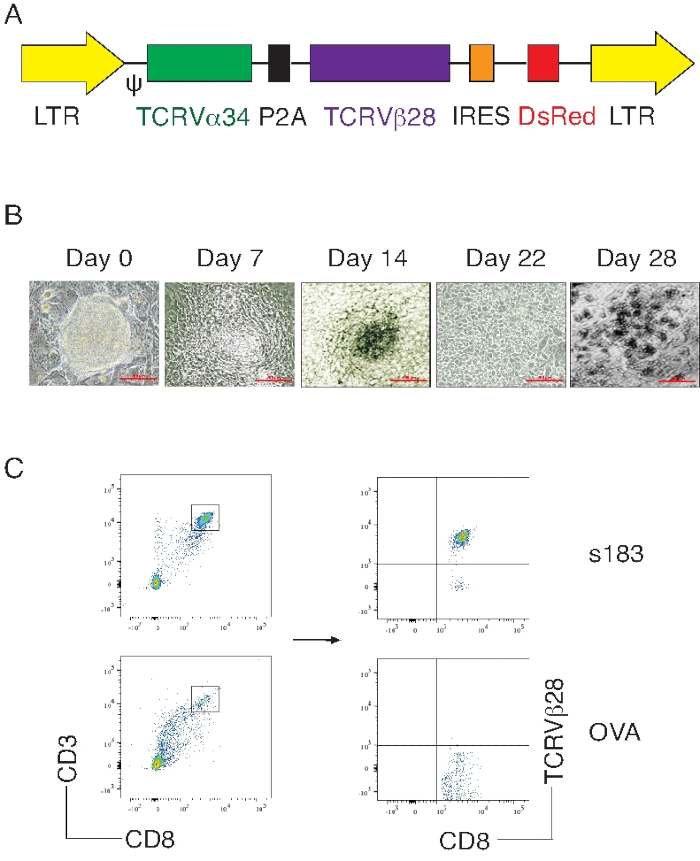
- Sub-clone HBs183-191 (FLLTRILTI)-specific A2-restricted human-murine hybrid TCR (s183 TCR) genes (Vα34 and Vβ28) into the MiDR to create the s183 MiDR construct (Figure 1A).
- Retroviral transduction
NOTE: The Platinum-E (Plat-E) cells are used for packaging retroviruses (carrying s183 TCR genes), which will be used for retroviral transduction. The Plat-E cells are effective retrovirus packaging cells underlying the 293T cells, which were developed by means of unique packaging constructs via the EF1α promoter to express retroviral structure protein, including gag, pol, and ecotropic env.
- On a 100 mm culture dish, seed 3 x 10⁶ Plat-E cells in 8 mL of DMEM culture medium containing 10% fetal calf serum (FCS) in an incubator at 37 °C with 5% CO2, 1 day before transfection.
- On day 0, transfect the s183 MiDR construct into the Plat-E cells using a DNA transfection reagent.
- On day 1, seed 1 x 10⁶ iPSCs (green fluorescent protein [GFP]+) into a gelatin pre-coated culture plate.
- On days 2–3, collect retroviruses-containing supernatant from Plat-E cell culture to transduce iPSCs with the s183 TCR in the presence of 1,6-dibromohexane solution.
- On day 4, trypsinize the s183 TCR gene-transduced iPSCs, centrifuge at 400 x g for 5 min, and seed 3 × 10⁵ iPSCs in a 100 mm culture dish pre-coated with 3 x 10⁶ irradiated SNL76/7 (irSNL76/7) feeder cells.
- On day 5 or 6 of confluence, trypsinize the cells, centrifuge at 400 x g for 5 min, and process for cell sorting. Gating on live cells, sort GFP and DsRED double-positive cells (the s183 TCR gene-transduced iPSCs) using a high-speed cell sorter. Similar to step 1.2.5, co-culture the sorted cells on irSNL76/7 feeder cells for future use.
- Differentiation of HBV-specific iPSC-CD8+ T cells
NOTE: The OP9-DL1/DL4 stromal cells overexpress both Notch ligands DL1 and DL4, and co-culturing iPSCs with the iPSCs can promote Notch signaling-mediated T cell differentiation.
- Grow s183 TCR gene-transduced iPSCs (s183/iPSCs) in the OP9-DL1-DL4 cell monolayer in α-minimum essential medium (MEM) media containing 20% fetal bovine serum (FBS). Include murine FMS‐like tyrosine kinase 3 (Flt3) ligand (mFlt-3L; final concentration = 5 ng/mL) in the culture.
- On day 0, seed 0.5–1.0 x 10⁵ S183/iPSCs in a 10 cm culture dish previously grown with OP9-DL1-DL4 cells. Validate that the OP9-DL1-DL4 cells are in a condition of 80%–90% confluency.
- On day 5, rinse the iPSCs with 10 mL of phosphate-buffered saline (PBS), aspirate off the PBS, add this to 4 mL of 0.25% trypsin, and incubate in a 37 °C incubator for 10 min. Afterward, add a supplementary 8 mL of iPSC media to end trypsin digestion. Accumulate all the digestive solutions containing the cells and centrifuge at 400 x g for 5 min at 15–30 °C.
- Aspirate the supernatant and resuspend cells in 10 mL of iPSC media. Transfer the cell suspension to a new 10 cm Petri plate and incubate in an incubator for 30 min at 37 °C.
- After 30 min, collect the iPSC media containing the floating cells. Pass the cell suspension through a 70 μm cell strainer and calculate the cell number.
- Seed 5 x 10⁵ cells in the culture dish previously grown OP9-DL1-DL4 cells with a condition of 80%–90% confluent, as described in step 1.3.1.
NOTE: For T cell differentiation, each 2–3 days, the iPSC-derived cells need to re-seed with a fresh layer of the OP9-DL1-DL4 cells.
- Evaluation
- Morphological changes of differentiating iPSCs
- Observe cells under a microscope on various days.
NOTE: By day 5, colonies have mesoderm-like characteristics, exhibiting a classic spindle-shaped morphology resembling human dermal fibroblasts and sustained growth in vitro. By day 8, small round clusters of cells begin to appear.
- Analysis of differentiating iPSCs by flow cytometry
- On various days of co-culture, analyze iPSC-derived cells as described previously (Figure 1B,C).
- Functional analysis of differentiating iPSCs
- On day 28 of co-culture, collect iPSC-CD8+ T cells from cultures by harvesting the floating cells, trypsinize the leftover cells with 0.25% trypsin, re-suspend in 8 mL of iPSC media, centrifuge for 5 min at 400 x g at 15–30 °C, remove the media, then resuspend the cells in 10 mL of media.
- Keep the re-suspended cells in a fresh 10 cm dish in a 37 °C incubator for 30 min and assemble the floating cells. Then rinse the cells one time with a cold PBS.
- Incubate 3 x 10⁶ T cell-depleted splenocytes (CD4-CD8-) from spleens of H-2 class I knockout, HLA-A2.1-transgenic (HHD) mice with 5 µM s183 peptide (FLLTRILTI) in 200 μL of media at 4 °C for 30 min.
- Produce a mixture of iPSC-CD8+ T cells with splenocytes pulsed with s183 peptide (T cells: splenocytes = 1:4; use 0.75 x 10⁶ T cells). Incubate the mixture of cells at 37 °C in a CO2 incubator for 40 h. During the last 7 h, add 4 µL of diluted brefeldin A into the culture (final concentration of 1,000x, which will be diluted in 1x culture media) to block transport processes during cell activation.
- Stain the cells and perform flow cytometric analysis of intracellular Interferon-gamma (IFN-γ) as described previously (Figure 2).
2. Induction of HBV replication through hydrodynamic delivery of HBV plasmid
NOTE: pAAV/HBV1.2 construct was generated as described previously. The HBV 1.2 complete DNA is incorporated in the vector pAAV.
- Hydrodynamic deliveries of HBV plasmid through the tail vein
- Heat HHD mice using a heat lamp for 5 min in the cage in order to dilate the tail vein.
- Detain the animal by means of a restrainer, and clean mouse tails with 70% ethanol spray.
- Delivery of plasmid into the liver.
- Measure the mouse's body weight by using a measuring scale.
- Dilute 10 μg of HBV plasmid in the 8% equivalent of body mass PBS (e.g., 1.6 mL for a 20 g mouse). Load diluted plasmid in a 3 mL syringe with a 26 G × 1⁄2″ (0.45 × 12 mm) hypodermic needle.
- Locate one of the two lateral tail veins in the middle third of the tail and position the needle into either lateral vein. Administer the injection containing HBV plasmid through the tail vein within 3 - 5 s.
- Quantification of viremia from blood serum of infected mice
NOTE: HBV replication occurs from day 3–35 in the mouse serum. The DNA replication peaks on day 7 and reduces gradually. The HBV DNA is not cleared from the serum until day 35.
- Collection of blood serum from infected mice
- Collect approximately 0.1 mL of blood in a microcentrifuge tube from each mouse on 3, 5, 7, and 10 days post-infection by retro-orbital bleeding in a 1.5 mL microcentrifuge tube, then incubate at room temperature (RT) for 20 min.
- Centrifuge the sample at 6,000 x g for 15 min at 4 °C and collect the serum supernatant after centrifugation.
- Purify the DNA from the blood serum using a commercially available DNA extraction kit following the manufacturer's recommendations. Briefly, lyse the cells in a silica-based column, perform the washes, and extract DNA by adding 100% ethanol to the elution column and centrifuging at 6000 x g for 1 min. Elute the DNA in 50 µL of RNase-free water.
- Use 100 ng of HBV DNA from the elution for real-time PCR analysis. Use the following primers and probes: forward 5' TAGGAGGCTGTAGGCATAAATTGG 3'; reverse 5' GCACAGCTTGGAGGCTTGT 3'; probe 5' TCACCTCTGCCTAATC 3'.
- Use the HBV genome containing plasmid (pAAV/HBV1.2) for the standard curve and perform real-time PCR in a total volume of 10 μL (Figure 3).
- Set up the PCR reaction in a total volume of 10 μL, as shown in Table 1.
- Set up the PCR program in the thermocycler, as shown in Table 2. The programmed temperature transition rate is 20 °C/s for denaturation/annealing and 5 °C/s for extension. Measure the fluorescence at the end of the annealing phase for each cycle for real-time PCR monitoring.
3. Reduction of HBV replication by ACT of viral Ag-specific iPSC-CD8+ T cells
- Adoptive cell transfer (ACT)
- Differentiate s183/iPSCs (1.3.7) upon the OP9-DL1-DL4 stromal cells in the presence of mFlt-3L and mIL-7 for 8 days, as described in section 1.3.
- On day 22, collect iPSC-CD8+ T cells from the 10 cm plate with trypsin, then wash and resuspend each 10 cm plate in 10 mL of fresh media. Add the cells to a fresh 10 cm plate and return to the incubator for 30 min, as done in section 1.3. After 30 min, collect floating cells.
- Use a 70 μm nylon strainer to pass cells to eliminate cell clusters and count the cell number. Adapt the cells to a concentration of 1.5 x 10⁷ cells/mL in cold PBS solution and use a 70 μm nylon strainer to pass cells to eliminate cell clusters again if needed. Keep cells on ice until the ACT.
- Inject 200 μL cell suspension (3 x 10⁶ cells) into 4–6 week-old HHD mice through the tail vein.
- Induction of HBV replication
- On day 14 after cell transfer, perform the hydrodynamic delivery of HBV plasmid through the tail vein as described in section 2.1.
Table 1: PCR reaction volume
| DNA template | 2 ml |
| DNA master hybridization mixture ((Taq DNA polymerase, PCR reaction buffer, 10 mM MgCl2, and dNTP mixture) | 1 ml |
| 25 mM MgCl2 | 0.8 ml |
| 0.3 μM the probe | 3 ml |
| 5 μM of each primer | 3.2 ml |
| Total | 10 ml |
Table 2: PCR program
| Denaturation | Temperature
95 °C | Time
5 s |
| Annealing | 53 °C | 10 s |
| Extension | 72 °C | 20 s |
| 5 °C | |