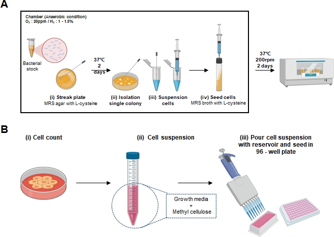
1. Bacterial cell cultures and preparation of Lactobacillus cell-free supernatant (LCFS)
NOTE: Steps 1.2 – 1.9 are conducted in an anaerobic chamber.
- Prepare a De Man–Rogosa–Sharpe (MRS) agar plate and broth containing L-cysteine and sterilize by autoclaving.
- Pre-incubate the MRS agar plate in H2 anaerobic chamber maintained at 37 ˚C with 20 ppm oxygen.
- Thaw Lactobacillus bacterial stock and inoculate the agar plate with the bacterial culture (Figure 1A (i)).
- Incubate bacteria for 2 - 3 days in H2 anaerobic chamber at 37 ˚C and 20 ppm oxygen until single bacterial colonies are obtained.
- Wash and dry the Hungate type anerobic culture tube. Autoclave the culture tube at 121 ˚C for 15 min.
- Then incubate the tube in H2 anaerobic chamber at 37 ˚C and 20 ppm oxygen to remove oxygen.
- Place 2 - 3 mL of MRS broth into the tube. Seal the tube with a butyl rubber stopper and screw the cap.
- Obtain a single colony with a loop and place it into the 1.5 mL culture tube with 500 µL of 1x PBS. (Figure 1A (ii)).
- Suspend the colony using a 1 mL syringe (Figure 1A (iii)). Do this by, inserting the needle of the 1 mL syringe in the center of the tube lid, aspirating the suspended colony and then resuspending it back into the MRS broth media. (Figure 1A (iv)).
- Incubate the MRS broth media in a shaker incubator for 2 days (37 °C, 5% CO2, 200 rpm).
- Measure the optical density (OD) using a spectrophotometer to monitor bacterial growth curves until the absorbance at OD620 reaches to 2.0.
- Separate the bacterial pellets and the conditioned media by centrifuging at 1,000 x g for 15 min. Wash the collected bacterial pellets with 1x PBS and resuspend in 4 mL of RPMI 1640 supplemented with 10% fetal bovine serum. Do not include any antibiotics in the medium.
- Maintain the bacterial pellets in RPMI and incubate in a shaker incubator for 4 h at 37 °C with 5% CO2 at a speed of 100 rpm.
- For the preparation of the probiotic supernatant, remove the bacterial pellet via centrifugation at 1000 x g, for 15 min at 4 °C. Sterile-filter the recovered supernatant using a 0.22 μm filter and store at −80 °C until use.
2. Generation of spheroids
- Preparing colorectal cancer cell lines
- Grow DLD-1, HT-29, and WiDr cell lines as monolayers until 70-80% confluency and incubate the plate at 37 °C in a 5% CO2 incubator (Growth medium: RPMI containing 10% fetal bovine serum (FBS) and 1% penicillin-streptomycin).
- For cells grown in 100 mm Petri dish, wash the plate twice with 4 mL of 1x PBS. Add 1 mL of 0.25% trypsin-EDTA and incubate the Petri dish for 2 min at 37 °C in a 5% CO2 incubator to dissociate the cells.
- After incubation, check for the cell dissociation under a microscope and neutralize trypsin-EDTA with 5 mL of growth medium.
- Transfer the dissociated cells to a 15 mL conical tube and centrifuge for 3 min at 300 x g.
- Discard the supernatant and resuspend gently with 3 mL of growth media.
- Count the cells with trypan blue to determine viable cells using a hemocytometer. (Figure 1B (i))
- Spheroid formation
- In a 15 mL conical tube, dilute the cells from 2.1.5 to obtain 1 - 2 x 10⁵ cells/mL (Figure 1B (ii))
- Add final concentration of 0.6% methylcellulose to the cell suspension and transfer the diluted cells to a sterile reservoir.
NOTE: For each cell line, the amount of methylcellulose needed should be titrated and determined accordingly.
- Use a multichannel pipette to dispense 200 µL of cells to each well of an ultra-low attachment 96-well round bottom microplate. (Figure 1B (iii))
- Incubate the plate at 37 °C in a 5% CO2 incubator for 24 - 36 h.
- After 24 - 36 h, observe the plate under a light microscope to ensure spheroid formation.
3. Treating 3D colorectal cancer cells with LCFS
- Generate spheroids as described in steps 2 and 3.
- Before performing the LCFS treatment, thaw the frozen LCFS at room temperature (RT) for 10 - 20 min.
- Inoculate the LCFS stock solution into a growth medium. Serially dilute to 25%, 12.5%, and 6% in the growth medium (i.e., 25% LCFS = 150 µL of growth medium + 50 µL of LCFS).
- Take out the cell culture plate containing spheroids from the incubator and remove as much of the growth medium as possible from each well using a 200 μL pipette.
- Add the growth media with LCFS on the cells and incubate at 37 °C in a 5% CO2 incubator for 24 - 48 h.
NOTE: The volume to be used will depend on the plate size as follows: 2 mL for 6-well cell culture plates; 200 μL for 96-well cell culture plates.