Methodenartikel

Direct Reprogramming of Hematopoietic Progenitor Cells into Neural Stem Cells

8 juli 2025

In dit artikel

Samenvatting

Loading...
$$\rightleftharpoonup{xx}$$ $$\longleftharp{xx}$$, $$\longrightharp{xx}$$,

Source: Wang, T., et al. Direct Induction of Human Neural Stem Cells from Peripheral Blood Hematopoietic Progenitor Cells. J. Vis. Exp, (2015)

This video demonstrates the direct reprogramming of genetically modified hematopoietic progenitor cells into neuronal stem cells. Transcription factors expressed by hematopoietic progenitor cells trigger a cascade of molecular events facilitating their transformation. Further specific media is used to select and grow induced neuronal stem cells.

Protocol

Loading...
$$\rightleftharpoonup{xx}$$ $$\longleftharp{xx}$$, $$\longrightharp{xx}$$,

1. Neural stem cell induction

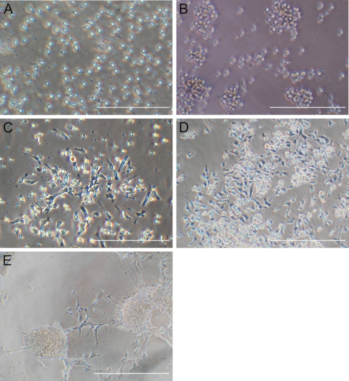
NOTE: Depending on cell conditions, about 5 or 7 days after transfection, monolayer adherent cells will reach 30 - 50% confluence (Figure 1C).

  1. Remove the supernatants containing floating cells and spheres and then add 1 ml of neural progenitor medium (Dulbecco's Modified Eagle Medium: Nutrient Mixture F-12 (DMEM/F12), containing 1x N2 supplement, 0.1% (w/v) bovine serum albumin (BSA), 1% (v/v) antibiotics, 20 ng/ml of basic fibroblast growth factor (bFGF), and 20 ng/ml of epidermal growth factor (EGF)) into the adherent cells.
  2. Optionally, dissociate the cell spheres by gently pipetting and centrifuging the cells at 170 x g for 10 min. Remove the supernatant and resuspend the cells in 1 ml of neural progenitor medium in a new 24-well plate coated with matrigel per instruction as a backup.
  3. Change the medium every other day till the cells reach 60 - 80% confluent, usually after 1 week.
  4. Discard the supernatant and add in 1 ml of neural stem cell medium (serum-free medium, NSC SFM). Dissociate the cells using a cell scraper, followed by very gentle pipetting.
  5. Remove the cells and seed all of them into one well of a 6-well plate. Add another 1 ml of neural stem cell medium to make 2 ml media for each well. Incubate the cells at 37 °C in a 5% CO2 incubator.
  6. Change the medium every other day.
  7. When cells reach 60% confluence, dissociate and replate the cells at a 1:3 ratio following the steps in 1.4 and 1.6.
Toegang beperkt. Log in of start een proefperiode om deze inhoud te bekijken.

Resultaten

Loading...
$$\rightleftharpoonup{xx}$$ $$\longleftharp{xx}$$, $$\longrightharp{xx}$$,

Microscope image of cell cultures at different stages of growth, showing cell density variations.
Figure 1. Morphological changes of CD34 cells after Sendai virus transfection. (A) Significant number of CD34 cells can be observed at the time of transfection. (B) Sphere-like cell aggregates can be observed 24 hr after S...

Toegang beperkt. Log in of start een proefperiode om deze inhoud te bekijken.

Openbaarmakingen

Loading...
$$\rightleftharpoonup{xx}$$ $$\longleftharp{xx}$$, $$\longrightharp{xx}$$,
No conflicts of interest declared.

Materialen

Lijst van materialen gebruikt in dit artikel
NaamBedrijfCatalogusnummerOpmerkingen
Cell scraperSarstedt83.183
DMEM/F12Life technologies12400-0241X
N2 supplementLife technologies17502-0481X
Bovine serum albuminSigmaA29340.1% (w/v)
bFGFPeprotech100-18B20 ng/ml
EGFPeprotechAF-100-15
B27 supplementLife technologies17504-0441X
NSC serum free mediumLife TechnologiesA10509011X

Herprints en machtigingen

Toestemming aanvragen om de tekst of afbeeldingen van dit JoVE-artikel te hergebruiken

Toestemming aanvragen

Trefwoorden

TranscriptiefactorenExtracellulaire MatrixNeuraal ProgenitormediumCelscraperMatrigel gecoate plaatCentrifugeprotocolCelconfluentie

Gerelateerde artikelen