Methodenartikel

Tissue Immunostaining for Imaging Mass Cytometry

15 januari 2026

In dit artikel

Samenvatting

Loading...
$$\rightleftharpoonup{xx}$$ $$\longleftharp{xx}$$, $$\longrightharp{xx}$$,

Source:
Jake Griner1, Arwen Wedgeworth2, Brenton Puckett2, Kacey Idouchi1, Slivia Guglietta2, Carsten Krieg1

1Department of Pathology and Laboratory Medicine, Medical University of South Carolina.

2Department of Regenerative Medicine and Cell Biology, Medical University of South Carolina.

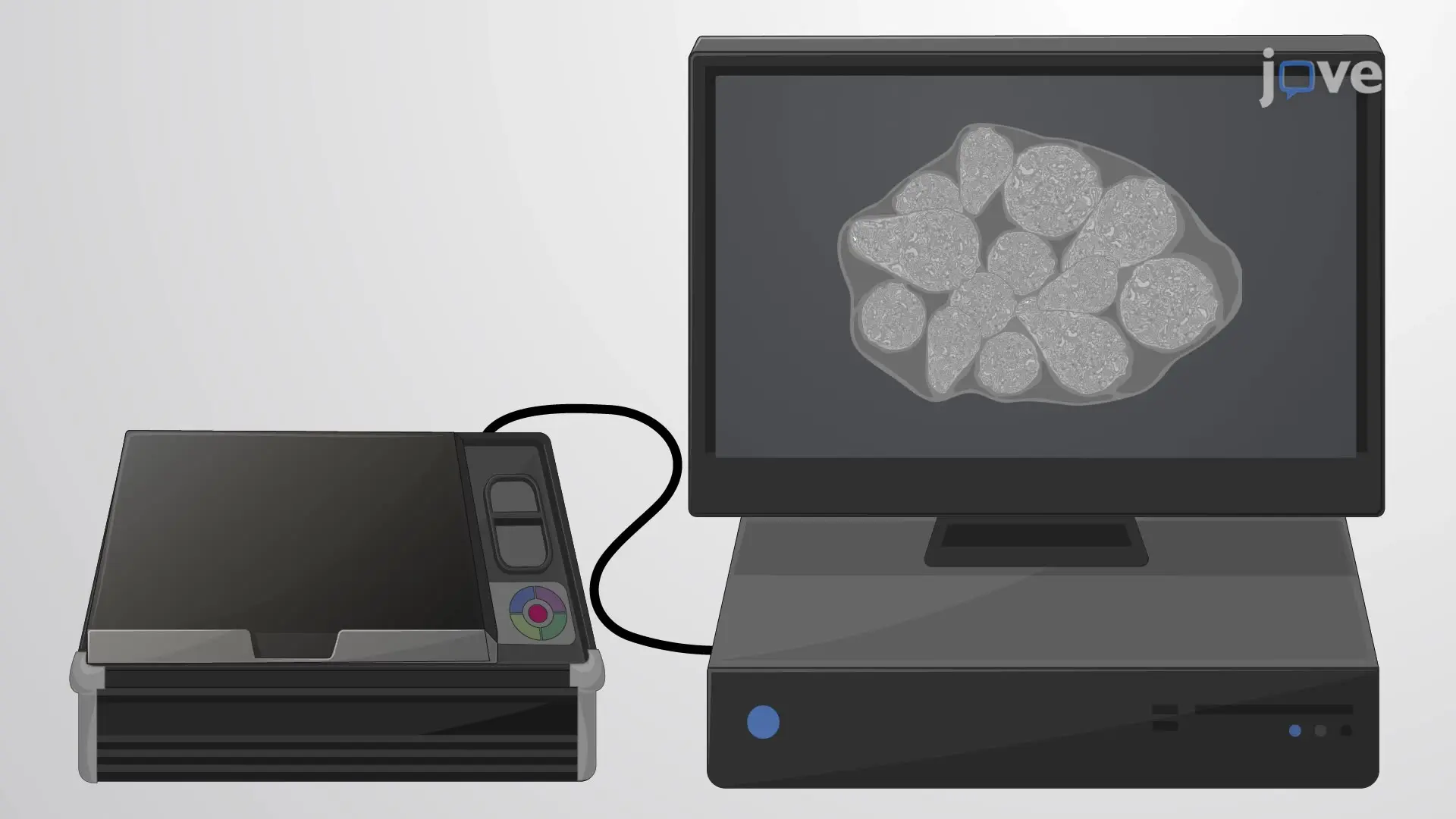
The video describes the preparation of a tissue sample for imaging mass cytometry. The tissue is first processed to unmask the antigen epitopes, permeabilize the cells, and block non-specific antibody binding sites. Then, the tissue is incubated with a metal-conjugated antibody cocktail and a nuclear stain. Finally, the treated tissue sample is imaged using an optical scanner.
Protocol

Loading...
$$\rightleftharpoonup{xx}$$ $$\longleftharp{xx}$$, $$\longrightharp{xx}$$,

1. Tissue pretreatment:

For Fresh Frozen tissues:

  1. Place a glass Coplin jar in the -20°C freezer for at least one hour before use.
  2. Remove the slides from the -80°C freezer and place them into the glass Coplin jar in the -20°C freezer for at least 15 minutes.
  3. Under the fume hood, use an ampule cutter to open the 16% paraformaldehyde (PFA) glass ampule. Discard the broken glass appropriately.
  4. Add 10 mL of 16% PFA and 30 mL phosphate-buffered saline (PBS) to a plastic Coplin jar. Ensure that the PBS is pre-chilled to 4°C.
  5. Transfer this solution to the 4°C fridge for at least 15 minutes.
  6. After 15 minutes, transfer the glass Coplin jar with the slides into the 4°C fridge for 10 minutes.
  7. Transfer the slides from the glass jar to the plastic Coplin jar containing the cold 4% PFA solution.
  8. Fix the slides with 4% PFA for 30 minutes inside the fridge.
  9. Rinse the slides thrice in room temperature PBS for 5 minutes each, ensuring thorough rinsing between cycles.

For FFPE tissues:

  1. Begin with the 5 µm Formalin-fixed paraffin-embedded (FFPE) tissue sections mounted on the positively charged standard microscope slides.
  2. Dewax the slides by heating them at 60°C for one hour. 
  3. Remove the slides and cool them to room temperature for approximately 5 minutes.
  4. Place the slides in a slide mailer and rinse them in a xylene bath for 3 minutes, twice. 
  5. Again, rinse the slides with 100% ethanol for 1 minute, twice. 
  6. Similarly, rinse the slides in 95% and 70% ethanol for 1 minute each. 
  7. Lastly, give a final rinse twice in distilled water for 3 minutes.
    NOTE: The Hyperion cannot ablate within 2mm of the slide edge, center tissue as much as possible. 

2. Heat-induced epitope retrieval (HIER)

  1. Prepare Tris-ethylenediaminetetraacetic acid (EDTA) pH9 buffer:
    1. Abcam ab93684 Tris-EDTA pH9 HIER Buffer is purchased at 100x and diluted to 1x into high-performance liquid chromatography (HPLC) water. 
    2. Ex: 2.5mL Abcam ab93684 into 247.5 mL HPLC water. 
      NOTE: Buffer older than six months should not be used.
  2. Set up a decloaking chamber for heating the slides. First, fill the bottom of the chamber with 500 mL of tap water. Then, add 250 mL tap water to both the side containers and 200 mL tap water to the central container. 
  3. Place up to 3 slides in a 5-place-slide mailer. 
  4. Fill the slide mailer with the HIER buffer until the tissue is completely submerged. NOTE: Ensure that the slide mailer has a small hole in the top to vent steam and prevent pressurization. 
  5. Run "Program 4" for 20 minutes at 95 °C. 
  6. Meanwhile, pre-warm the incubation chambers for subsequent steps (refer to step 3.5)
  7. After incubation, remove the mailer and place it in a tub with cold water from the faucet. 
  8. Allow it to cool for 5 minutes. 
  9. Remove half the buffer from the mailer and replace it with distilled water. Allow it to cool for 5 minutes.
  10. Gradually remove the buffer and increase the water volume twice, each with a 5-minute cooling period.
  11. Lastly, rinse the slides in 100% distilled water.

3. Blocking the non-specific binding

  1. Prepare Blocking Buffer (Tris-buffered saline or TBS, 0.1% Triton X-100, 3% Bovine serum albumin or BSA) by adding 150uL of TBS with 10% BSA to a 350uL of TBS with 0.1% Triton X-10.
    NOTE: TBS with 10% BSA is stored at -20 °C and should be thawed fresh each time.
  2. Using a slide mailer or Coplin jars, wash slides for 5 minutes in TBS, 0.1% Triton X-100 twice.
  3. Remove the slide from the mailer and use a Kim wipe to remove any buffer outside the PAP circle. Allow ~1 minute to dry slightly. 
  4. Using a P200, add a few drops of the blocking buffer over the tissue, just enough to cover it. The PAP line should keep the liquid over the tissue.
    NOTE: If the buffer is not confined in the PAP circle, re-draw the PAP circle around the tissue to prevent antibody loss later. 
  5. Incubate the slide at room temperature in a humidity chamber for 1 hour. 
    NOTE: An empty pipette tip box works well for a humidity chamber. Ensure a small amount of tap water is present at the bottom. 

4. Staining with metal-conjugated antibodies

  1. Remove the metal-conjugated antibodies from 4°C and centrifuge at 10,000 x G for 1 minute.
  2. Preparing optimal dilution of antibody master mix in TBS, 0.1% Triton X-100, and 1% BSA mixture.
    NOTE: a) Optimal dilution of each antibody must be determined experimentally and is one of the most critical steps for successful imaging mass cytometry experiments. Use an Excel spreadsheet to calculate volumes. Make only slightly more than required due to antibody cost. e.g., if it took 200 uL of blocking buffer, make 250uL master mix. b) The master mix should be prepared fresh for each experiment during the blocking step. 
  3. After incubation (refer to step 3.5), remove the slide from the incubator.
  4. Remove excess blocking buffer by tapping the slide vertically and horizontally on Kim wipes. This should leave some blocking buffer on the tissue but will remove most of it. 
  5. Allow ~1 minute to dry slightly.
  6. Place the slide in the humidity chamber at 4°C and allow it to equilibrate.
  7. Then, carefully pipette the antibody master mix onto the tissue, extremely careful that the solution is contained within the PAP circle. 
  8. Close the humidity chamber and incubate overnight at 4°C. 
  9. Following overnight incubation, remove the slide from 4°C and remove excess master mix by tapping the slide vertically and horizontally on Kim wipes. 
  10. Place the slide in the mailer or Coplin jar; wash for 5 minutes with phosphate-buffered saline or PBS, supplemented with 0.2% Triton X-100, twice.
  11. Then, wash the slide in PBS for 5 minutes, twice. Remove the slide and allow it to dry for approximately 1 minute.

5. DNA Intercalation 

  1. For DNA Intercalation, dilute the Intercalator-Ir 1:1000 in PBS. 
  2. Carefully pipette the diluted intercalator onto the tissue, being extremely careful that the solution is contained within the PAP circle.
  3. Incubate for 30 minutes at room temperature in the humidity chamber used for blocking.
  4. After that, tap the slides vertically and horizontally on Kim wipes to remove excess Intercalator-Ir dye. 
  5. Place the slide in the mailer or Coplin jar and wash twice with HPLC water for 5 minutes each.
  6. Dry the slides in a desiccator for 5 minutes before scanning.

6. Image scanning

  1. For slide scanning, use a carbide tip glass etcher to scratch four faint Xs into the slide. Precise teaching points will allow for the best image registration.
    NOTE: Do not get too close to the edge.
  2. Scan using a flatbed document scanner at no less than 1200 dots per inch 
  3. Crop this image as closely as possible to the slide borders and save it to a flash drive. 
  4. For image co-registration, scan the slide on a Hamamatsu NanoZoomer with "Pre_IMC" in the file name. 

Toegang beperkt. Log in of start een proefperiode om deze inhoud te bekijken.

Herprints en machtigingen

Toestemming aanvragen om de tekst of afbeeldingen van dit JoVE-artikel te hergebruiken

Toestemming aanvragen

Trefwoorden

Antigeenherstelpermeabilisatieblokkeringmetaalgeconjugeerd antilichaamkernkleuringoptische scannerbevochtigingskamerhydrofobe barri reobjectglaasjesverzenddoos

Gerelateerde artikelen