Methodenartikel

Recording Changes in Renal Sympathetic Nerve Activity in Response to Arterial Pressure in Rats

8 juli 2025

In dit artikel

Samenvatting

Loading...
$$\rightleftharpoonup{xx}$$ $$\longleftharp{xx}$$, $$\longrightharp{xx}$$,

Source: Fink, A. M., et al. Quantifying Acute Changes in Renal Sympathetic Nerve Activity in Response to Central Nervous System Manipulations in Anesthetized Rats. J. Vis. Exp. (2018).

This video demonstrates a technique of studying the activity of renal sympathetic nerves (RSNs) in response to changes in arterial pressure in rats. Upon attaching a pressure-sensing catheter to the femoral artery, the renal sympathetic nerves are exposed. The nerves are then lifted onto a recording electrode, and their electrical activity is recorded. Next, a vasoconstricting drug is injected to elevate blood pressure, and corresponding changes in RSN activity are monitored.

Protocol

Loading...
$$\rightleftharpoonup{xx}$$ $$\longleftharp{xx}$$, $$\longrightharp{xx}$$,

All procedures involving animal models have been reviewed by the local institutional animal care committee and the JoVE veterinary review board.

1. Create Bipolar RSNA Electrodes

  1. To create the electrode, cut two pieces of stainless steel wire each approximately 18 mm long. Cut one piece of polyethylene (PE-50) tubing approximately 15 mm long. Feed both pieces of wire into the tubing, leaving the wire protruding from both ends.
  2. Remove the insulation from the ends of the wires; trim the wires leaving 2-3 mm of exposed wire. On one end, crimp male pins over the exposed wire. Solder the pins securely to the wire, secure the pins inside a connector strip, and cover the connection with epoxy.
    NOTE: An alternative approach that avoids soldering is to use quick connect/release alligator clips.
  3. On the opposite end of the electrode, remove the insulation from the ends of the wires, leaving 2-3 mm of exposed wire. Bend this portion of the wires to create small "V" shaped hooks in the uninsulated wire.
    NOTE: This is the portion of the electrode that will be in contact with the renal sympathetic nerve. It is important to seal this end to prevent fluids from entering the tubing. Silicone or epoxy effectively can be used.

2. Administer Anesthesia and Prepare Surgical Sites

  1. Administer anesthesia to a male Sprague Dawley rat (age 9-11 weeks, weighing 150-400 g). Administer pentobarbital sodium 50 mg/kg via an intraperitoneal (IP) injection. To assess a stable plane of anesthesia during surgery, check toe-pinch reflex every 15 min and re-dose anesthesia as needed.
    NOTE: Pentobarbital sodium (Nembutal) was used in previous studies to achieve a sustained plane of anesthesia without interfering with the modulation of RSNA. This protocol is for non-survival surgery, so there is no recovery/post-operative monitoring period.
  2. Prepare the surgical site according to institutional animal care guidelines (i.e., shave the rat's abdomen, back, and head; cleanse the skin with 10% povidone-iodine solution; apply eye lubricant; and place the rat on a heating pad). Maintain the body temperature at 37 °C during the experiments.

3. Cannulate the Femoral Artery for Mean Arterial Pressure Monitoring

  1. Visualize the artery under the microscope. Similar to the method used for venous cannulation, tie the distal suture to occlude the artery; secure the edges of this suture to the surgical field with silk tape and position the artery perpendicular to the surgeon's dominant hand.
  2. Arterial access if using telemetry
    1. Inspect the pressure-sensing catheter under high magnification before arterial cannulation. Ensure that the catheter is free of bubbles/ debris and has an intact meniscus between the fluid-filled (proximal) and gel-filled (distal) components. Prior to each implantation, refill the gel at the distal tip of the catheter. Turn on the transmitter using a magnet; monitor blood pressure (BP) during the surgery to endure perfect placement.
    2. Use a loose overhand half knot in the proximal suture to briefly occlude the femoral artery. Hold the catheter introducer in the non-dominant hand. Hold the tip of the cannula of the telemetry unit with vessel cannulation forceps to avoid displacing gel from the tip.
    3. Puncture a hole in the artery with the bent 22G needle and insert the cannula into the artery using vessel cannulation forceps to avoid displacing gel from the tip. Advance the cannula as far as possible. Using the proximal and distal suture ties, secure the pressure catheter.
    4. Tuck the body of the telemetry implant inside the flank adjacent to the incision and close this incision using 4-0 nylon suture on a cutting needle. Turn the telemetry devices off by magnet at the conclusion of the recording period to conserve battery life.

4. Position the Rat in the Stereotaxic Surgery Frame to Access the Brain

  1. Gently move the rat into the prone position in the stereotaxic surgery frame.
  2. Position the rat between the ear bars and adjust the incisor bar to equalize the height of lambda and bregma. Positioning may depend on the rat strain, weight, and locations of central nervous system (CNS) targets.
  3. Make a 2 cm rostrocaudal scalpel incision through the midline of the scalp. Using cotton-tipped applicators, firmly remove connective tissue from the skull surface. Apply hydrogen peroxide to the skull to assist in visualizing the bregma, lambda, and midline sutures.
  4. Using an atlas of the rat brain to guide targeting, drill a burr hole osteotomy, sized for electrode access, through the skull.

5. Isolating the Renal Sympathetic Nerves

  1. Connect the wire RSNA electrode (Steps 1.1-1.3) to a 10X pre-amplifier and microelectrode amplifier.
  2. Isolate the renal nerves through a retroperitoneal incision prior to, or after, the rat is secured in the stereotaxic frame. Position the RSNA electrodes once the rat is in the stereotaxic frame. Make a scalpel incision extending from 4-5 cm below the ribs in the caudal direction, slightly lateral to the spine. Blunt dissects the incision to visualize the paraspinal muscles.
  3. Use scissors to make a very superficial 1-2 cm rostrocaudal incision where the fat meets the muscle. Using cotton-tipped applicators, spread the fat away from the muscle to visualize the kidney. It is important not to enter peritoneal space.
  4. Use retractors to gently separate the kidney from the paraspinal muscles to visualize the renal artery and abdominal aorta. Do not stretch the vessels excessively to avoid damaging the renal nerves. Use a 2" x 2" cotton gauze pad soaked in saline to protect the kidney from injury.
  5. Under high magnification, identify the renal nerves in the incision pocket. The nerve bundles are most easily visible in the right angle formed by the aorta and the renal artery. The renal nerves closely follow the renal artery from the aorta to the kidneys.
  6. Select a segment of the nerve bundle that will be placed on the recording electrode. Gently dissect the nerve fibers from the surrounding tissue/vessel using micro-dissecting tweezers.
  7. Secure the wire RSNA electrode in a holder (e.g., an alligator clip attached to a support stand). Lower the electrode to the level of the nerve segment. Use a nerve hook to gently lift the segment of the renal nerve onto the electrode without stretching the nerve.
    NOTE: The nerve should rest inside both "V" shaped hooks in the uninsulated wire, parallel to the nerve. The electrode wires must not touch any other tissue, blood, or lymph fluid.
  8. Fill the incision with mineral oil to prevent the exposed renal sympathetic nerve from becoming dry. Use a grounding clip with one end on the skin of the incision and the other attached to the Faraday cage.
  9. Direct the signal to the amplifiers using high- and low-pass filtering (10 Hz and 3 kHz). Adjust the gain up to 10 K. Include an audio monitor to assess the bursting pattern of the RSNA. Use sampling rates ranging between 2,000-10,000 Hz. Use an increased sampling rate when a CNS manipulation is hypothesized to cause rapid/brief sympathetic responses.

6. Record Data

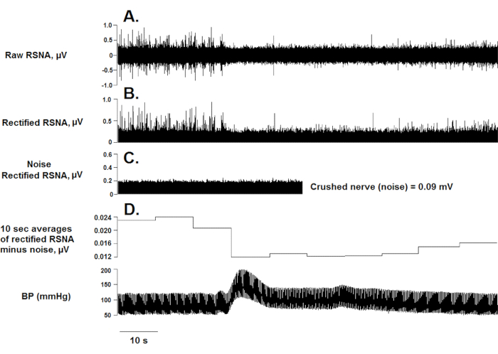
  1. Assess the quality of the RSNA recording by evoking the baroreflex with a bolus injection of 1 mL of saline or 10 µg/mL phenylephrine (in 0.1 mL) intravenously. As illustrated in Figure 1, the infusion should increase BP and inhibit RSNA. An increase in mean arterial pressure of 60-80 mmHg is sufficient for renal sympathoinhibition.
  2. Adjust the position of the electrodes to improve the signal if necessary. Repositioning is required if the nerve is not in contact with both hooks on the electrode or if any tissue, blood, or lymph fluid is in contact with the wires.
    NOTE: The need for repositioning is based on the auditory characteristics of the nerve discharges.
  3. Once a clear signal is obtained, secure the RSNA electrode in place by withdrawing mineral oil and applying a silica gel to cover the nerve/ electrode connection in the incision pocket. Do not move the rat before the gel has set completely.
  4. Perform CNS manipulation protocols while continuously recording RSNA and mean arterial pressure. If a microinjector/pulser is used for brainstem manipulations, a logic signal from this device can be introduced into the RSNA/BP recordings to document the timing of CNS manipulations.
  5. When the experiment is complete, determine the noise level by crushing the nerve proximal to the recording electrodes between the silica gel and the spinal muscle. Record at least 30 s of this "zero" value for RSNA. As an alternative approach for quantifying noise, administer a short-acting ganglionic blocker such as atropine, hexamethonium, chlorisondamine, or pentolinium tartrate.
  6. Carefully remove the RSNA electrode and remove any traces of silica gel from the wire electrodes. Save the electrode for re-use. Turn off the telemetry transmitter and remove it, taking care not to damage the tip of the catheter.

Toegang beperkt. Log in of start een proefperiode om deze inhoud te bekijken.

Resultaten

Loading...
$$\rightleftharpoonup{xx}$$ $$\longleftharp{xx}$$, $$\longrightharp{xx}$$,

Renal sympathetic nerve activity graph; displays raw, rectified RSNA, noise, blood pressure over time.
Figure 1: RSNA and BP in response to phenylephrine injection IV. The raw RSNA (A) was full-wave rectified (B); rectified crushed "zero" RSNA is shown in inset C. Non-overlapping 10 s averages (minus n...

Toegang beperkt. Log in of start een proefperiode om deze inhoud te bekijken.

Openbaarmakingen

Loading...
$$\rightleftharpoonup{xx}$$ $$\longleftharp{xx}$$, $$\longrightharp{xx}$$,
No conflicts of interest declared.

Materialen

Lijst van materialen gebruikt in dit artikel
NaamBedrijfCatalogusnummerOpmerkingen
Stainless steel wireA-M Systems; Sequim, WA791000RSNA electrode
Polyethylene (PE-50) tubingVWR; Radnor, PA63019-048RSNA electrode; vessel cannulation
Miniature pin connectorA-M Systems; Sequim, WA520200RSNA electrode
Crimping toolDaniels Manufacturing Corp.; Orlando, FLM22520RSNA electrode
Connector stripAmphenol; Clinton Township, MI221-2653RSNA electrode
J-B Kwik EpoxyJ-B Weld, Sulphur Springs, TX8270RSNA electrode
SiliconePermatex; Hartford, CT2222RSNA electrode
Heparin sodium; Injectable (10 mL vial, 1000 U/mL)KV Veterinary Supply; David City, NEP03466Venous line patency
Phenylephrine HCl; Injectable (1 mL vial; 10 mg/mL)ACE Surgical Supply; Brockton, MA950-6312Testing renal sympathoinhibition
Single-hook elastic surgical staysHarvard Apparatus; Holliston, MA72-2595Incision
Silk surgical tape3M, Minneapolis, MN1538-0Secure surgical stays
Needles, 20 GSigma-Aldrich; St. Louis, MOZ192554-100EAVessel cannulation
Dumont #7 curved forcepsFine Science Tools; Foster City, CA11274-20Vessel cannulation
5-0 silk suture tiesBraintree Scientific; Braintree, MASUT-S 106Vessel cannulation
Delicate hemostatic forcepsRoboz Surgical Instrument Co.; Gaithersburg, MDRS-7117Vessel cannulation and RSNA surgery
Crile Hemostatic forcepsFine Science Tools; Foster City, CA13004-14Needle bending
Telemetry transmitterData Sciences International; Minneapolis, MNPA-10Mean arterial pressure monitoring (telemetry)
Re-gel syringeData Sciences International; Minneapolis, MN276-0038-001Transmitter reuse (telemetry)
Disposable pressure transducerTranspac; San Clemente, CAMI-1224Mean arterial pressure monitoring
Clear-Cuff pressure infuserMILA International Inc.; Florence, KY2281339Mean arterial pressure monitoring
Vessel cannulation forcepsFine Science Tools; Foster City, CA00574-11Catheter insertion
Black monofilament nylon 4-0 suture on reverse cutting needleMcKesson Medical-Surgical; San Francisco, CAS661GXSecure telemetry transmitter
Telemetry receiverData Sciences International; Minneapolis, MNRPC-1Mean arterial pressure monitoring (telemetry)
LabChart Pro (software), PowerLab (acquisition hardware)AD Instruments; Colorado Springs, COML846, MX2 matrix 2.0 (Compatible with Data Science International telemetry)3 options for software/acquisition hardware
SciWorks (software), DataWave (acquisition hardware)DataWave Technologies, Loveland, CON/A
Spike 2 (software), Micro1401-3Cambridge Electronic Design Ltd., London UK1401-3
Micro-drillRoboz Surgical Instrument Co.; Gaithersburg, MDRS-6300CNS surgery
Stereotaxic surgery frameStoelting; Wood Dale, IL51600CNS surgery
Microelectrode amplifier with 10X pre-amplifierA-M Systems; Sequim, WA1800-2RSNA recording
RetractorsFine Science Tools; Foster City, CA17009-07RSNA recording
Micro-dissecting tweezersFine Science Tools; Foster City, CA11251-10RSNA recording
Micro-hookFine Science Tools; Foster City, CA10064-14RSNA recording
Mineral oilFisher Scientific; Waltham, MA8042-47-5RSNA recording
Audio monitorA-M Systems; Sequim, WA3300RSNA recording
Silica gelWacker, Munchen; GermanyRT601A-BRSNA recording
Electrical clipsTyco Electronics; Schaffhausen, SwitzerlandEB0283-000Grounding or securing perfusion needle
Bonn scissors, straight/sharp pointsRoboz Surgical Instrument Co; Gaithersburg, MDRS-5840Perfusion
Gavage needleHarvard Apparatus; Holliston, MA75-0286Perfusion
Masterflex perfusion pumpCole-Parmer; Vernon Hills, IL7524-10Perfusion
Masterflex platinum-cured silicone tubingCole-Parmer; Vernon Hills, IL96410-15Perfusion
Formalin (10% buffered solution; 4 L)Sigma-Aldrich; St. Louis, MOHT501128Perfusion
SucroseSigma-Aldrich; St. Louis, MOS0389Cryoprotection

Herprints en machtigingen

Toestemming aanvragen om de tekst of afbeeldingen van dit JoVE-artikel te hergebruiken

Toestemming aanvragen

Trefwoorden

Meting van de arteri le drukcanulatie van de arteria femoraliszenuwintering elektrodefenylefrine injectiebaroreflexresponsstereotaxisch chirurgisch framehydratatie met minerale oliesilicagel elektrodebloeddrukmonitoring

Gerelateerde artikelen