All procedures involving animal models have been reviewed by the local institutional animal care committee and the JoVE veterinary review board.
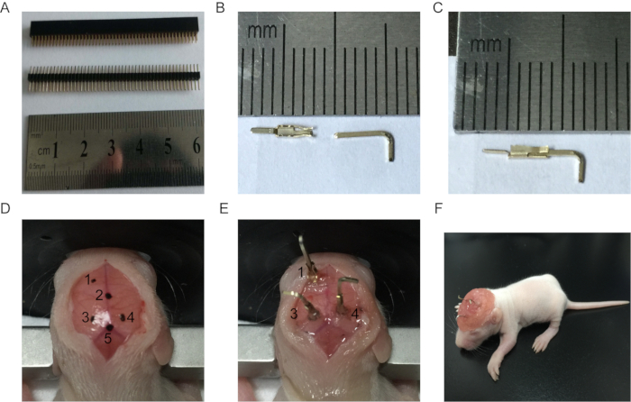
1. Electrode Preparation (Figure 1A-C)
NOTE: Computer pin loci is simply a pronged contact as a part of signal interface in communication devices. It consists of a male connector that plugs into the female connector.
- Carefully separate the male and female pins from the computer pin loci (Figure 1A) with the help of a pincer. Connect the male and female pins together to form an electrode and apply cyanoacrylate to create a strong adhesive bond (Figure 1C).
- Put the electrodes into a beaker filled with distilled water and place it to ultrasonic cleaner for 10 min. Move them to a drying oven at 45 °C for 30 min. Sterilize the electrodes using ultraviolet (UV) light for 30 min.
2. Surgical procedure (Figure 1D-F)
- Prepare the sterilized surgical instruments and stereotaxic apparatus. Anesthetize the neonatal rat pup using isoflurane anesthesia (2.5 %) with air. When the pup is deeply anesthetized, adjust the dose of isoflurane to 1.0%. Perform a tail or toe pinch prior to the surgery to ensure the proper depth of anesthesia.
- Fix the head of the pup in the stereotaxic apparatus by placing the ear bars into the ear canals and slightly tightening them.
NOTE: Do not excessively tighten the ear bars, as the neonatal skull is very soft.
- Maintain the sterile surgical field by spraying all equipment with 70% ethanol. Make a 15-mm incision on the head using a scalpel. Using forceps, gently pull the scalp away from the midline at the four corners. Put some saline-soaked cotton below the skin to keep the incision wide open (Figure 1D).
- Find the bregma and lambda points on the skull and mark them with a pencil. Use a syringe needle (26 G) to make two burr holes into the prefrontal cortex (PFC) and hippocampus.
NOTE: The PFC is located at +1.8 mm posterior to bregma and -0.5 mm lateral to the midline, while the hippocampus is located at -2.0 mm anterior to bregma and ±0.5 mm lateral to the midline (Figure 1D and E). The depth of the electrode should not be more than 2 mm below the cortical surface to minimize brain damage.
- Use forceps to hold the electrodes and insert the reference and recording electrodes into the PFC and hippocampus, respectively. Apply erythromycin ointment around the electrode to avoid any possible infection. Fix the electrode using cyanoacrylate.
- Prepare dental acrylic cement so that it has a gluey and viscous consistency. Apply the dental cement to cover the electrodes and the rest of the skull.
NOTE: Thoroughly dry the skull before applying the dental cement.
- Apply 5% picric acid onto the electrodes to protect them.
NOTE: The entire procedure should be done in a biosafety hood to maintain sterile conditions.
- Remove the animal from the stereotaxic frame and inject 300 µL of 10% glucose subcutaneously. Place it on a heated blanket for recovery. Make sure that the animal is warm (37 °C) and ambulatory (i.e., completely recovered). Administer buprenorphine intraperitoneally (0.05 mg/kg) for post-surgical pain.
NOTE: Do not leave an animal unattended until it has regained sufficient consciousness (i.e. normal behavior and movement).
- Return the pup to its home cage with their dam after it regains consciousness. Wait for two days until the animal is fully recovered.
3. EEG Recording
- After full recovery, connect the electrodes implanted on the skull of the pup to the amplifier in its own cage. Connect the amplifier to an analogue-to-digital converter and attach the converter to a computer; the connecting lines should be treated carefully so that they do not get tangled.
- Select at least 10,000 Hz sampling rate on the data acquisition unit for recording (the bandwidth of the transmitter is 1-100 Hz). Ensure that the data is sampled properly.
- After getting the baseline recording, inject the pup intraperitoneally with kainic acid (KA, 2 mg/kg) to induce epileptic seizures. 15 min after the KA injection observe and record the epileptic discharges. Seizure produce through KA are usually physical.
NOTE: The ictal-tonic duration is about 15.2 ±0.9 s, seizure duration is about 62±5 s. Seizures can be prevented in the neonatal rat by giving IP injection of chloral hydrate (400 mg/kg).
- Save the digitized data and analyze it using signal-processing software packages, such as spike2. Reveal the power level of different frequency components in the neonatal EEG signal by performing a power spectrum analysis. Calculate the power in a 1-min timeframe by finding the root-mean-square amplitude from 1 to 100 Hz (EEG band).