Een abonnement op JoVE is vereist om deze inhoud te bekijken. Log in of start vandaag met uw gratis proefperiode.

Methodenartikel

Immunostaining of Neurons Treated with Alpha-Synuclein Aggregates

946 weergaven

8 juli 2025

In dit artikel

Samenvatting

Source: Panattoni, G., et al. Exogenous Administration of Microsomes-associated Alpha-synuclein Aggregates to Primary Neurons As a Powerful Cell Model of Fibrils Formation. J. Vis. Exp. (2018)

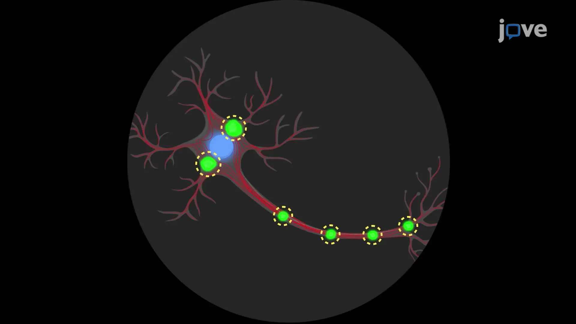
This video demonstrates the immunostaining of neurons treated with alpha-synuclein (αS) aggregates. The neurons are first treated with αS aggregates. They are then fixed, permeabilized, and treated with a blocking solution. Primary antibodies are added, followed by a fluorescently labeled secondary antibodies and a nuclear stain. The sample is then mounted and observed under a microscope.

Protocol

1. Neurons Treatment

Note: The treatment has been performed at Division (DIV) 7. All the steps are carried on under a cell culture hood in sterile conditions. An example of cortical neuronal culture density at DIV 7 is illustrated in Figure 1.

  1. Pool microsomes-associated αS aggregates obtained from the spinal cord of three different diseased Transgenic mice to have a 1 µg/µL solution, using the original homogenization buffer [composed of 250 mM sucrose, 20 mM HEPES (4-(2-hydroxyethyl)-1-piperazineethanesulfonic acid), 10 mM KCl (Potassium Chloride), 1.5 mM MgCl2 ( (Magnesium Chloride), 2 mM EDTA (Ethylenediaminetetraacetic acid), and 1x phosphatase/protease-inhibitors] for the dilution.
  2. Take a neuron culture on poly-D-Lysine coverslips in 24 well plates containing medium (2% B27, 10x gentamicin, and 2 mM glutamine). Remove 1/3 of the medium and replace it gently with the fresh medium containing 2% B27, 1x gentamicin, and 2 mM glutamine.
  3. Add 1 µg of pooled microsomes-associated alpha-synuclein (αS) aggregates to the cell medium. Return neurons to the incubator at 37 °C.
  4. Every 3 days for 1 week, add 1/3 of fresh medium. Do not replace the medium. Just add it.
  5. After 1 week of treatment, remove 1/3 of the medium and replace it gently with the fresh medium containing 2% B27, 10 µg/mL gentamicin, and 2 mM glutamine. Repeat every 3 days.
  6. Fix neurons after 2 weeks of treatment (DIV 21).

2. Immunofluorescence

  1. Fix neurons with 2% paraformaldehyde in Phosphate-Buffered Saline (PBS) 1x and 5% sucrose solution for 15 min at room temperature (RT) without shaking under a chemical fume hood.
  2. Remove the fixing solution. Briefly wash with PBS 1x, 3 times.
    NOTE: Perform all the washing steps gently because primary neurons do not stick firmly to poly-lysine coverslips.
  3. Permeabilize neurons with 0.3% of non-ionic surfactant in PBS 1x for 5 min at RT.
  4. Briefly wash with PBS 1x, 3 times.
  5. Incubate neurons with 3% Fetal Bovine Serum (FBS) in PBS 1x for 30 min at RT to block unspecific binding sites on an orbital shaker.
  6. Incubate neurons with appropriate primary antibody dissolved in 3% FBS in PBS 1x, O/N at 4 °C on an orbital shaker.
    NOTE: syn303 (1:1,000), mouse αS (1:200), pser129-αS (1:1,000), and Tau (1:10,000) antibodies were used.
  7. Remove the antibody solution and wash, briefly, with PBS 1x, 3 times.
  8. Incubate neurons with appropriate fluorescent secondary antibody dissolved in 3% FBS in PBS for 1 h at RT in the dark on an orbital shaker.
  9. Remove the antibody solution and wash, briefly, with PBS 1x, 3 times.
  10. Stain neurons with DAPI (4′,6-diamidino-2-phenylindole) solution (0.1 µg/mL in PBS 1x) for 15 min at RT in the dark on an orbital shaker.
  11. Remove the antibody solution and wash, briefly, with PBS 1x, 3 times.
  12. Mount coverslips on a slide using an antifade mounting medium.

Toegang beperkt. Log in of start een proefperiode om deze inhoud te bekijken.

Resultaten

Cell culture under microscope; neuron-like network; biological research; microscopy study.
Figure 1. Cortical neuronal cultures. Representative image showing density at DIV 7 of cortical neuronal cultures. Images were taken with an inverted light microscope, 10X objective. Scale bar = 100 µm.

Toegang beperkt. Log in of start een proefperiode om deze inhoud te bekijken.

Openbaarmakingen

No conflicts of interest declared.

Materialen

Lijst van materialen gebruikt in dit artikel
NaamBedrijfCatalogusnummerOpmerkingen
SucroseSigma-Aldrich84097-1KG
HepesSigma-AldrichH0887-100ML1M pH=7-7.6
EDTASigma-Aldrich0390-100mlpH=8 0.5M
Magnesium chlorideSigma-AldrichM8266-100G
Sodium carbonateSigma-AldrichS7795-500G
Sodium bicarbonateSigma-AldrichS5761-500G
MethanolSigma-Aldrich322415-6X1L
KClSigma-AldrichP9541-500G
cOmplete MiniRoche11836170001protease inhibitor
PhosStopRoche4906837001phosphatase inhibitor
BCA Protein Assay KitEurocloneEMPO14500
Criterion TGX 4-20% Stain Free, 18 wellsBiorad5678094
Supported Nitrocellulose membraneBiorad16200970.2 μm
Blotting-Grade BlockerBiorad1706404Non-fat dry milk
SuperSignal West Pico Chemiluminescent SubstrateTermo Fisher Scientific34077
Nitric acidSigma-Aldrich100441100065%
Glass CoverslipsTermo Fisher Scientific1014355118NR118 mm x
Poly-D-LysineSigma-AldrichP7280
Hank's Balanced Salt SolutionTermo Fisher Scientific14170-500 mL
Penicillin/StreptomycinTermo Fisher Scientific1514012210,000 U/mL, 100 mL
Dulbecco's Modified Eagle's MediumTermo Fisher ScientificD5796-500 mL
Trypsin-EDTATermo Fisher Scientific154000540.50%
B27 SupplementTermo Fisher Scientific1750404450X
GlutamaxTermo Fisher Scientific35050-038100x
DNAseSigma-AldrichD5025
Fetal bovine serumEurocloneEC50182L
GlutamateSigma-Aldrich1446600-1G
GentamicinTermo Fisher Scientific1571010 mg/ml
Neurobasal MediumTermo Fisher Scientific10888-022
Cytosine arabinoside (AraC)Sigma-AldrichC3350000
VECTASHIELD antifade mounting mediumVector LaboratoriesH-1000
DAPITermo Fisher Scientific62247
90 Ti rotorBeckmanN/AUltracentrifuge rotor
Optima L-90K UltracentrifugeBeckmanN/A
Syn-1 antibody, clone 42BD Biosciences610786anti-mouse WB: 1:5000
Syn303 antibodyBioLegend824301anti-mouse IF: 1:1000
Tau antibodySynaptic Systems314 002anti-rabbit IF: 1:10,000
pser129-αS antibodyA gift from Fujiwara et al, reference 19anti-rabbit WB: 1:5000
pser129-αS antibodyAbcamab51253anti-rabbit IF: 1:1000
Mouse αS (D37A6) XPCell Signaling4179anti-rabbit IF 1:200
Alexa fluor 555-conjugated anti-rabbit antibodyTermo Fisher ScientificA27039
Alexa fluor 488-conjugated anti-mouse antibodyTermo Fisher ScientificA-11029
Microson XL-2000MisonixSonicator
Ultra Bottles (Oakridge Bottles), PCB, 16x76mm, Assembly, Noryl Cap, Beckman-typeScience Service EUS4484Ultracentrifuge tubes
AXIO Observer Inverted Light MicroscopeZeissN/A
TCS SP2 laser scanning confocal microscopeLeicaN/A
Inverted epi-fluorescence microscopeNikonN/A
Triton x-100Sigma-AldrichX100-500MLNonionic surfactant

Tags

Immunokleuring van neuronenIncubatie van primaire antilichamenFluorescerend secundair antilichaamDAPI kernkleuringFixatie met paraformaldehydePermeabilisatie van membranenBehandeling met blokkeringsoplossingFluorescentiemicroscopische observatieProcedure voor het dekglasmontage