Methodenartikel

Modeling Retinal Degeneration and Regeneration in Zebrafish Using a Focal Laser Injury

8 juli 2025

In dit artikel

Samenvatting

Loading...
$$\rightleftharpoonup{xx}$$ $$\longleftharp{xx}$$, $$\longrightharp{xx}$$,

Source: Conedera, F. M., et al. Müller Glia Cell Activation in a Laser-induced Retinal Degeneration and Regeneration Model in Zebrafish. J. Vis. Exp. (2017).

This video demonstrates a procedure to induce retinal degeneration in adult zebrafish using laser injury. The laser targets photoreceptor cells in the retina, triggering cell death and a degenerative process. This damage activates Müller glia (MG) cells, leading to their dedifferentiation into stem cell progenitors that aid in retinal regeneration.

Protocol

Loading...
$$\rightleftharpoonup{xx}$$ $$\longleftharp{xx}$$, $$\longrightharp{xx}$$,

All procedures involving animal models have been reviewed by the local institutional animal care committee and the JoVE veterinary review board.

1. Animals

  1. Maintain TgBAC (gfap:gfap-GFP) zebrafish 167 (AB) strain aged between 6 - 9 months under standard conditions in water with a temperature of 26.5 °C and a 14/10 h light/dark cycle.

2. Reversible Systemic Anesthesia

  1. Prepare the stock solution of ethyl 3-aminobenzoate methanesulfonate salt (tricaine) by dissolving 400 mg of tricaine powder in 97.9 mL of tank water and 2.1 mL of 1 M of Tris buffered saline (TBS). Adjust to pH 7.0 with 1 M Tris (pH 9) and store at 4 °C in the dark up to one month.
    NOTE: Tricaine should be prepared in water like the natural living conditions of the animal, preferably original tank water should be used.
  2. Dilute the stock solution to 1:25 in tank water and use immediately.
  3. Place the zebrafish into a 10 cm Petri dish containing 50 mL of anesthesia solution until they become immobile and do not respond to external stimuli (approximately 2 - 5 min, depending on weight and age).
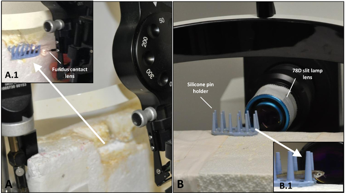
  4. Transfer each fish by hand to a custom-made silicone pin holder for laser treatment (Figure 1A).
    CAUTION! The fish can remain anesthetized outside of the tank for up to 10 min only.
  5. To reverse anesthesia after the treatment and/or imaging, place the zebrafish in container containing tank water.
  6. To support recovery, create a flow of fresh tank water over the gills by moving the zebrafish back and forth in the water.

3. Laser Focal Injury on Retina

NOTE: A 532 nm diode laser is used to create focal light damage on the retina of the zebrafish. The experimental set-up of the laser enables the establishment of a reproducible focal retinal injury in adult zebrafish.

  1. Set up the output power of the laser: 70 mW; aerial diameter: 50 µm; pulse duration: 100 ms.
    CAUTION! The use of laser light requires appropriate personal protection and labelling of the area.
  2. Apply 1 - 2 drops of 2% hydroxypropylmethylcellulose topically in the eye before treatment and use a 2.0 mm fundus laser lens to focus the laser-aiming beam on the retina.
    CAUTION! hydroxypropylmethylcellulose drops are viscous and may cause problems in breathing if it goes on the gills.
  3. Place four laser spots around the optic nerve on the left eye and use the right, untreated eye as internal control.
Toegang beperkt. Log in of start een proefperiode om deze inhoud te bekijken.

Resultaten

Loading...
$$\rightleftharpoonup{xx}$$ $$\longleftharp{xx}$$, $$\longrightharp{xx}$$,

Optical setup with fundus contact lens and 78D slit lamp lens for ocular diagnostics analysis.
Figure 1: Setup for the induction of the laser injury of zebrafish retina and following OCT analysis. (A) Arrangement of the laser system before treatment. (A.1) Zebrafish placed on silicone pin holder with fundus contact len...

Toegang beperkt. Log in of start een proefperiode om deze inhoud te bekijken.

Openbaarmakingen

Loading...
$$\rightleftharpoonup{xx}$$ $$\longleftharp{xx}$$, $$\longrightharp{xx}$$,
No conflicts of interest declared.

Materialen

Lijst van materialen gebruikt in dit artikel
NaamBedrijfCatalogusnummerOpmerkingen
Hydroxypropylmethylcellulose 2%OmniVision, Neuhausen, Switzerlandn.a.Methocel 2%
Ethyl 3-aminobenzoate methanesulfonateSigma-Aldrich, Buchs, SwitzerlandA5040Tricaine, MS-222
Visulas 532sCarl Zeiss Meditec AG, Oberkochen, Germanyn.a.532 nm laser
Silicone pin holderHuco Vision AG Switzerlandn.a.Cut by hand from silicone pin mat of the sterilization tray accordingly.
TgBAC (gfap:gfap-GFP) zf167 (AB) strainKIT, Karlsruhe, Germany15204http://zfin.org/ZDB-ALT-100308-3
Tris buffered saline (TBS)Sigma-Aldrich, Buchs, SwitzerlandP5912
Ocular fundus laser lensOcular Instruments, Bellevue, WA, USAOFA2-0

Herprints en machtigingen

Toestemming aanvragen om de tekst of afbeeldingen van dit JoVE-artikel te hergebruiken

Toestemming aanvragen

Trefwoorden

ZevismodelM ller gliacellenretinale regeneratiefotoreceptorcellenanesthetische oplossinghydroxypropylmethylcellulosefunduslaserlenstricaine anesthesie

Gerelateerde artikelen