All procedures involving animal samples have been reviewed and approved by the appropriate animal ethical review committee.
1. Determination of infarct zone
- Measure infarct zone (IZ) at 24 h after middle cerebral artery occlusion (MCAO).
NOTE: Rats that lost more than 20% of their weight or developed seizures or hemiplegia are excluded from the experiment.
- Euthanize the rat by replacing the inspired gas mixture with 20% oxygen and 80% carbon dioxide until the rat ceases to breathe spontaneously.
- Open the chest with a 5-6 cm lateral incision through the abdominal wall under the rib cage using scissors and surgical forceps.
- Perform a diaphragmatic incision along the entire length of the rib cage with scissors and surgical forceps.
- Carefully displacing the lungs, cut through the rib cage up to the collarbone on the right and left sides.
- Perfuse with 200 mL of normal saline through the left ventricle of the heart.
- Puncture or incise the right atrium of the heart with scissors.
- Perform decapitation using a guillotine and collect brain tissue.
- Using iris scissors, cut from the foramen magnum to the distal edge of the posterior skull surface on both sides.
- Separate the olfactory bulbs, nervous connections along the ventral surface and dorsal surface of the skull from the brain.
- Remove the brain from the head.
- Produce 6 brain slices by creating 2 mm thick horizontal sections with a .009" stainless steel, uncoated, single edge razor blade.
- Incubate for 30 min at 37 °C in 0.05% TTC.
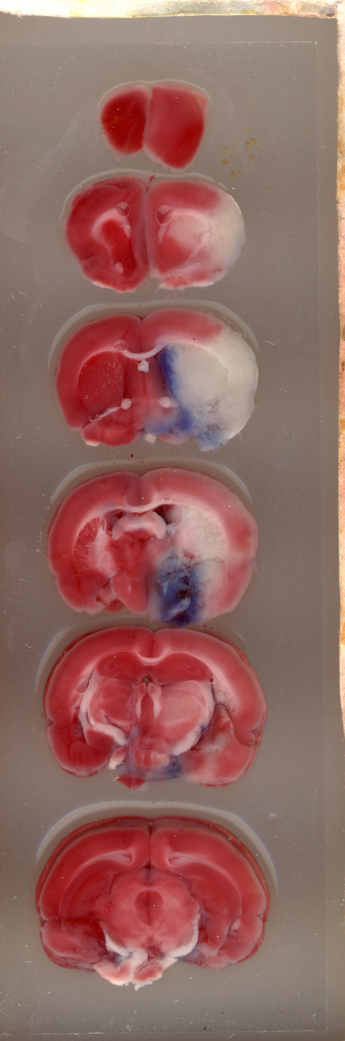
- Place the brain tissue on the microscope slides and perform optical scanning of these 6 brain-slices with a resolution of 1600x1600 dpi (see Figure 1 for example).
- Add a blue filter with a photo editor (e.g., Adobe Photoshop CS2) using the Channel Mixer function (Image > Adjustments > Channel Mixer) and save the image as a JPEG file format.
NOTE: After applying the blue filter, the image will appear greyscale.
- Open the saved image in ImageJ 1.37v.
NOTE: This computer program uses a threshold function to isolate and calculate the pixels that are either black or white (see Figure 2).
- For each of the 6 brain slices of the image, select and save each hemisphere (right injured ipsilateral and left uninjured contralateral) as a separate image file using the "polygon selection" tool from the main menu.
- Set the cut-off for determining IZ by using an auto threshold function from the main menu of the ImageJ software by selecting Image > Adjust > Threshold and measure the number of pixels in each hemisphere of a single brain set.
NOTE: Macros may be used for this step in ImageJ software (see Supplementary document 1 for the code). The cut off is a critical parameter for determining which pixels to convert to white and which to convert to black depending on the shade of gray (see Figure 3 and Figure 4 as examples). ImageJ then compares white and black pixels to determine IZ. Based on the staining protocol and scanner settings, we used a constant cut-off value of 0.220.
- Perform measurement of IZ correcting for tissue swelling using the Ratios of Ipsilateral and Contralateral Cerebral Hemispheres (RICH) method (see example in Figure 5).

NOTE: Infarct size is assessed as a percentage of the contralateral hemisphere.
2. Determination of brain edema
NOTE: Use ImageJ 1.37v for measurement of brain edema (BE).
- Measure BE 24 h after MCAO. For calculation of BE, use the data from left and right hemisphere volume (in units).
- Perform optical scanning with a resolution of 1600x1600 dpi (see Figure 1 for example).
- Select brain hemispheres and set the cut-off for determining BE with ImageJ 1.37v, as described above in sections 1.17-1.19.
- Express the BE area as a percentage of the standard areas of the unaffected contralateral hemisphere, calculated by the RICH method using following equation (see example in Figure 5).

NOTE: Extent of BE is assessed as a percentage of the contralateral hemisphere.