Een abonnement op JoVE is vereist om deze inhoud te bekijken. Log in of start vandaag met uw gratis proefperiode.

Methodenartikel

Assessing Transendothelial Electrical Resistance in an In Vitro Blood-Brain Barrier Model

1K weergaven

8 juli 2025

In dit artikel

Samenvatting

Source: Kuzmanov, I., et al. An In Vitro Model of the Blood-brain Barrier Using Impedance Spectroscopy: A Focus on T Cell-endothelial Cell Interaction. J. Vis. Exp. (2016)

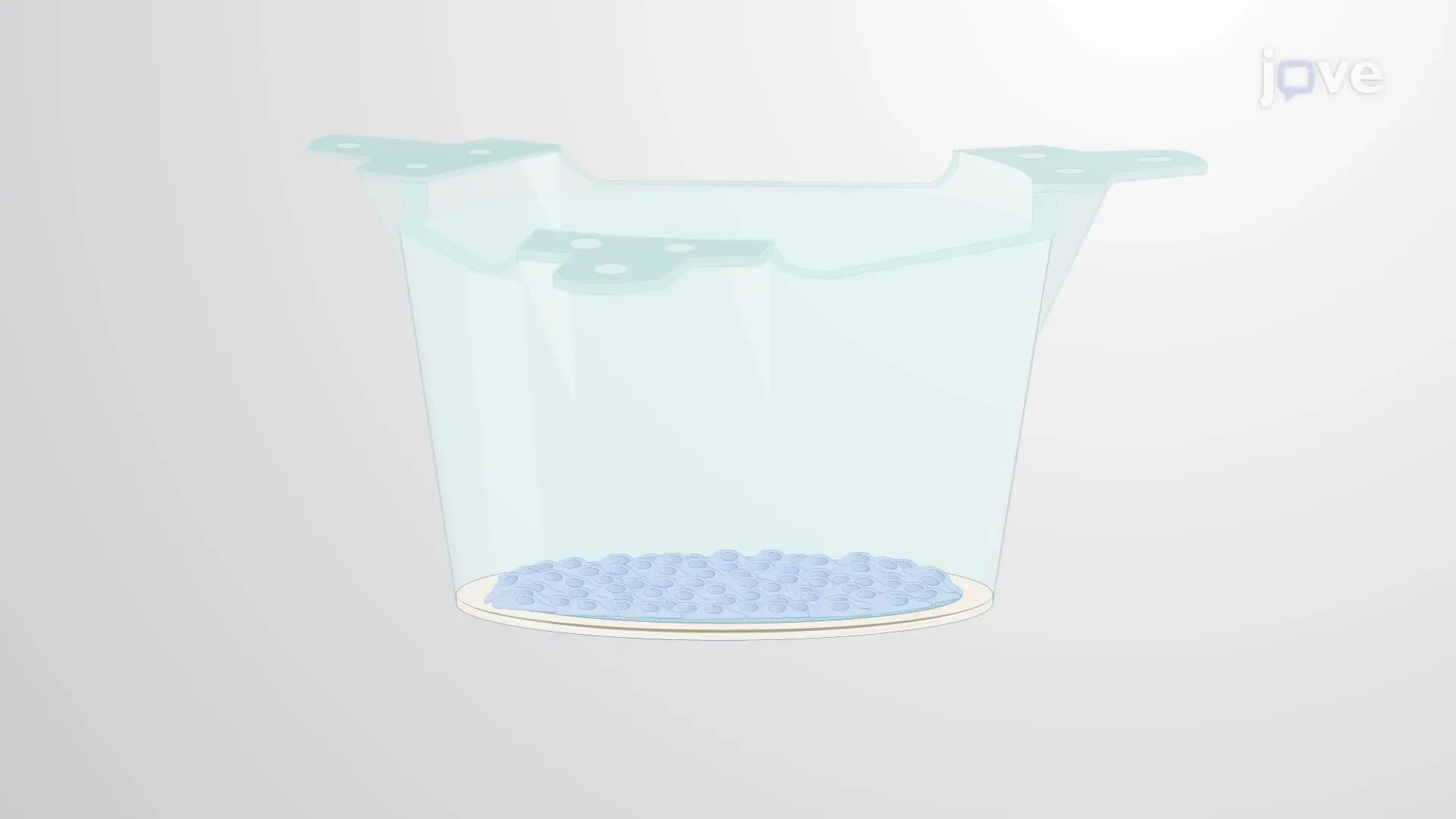
This video demonstrates a method for assessing transendothelial electrical resistance (TEER) in mouse brain microvascular endothelial cells in vitro. A high TEER value indicates intact endothelial cell junctions, while the addition of activated T cells lowers the TEER value, reflecting disruption of the junctional barrier.
Protocol

1. Setting Up and Performing Transendothelial Electrical Resistance (TEER) Measurement

  1. Place the 24-well module of the transendothelial electrical resistance (TEER) instrument under the laminar flow hood. Remove the lids and place inserts with primary mouse brain microvascular endothelial cells (MBMECs) in the instrument using forceps.
  2. Pipette 810 µl of fresh medium to the lower compartment of the module wells: add it carefully between the insert and the wall of the module well.
  3. Close the lids and place the instrument in the incubator. Connect the instrument to its computer; turn on the instrument controller and open the software.
  4. Select 'new measurement' in the pop-up window. Check the 'show TEER' and 'show Ccl' boxes; then press 'start.' After completing the first measurement, select 'check all wells' in the 'results' tab to see all TEER and cell layer capacitance (Ccl) values.
  5. Save the file: File>Save as>' the name of your file'.
    NOTE: As TEER and Ccl are continuously measured in an automated fashion, monitor their values over a period of three to five days. Changing the medium is not necessary unless cell viability is suboptimal, as visualized by a lack of increase in TEER.
  6. Choose the time point to co-culture MBMECs with T cells when Ccl is stable and lower than 1 µF/cm2 and TEER has reached its maximum level.
  7. Carefully inspect absolute TEER and Ccl values and exclude the wells in which MBMECs have not developed confluent enough monolayers by unchecking such wells.
  8. Group the rest of the wells: right-click on a well and select 'add well to new average well'. Name it in the pop-up window; do the same for all individual wells to be grouped.
  9. Check all average wells to confirm that all of them have the same initial conditions before the co-culture. If some have significantly different absolute TEER values or TEER slopes, or the standard errors are too large, redo the grouping.
    NOTE: The optimal grouping of wells provides minimal variation in TEER values within and between experimental groups.
  10. In the 'experiment' tab, press 'pause', disconnect the instrument and take it out of the incubator. Remove the lids under the laminar flow hood.
  11. Prepare pre-activated and/or naïve T cells, with or without specific cytokines, antibodies, or other substances, as desired. For example, mix purified native/low endotoxin (NA/LE) rat anti-mouse interferon-gamma (IFN-γ) antibody (clone XGM1.2) with prepared T cells at 20 µg/ml per well of the TEER instrument.
    NOTE: If substances such as granzyme B inhibitor are used, they are added to T cell culture at the beginning of T cell stimulation: e.g., Granzyme B Inhibitor II (Calbiochem) is mixed with isolated T cells at a final concentration of 10 µM in dimethyl sulfoxide (DMSO).
  12. Remove some of the media from the insert (the upper well compartment). Example: carefully pipette out 150 µl (removing all the medium should be avoided as it could disturb the MBMEC monolayer).
  13. Add T cells to MBMECs by carefully pipetting 150 µl of medium containing 2 x 10⁵ cells/insert.
    NOTE: When harvesting pre-activated T cells, count only blasting Trypan Blue-free cells.
  14. Close the lids and place the instrument back in the incubator. Reconnect the instrument and press 'resume measurement'. After 24 hours, press 'stop' in the 'experiment' tab and save the file (File>Save).

Toegang beperkt. Log in of start een proefperiode om deze inhoud te bekijken.

Openbaarmakingen

No conflicts of interest declared.

Materialen

Lijst van materialen gebruikt in dit artikel
NaamBedrijfCatalogusnummerOpmerkingen
cellZscopenanoAnalytics GmbHwww.nanoanalytics.comincluding: 24-well Cell Module, Controller, PC with cellZscope software v2.2.2
Transwell membrane inserts - pore size 0.4 µmCorning3470for TEER measurement as the main readout
Transwell membrane inserts - pore size 3 µmCorning3472for TEER measurement as the quality control prior to T-cell transmigration assay
24-well cell culture plateGreiner650 180flat-bottom; for MBMEC culture
96-well cell culture plateCostar3526round-bottom; for immune cell culture
Neubauer counting chamberMarienfeldMF-0640010for cell counting
Collagen type IV from human placentaSigmaC5533for MBMEC coating solution
Fibronectin from bovine plasmaSigmaF1141-5MGfor MBMEC coating solution
DMEM (+ GlutaMAX)Gibco31966-021for MBMEC isolation and MBMEC culture medium
Penicillin/StreptomycinSigmaP4333for MBMEC isolation and MBMEC culture medium
HeparinSigmaH3393for MBMEC culture medium
Human Basic Fibroblast Growth Factor (bFGF)PeproTech100-18Bfor MBMEC culture medium
IMDM + 1% L-GlutaminGibco21980-032for T cell culture medium
Recombinant Murine IFN-γPeproTech315-05for T-cell transmigration assays
Recombinant Murine TNF-αPeproTech315-01Afor T-cell transmigration assays
NA/LE purified anti-mouse IFN-γ antibodyBD Biosciences554408clone XMG1.2; recommended final concentration: 20 µg/ml
Granzyme B Inhibitor IICalbiochem368055recommended final concentration: 10 µM
PE anti-mouse CD4 antibodyBiolegend116005clone RM4-4; for analysis of T cell transmigration
Trypan Blue solution, 0.4%Thermo Fisher Scientific15250061for cell counting

Trefwoorden

Microvasculaire endotheelcellen uit de muizenhersenenT cel endotheelcelinteractieimpedantiespectroscopieendotheelcelverbindingenverstoring van de junctionele barri reTEER meetprotocolin vitro BBB modelgeactiveerde T cellen