This video demonstrates the induction and monitoring of elevated intraocular pressure (IOP) in a rat model. The process involves cauterizing the limbal blood vessels, resulting in vessel occlusion and elevated IOP. The rat is then allowed to recover, and IOP is regularly monitored in both the experimental and control eyes for comparison.
Een abonnement op JoVE is vereist om deze inhoud te bekijken. Log in of start vandaag met uw gratis proefperiode.
Methodenartikel
Induction of Elevated Intraocular Pressure in a Rat Model
1K weergaven
⸱
28 april 2025
In dit artikel
Samenvatting
Protocol
All procedures involving animal models have been reviewed by the local institutional animal care committee and the JoVE veterinary review board.
In the present work, Lister Hooded rats of both genders were used, aged 2-3 months and weighing 180-320 g. However, the procedure can be adapted in different rat strains of various age ranges.
Ocular hypertension surgery and clinical follow-up
- House animals in a controlled temperature environment and 12 h light/dark cycle (6 am: light on/ 6 pm: light off) with standard pelletized gamma-irradiated food (Nuvilab® CR-1, Quimtia S/A, Brazil) and water (triple filter, with non-toxic activated charcoal) available ad libitum.
- Prepare a stock anesthetic cocktail mixture composed of three parts of 10% ketamine hydrochloride and one part of 2% xylazine hydrochloride (diluent: sterile water).
- Gently restrain the animal by holding the dorsal skin and induce anesthesia by intraperitoneal administration of 1 µL/g of body weight of the mixture (ketamine: 75 mg/kg; xylazine: 5 mg/kg). Check for proper anesthetization after approximately 5 min by performing a toe pinch response.
- Topically anesthetize both eye surfaces by instilling proxymetacaine hydrochloride 0.5% eye drop. Wait for 30-60 s and remove the remaining solution from the anterior aspect of the globe, by gently touching the nasal or lateral bulbar conjunctiva with a small sterile cotton swab.
- Measure baseline IOP of both experimental and contralateral control eyes by placing the animal in a ventral decubitus position on the benchtop such that the corneal surface is easily accessible to the tip of the tonometer.
- Use either applanation or rebound handheld tonometer (Figure 1A). Position the tonometer tip such that it slightly touches the central corneal zone perpendicularly. Acquire and average 3-5 reliable machine-generated averages. Each mean is automatically calculated by the device after six successful individual measurements.
- Load the rebound tonometer with a probe and press the measurement button once to turn it on, while placing the tip of the device upwards to avoid the probe falling. After turning it on, the display will show 00 indicating that the equipment is ready to measure.
- Position the device with the tip of the probe at 1-4 mm from the cornea and acquire measurements by rapidly and carefully pressing the measurement button, without moving the equipment. Each successful measure is identified by a short beep and after six times, the mean is displayed on the device's screen.
NOTE: Despite a long experience with applanation tonometer, to optimize the entire procedure the authors recommend the use of a rebound tonometer, which provides easier acquisition of reliable IOP measurement.
- Place the animal on a slight lateral decubitus position under a stereo microscope, and carefully plan the surgery by inspecting the experimental eye at 40x magnification. Do not forget to maintain the contralateral control eye lubricated during the procedure by instilling a drop of carmellose sodium or sodium hyaluronate.
- With the aid of curved forceps, gently push forward the experimental eyeball so as to expose the vasculature that surrounds 360° of the limbus. With the other hand, gently cauterize the vessels all around the cornea with a low-temperature ophthalmic cautery (1,300 °F; Bovie Medical, USA) (Figure 1B-E).
NOTE: The mentioned cautery has a round tip that should touch limbal vasculature longitudinally. - Be careful not to cauterize the corneal periphery, as this may result in post-operative corneal opacification, which precludes in vivo assessment of retinal function.
- Observe the emergence of small circular marks of cauterization on the scleral limbus, the obliteration of limbal vasculature, and pupil dilation in the operated eye, which are signs of a successful surgical procedure.
NOTE: As the limbal vasculature of rats and mice is anatomically similar and the cautery used has a gentle small tip, this protocol can work for the mouse eye as well without any adaptation. - After surgery, check the immediate post-operative IOP in both eyes such as described in step 1.5.
- Apply a drop of ophthalmic prednisolone acetate (1.2 mg/mL) and maintain this in contact with the anterior surface of the experimental eye for around 40 s, then replace it with an ophthalmic ointment of antibiotics (oxytetracycline hydrochloride 30 mg/g plus polymyxin B 10,000 U/g; or ciprofloxacin 3.5 mg/g). Perform intramuscular injection of tramadol hydrochloride (single dose; 2 mg/kg) to prevent pain after the procedure.
Toegang beperkt. Log in of start een proefperiode om deze inhoud te bekijken.
Resultaten
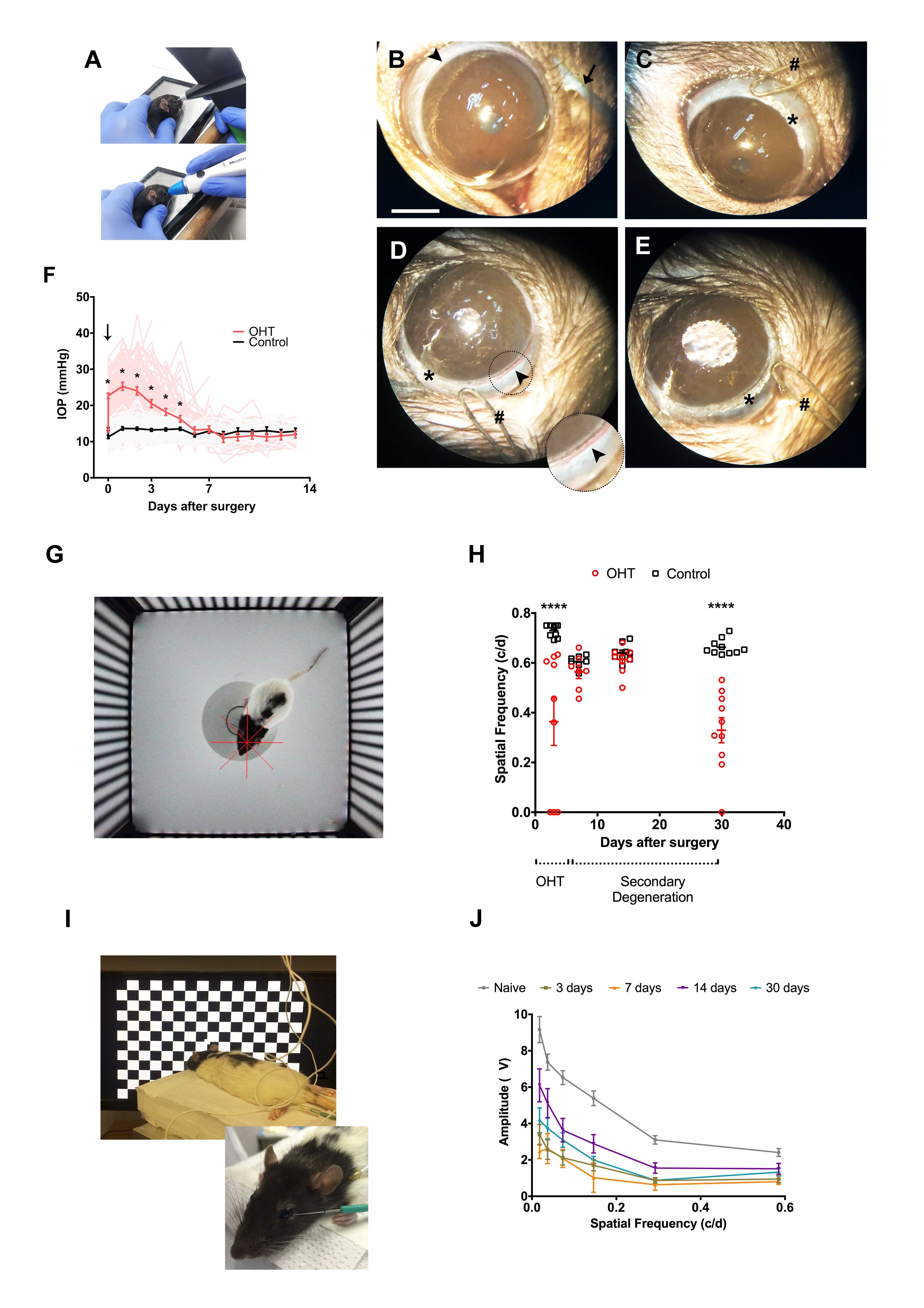
Figure 1: Thermal cauterization of limbal vascular plexus and consequences to retinal function in vivo. (A) Alternative methods to measure IOP in rats: rebound tonometry (superior), and applanation tono...
Toegang beperkt. Log in of start een proefperiode om deze inhoud te bekijken.
Materialen
| Naam | Bedrijf | Catalogusnummer | Opmerkingen |
|---|---|---|---|
| Cautery Low Temp Fine Tip 10/bx | Bovie Medical Corporation | AA00 | Low-temperature ophthalmic cautery |
| Cetamin | Syntec do Brasil Ltda | 000200-3-000003 | Ketamine hydrochloride 10% |
| Ecofilm | Cristália Produtos Químicos Farmacêuticos Ltda | MS-1.0298.0487 | Carmellose sodium 0.5% |
| Hyabak | União Química Farmacêutica Nacional S/A | MS-8042140002 | Sodium hyaluronate 0.15% |
| Icare Tonolab | Icare Finland Oy | TV02 (model number) | Rebound handheld tonometer |
| Maxiflox | Cristália Produtos Químicos Farmacêuticos Ltda | MS-1.0298.0489 | Ciprofloxacin 3.5 mg/g |
| Ster MD | União Química Farmacêutica Nacional S/A | MS-1.0497.1287 | Prednisolone acetate 0.12% |
| Terolac | Cristália Produtos Químicos Farmacêuticos Ltda | MS-1.0497.1286 | Ketorolac trometamol 0.5% |
| Terramicina | Laboratórios Pfizer Ltda | MS-1.0216.0024 | Oxytetracycline hydrochloride 30 mg/ g + polymyxin B 10,000 U/g |
| Tono-Pen XL | Reichert Technologies | 230635 | Digital applanation handheld tonometer |
| Carl Zeiss | - | Stereo microscope for surgery and retinal dissection |
