This video demonstrates the use of Doppler ultrasound as a functional imaging tool to measure cerebral blood flow velocity, or CBFV in real time. It highlights how frequency shifts in the middle cerebral artery during tasks like breath-holding reveal changes in CBFV, offering insights into brain activity and vascular dynamics.
Een abonnement op JoVE is vereist om deze inhoud te bekijken. Log in of start vandaag met uw gratis proefperiode.
Methodenartikel
Functional Imaging of Cerebral Blood Flow Using Transcranial Doppler Ultrasound
1.7K weergaven
⸱
29 mei 2025
In dit artikel
Samenvatting
Protocol
All procedures involving human participants have been performed in compliance with the institutional, national, and international guidelines for human welfare and have been reviewed by the local institutional review board.
1. Locating the middle cerebral artery (MCA) signal by freehand transcranial Doppler ultrasound (TCD)
NOTE: “Freehand” TCD refers to an operation of TCD with a handheld transducer to find a cerebral blood flow velocity (CBFV) signal before beginning a functional transcranial Doppler ultrasound (fTCD) experiment.
- Setting TCD parameters
- Keep the power at a reasonably high value (e.g., 400 mW) during the initial search for the MCA. Once the MCA signal is located, reduce the power as much as possible while still maintaining a “good” signal (see step 2.2.7).
NOTE: Using a reasonably high power during the initial search does not violate the “As Low As Reasonably Achievable” (ALARA) principle of exposure to acoustic radiation because higher power will allow the MCA signal to be discovered more quickly. - Set the sample volume to 8–12 mm during the initial search for the MCA signal. If the signal is difficult to find, increase the gate size to increase the intensity of the signal, but note that this may incorporate the signal from one or more nearby arteries into the signal from the MCA.
- Set the gain at a medium level, with the goal of “keeping background noise at a minimum, but present”.
- Set the high-pass filter cutoff (normally termed “threshold”) to 50–150 Hz.
- If the subject is an adult, set the depth to 50 mm, which is the average mid-point depth of the M1 segment of the MCA (Figure 1).
NOTE: This setting will be discussed in more detail in subsequent steps. Depth settings for children are given in Table 1.
- Keep the power at a reasonably high value (e.g., 400 mW) during the initial search for the MCA. Once the MCA signal is located, reduce the power as much as possible while still maintaining a “good” signal (see step 2.2.7).
2. Locating the temporal window
NOTE: The temporal window, also called the transtemporal acoustic window, is a part of the skull where the bone is thinnest, thus allowing transmission of low-frequency ultrasound energy through the cranium (Figure 2).
- For infants and small children, locate the temporal window just in front of the ear (the “intertragal space”) and above the rostral edge of the zygomatic arch, which can be easily felt under the skin.
- For teenagers and young adults, locate the temporal window via any of the subwindows.
NOTE: The posterior subwindow usually provides the best signal (Figure 2). - For adults aged 30 years or older, locate the temporal window just in front of the ear.
NOTE: The acoustic window decreases in size as people age due to increasing porosity of the bone of the cranium, causing some older people to have a very limited temporal window. In such individuals, bilateral insonation of the MCA is sometimes impossible.
3. Applying the transducer
- Apply enough ultrasound gel to cover the surface of the transducer.
NOTE: When placed on the head, the gel should cover sufficient space to maintain a seal between the scalp and the Doppler probe’s surface, thus preventing signal interruption from air coupling underneath the probe’s surface. - Alert the subject that the gel may feel cold (if at room temperature).
- Place the transducer on the temporal window, which was located in section 1.2.
4. Searching for the MCA
- After placing the transducer on the scalp, search for the MCA signal, which will generally be located slightly anterior (forwards) and rostral (towards the head) from the location of the initial transducer scalp placement.
- If the TCD spectral signal is not immediately obvious, adjust the angle of the transducer while keeping it in the same location relative to the scalp. Slowly angle the probe from rostral to caudal (towards feet) and posterior to anterior.
NOTE: Figure 3 shows two spectra taken from the same position, but at different angles. - If a signal is still absent after performing step 1.4.2, check the color M-mode display for flow in the MCA at different depths (indicated by red coloring). Increment or decrement the signal depth in 5 mm steps and search as described in step 1.4.2. If flow is visible in M-mode but not in the Doppler spectrum, increase or decrease the depth until the flow signal is visible in the Doppler spectrum.
- If a satisfactory signal is still not obtained, move the transducer to a nearby position on the scalp, which is slightly more anterior, and repeat steps 1.4.1–1.4.3.
- When an optimal MCA signal is obtained, note the depth and maximum velocity.
- Using a washable makeup pen, place a mark on the scalp (trace part of the transducer edge) where the optimal signal was found.
5. Searching for the bifurcation
NOTE: Finding the bifurcation of the internal carotid artery (ICA) is important to help confirm that the MCA is the artery being monitored. This step should be performed on both sides if bilateral monitoring will be performed, as the bifurcation may not be at the same depth on both sides.
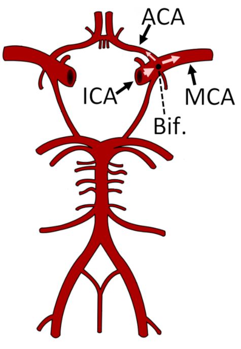
- Increase the depth until the signal from the bifurcation of the ICA into the MCA and anterior cerebral artery (ACA) is noted (Figure 4), typically at a depth of 51–65 mm.
- Search for the optimum bifurcation spectral signal using the procedure described in step 1.4.2. Always strive for the highest-velocity spectral signal possible.
- When an optimal bifurcation signal is obtained, note the depth of the bifurcation.
- For bilateral monitoring, repeat sections 1.1–1.4 and steps 1.5.1–1.5.3 on the other side of the head.
2. Relocating the MCA after placing a fixation device
NOTE: For fTCD experiments, it is necessary to monitor CBFV for 10–90 min or longer. Therefore, a fixation device (Figure 5) is crucial to provide stability.
- Placing the fixation device
- By visual inspection, adjust the fixation device (Figure 5) to the subject’s approximate head size.
- Alert the subject before placing the headset on his or her head. Place the headset on the subject’s head.
NOTE: If the subject has long or thick hair, it may be necessary to tie the subject’s hair back, depending on the fixation device being used. - Adjust the fixation device’s fit, and ask the subject if the device is too tight.
NOTE: The device should be tight enough that it does not move when bumped slightly, but loose enough that the subject is not uncomfortable.
- Locating the MCA signal
- Loosen the mechanism of the fixation device holding the transducer in place (e.g., loosen the mechanism, shown in in Figure 5, by turning a knob counterclockwise) so that the transducer can move freely.
- Alert the subject before applying gel to the transducers (which should already be in place from section 2.1), and that the gel may be cold (if it has been stored at room temperature).
- Apply enough ultrasound gel to the transducer to cover the face of the transducer.
- Adjust the fixation device so that the transducer is located over the top of the mark made in step 1.4.6.
- Search for the optimal MCA spectral signal using the procedure described in steps 1.4.1–1.4.3. Always strive for the highest-velocity spectral signal possible.
NOTE: When compared to freehand TCD, the optimal depth at which the MCA is located using the fixation device may differ slightly (at most 1–2 mm) from the depth for the freehand device. This is because the fixation device may hold the transducer slightly further away from the scalp while still maintaining a coupling gel seal. - When the optimal MCA spectral signal is found, tighten the mechanism of the fixation device to lock the transducer in place. Note the depth and all other settings.
- Decrease the power (see step 1.1.1) as much as possible while still maintaining a spectral envelope that traces the maximal velocity accurately.
- For bilateral monitoring, repeat steps 2.2.1–2.2.7 on the other side.
3. Performing a breath-hold maneuver
NOTE: This section is given as an example of a functional experiment that may be performed using the experimental setup described in section 1 and section 2.
- Perform all steps described in section 1 and section 2.
- Begin recording on the TCD software.
- Breathe normally for 3 min to achieve a good baseline recording, and allow CBFV to stabilize from any previous experiments or stimuli.
- Count down slowly from three. On the count of one, ask the subject to begin breath-holding following a normal inspiration.
NOTE: The subject should not inhale deeply, as this would decrease carbon dioxide in the lungs and decrease the likelihood of observing the increase in CBFV due to cerebrovascular reactivity. The subject should also avoid performing a Valsalva maneuver, in which intrathoracic pressure is substantially increased against a held inspiration. - Place a marker in the TCD recording to signify the start of breath-holding.
- Have the subject hold their breath for 30 s, or until they are no longer comfortable holding their breath.
- When the subject inhales, place a marker in the TCD recording to signify the end of breath-holding.
- Continue monitoring CBFV using TCD and recording for at least 30 s following the end of breath-holding to ensure that CBFV returns to baseline values.
Table 1: MCA depths at various ages.
Toegang beperkt. Log in of start een proefperiode om deze inhoud te bekijken.
Resultaten
Toegang beperkt. Log in of start een proefperiode om deze inhoud te bekijken.
Materialen
| Naam | Bedrijf | Catalogusnummer | Opmerkingen |
|---|---|---|---|
| Aquasonic | |||
| Doppler Box X | |||
| Kimwipes | |||
| Transeptic |
