This video demonstrates time-lapse imaging of neural stem cells (NSCs) from a transgenic mouse brain using a confocal laser scanning microscope. NSCs fluoresce red during the G1 phase and are non-fluorescent in other cell-cycle phases. An adhered NSC culture is placed in the imaging chamber, imaging parameters are set, and images are captured at regular intervals for the desired duration.
Een abonnement op JoVE is vereist om deze inhoud te bekijken. Log in of start vandaag met uw gratis proefperiode.
Methodenartikel
Time-Lapse Imaging of Mouse Neural Stem Cell Division Using Confocal Microscopy
751 weergaven
⸱
18 juni 2025
In dit artikel
Samenvatting
Protocol
1. Basic Setup Prior to Culture and Videomicroscopy
- Use glass-bottom culture plates or µ-Plates for confocal video microscopy. For 1 - 5 x 10³ cells/well, use 96-well plates, and for more than 5 x 10³ cells/well, use 24-well plates.
- At least one day prior to commencing the experiment, prepare sterile Poly-D-lysine (PDL) coated plates for adherent monolayer cultures. Add enough PDL (10 µg/ml in distilled water or dH2O) to coat the bottom of each well and incubate overnight (O/N) at 37 °C. Remove the PDL solution and rinse three times with dH2O before allowing the plate to dry in the hood during a....
Toegang beperkt. Log in of start een proefperiode om deze inhoud te bekijken.
Resultaten
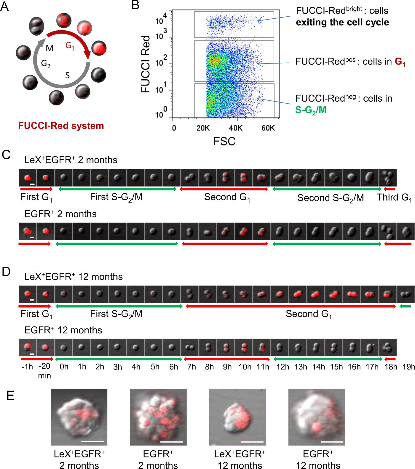
Figure 1: Live analyses of cell cycle using FUCCI-Red mice. (A) Schematic representation of FU.......
Toegang beperkt. Log in of start een proefperiode om deze inhoud te bekijken.
Materialen
| Naam | Bedrijf | Catalogusnummer | Opmerkingen |
|---|---|---|---|
| 96-well uncoated glass bottom culture plates | MatTek Corp. | P96G-1.5-5-F | |
| µ-Plates 96-well | Ibidi | 89626 | |
| Poly-D-Lysine | Millipore | A003E | |
| NeuroCult NSC Basal Medium | STEMCELL Technologies | 5700 | |
| NeuroCult NSC Proliferation Supplement | STEMCELL Technologies | 5701 | |
| Heparin | STEMCELL Technologies | 7980 | |
| EGF | Millipore | GF144 | |
| FGF-2 | Millipore | GF003 | |
| Papain | Worthington | LS003119 | |
| EBSS | Invitrogen | 24010-043 | |
| L-cysteine | Sigma | C-7352 | |
| 0.5M EDTA, pH 8.0 | Promega | V4231 | |
| DNase I | Sigma | D5025-15KU | |
| Trypsin inhibitor type II | Sigma | T9128 | Ovomucoid |
| DMEM:F12 medium | Life Technologies | 31330-038 | |
| 20 µm filter | BD Medimachine Filcon | 340622 | |
| Eppendorf microtubes 3810X | Sigma | Z606340-1000EA | |
| BSA | Sigma | A1595 | Bovine serum albumin solution |
| CD24 phycoerythrin-cyanine7 conjugate [PC7] | Life Technologies | A14776 | Mouse IgM; clone MMA. 1:50 |
| CD15/LeX fluorescein isothiocyanate [FITC] - conjugated | BD Biosciences | 332778 | Rat IgG2b; clone M1/69. 1:50 |
| Mouse anti-human LeX antibody | BD Pharmingen | 559045 | Mouse IgM; clone MAM. 1:50 |
| Alexa647 - conjugated EGF ligand | Life Technologies | E35351 | 1:200; Ax647-conjugated EGF ligand |
| CompBeads | BD Biosciences | 644204 | |
| MACS LS separation columns | Miltenyi Biotec | 130-042-401 | Separation columns |
| Anti-mouse IgM microbeads | Miltenyi Biotec | 130-047-301 | |
| Hoechst 33258 | Sigma | 861405 | |
| INFLUX cell sorter | BD Biosciences | FACS sorter | |
| Nikon A1R confocal laser scanning microscope system attached to an inverted ECLIPSE Ti | Nikon Corp. | Confocal laser scanning microscope | |
| NIS-Elements AR.4.13.01 64-bit software | Nikon Corp. | NIS-Elements software | |
| Plan Apo VC 20x DIC objective (NA: 0.75) | Nikon Corp. | ||
| ECLIPSE Ti thermostated chamber | Nikon Corp. | Thermostated chamber | |
| ImageJ | RBS | ||
| FlowJo | Tree Star, Ashland, OR | ||
| FUCCI mice | RIKEN BioResource Center, JAPAN |
