This video demonstrates near-infrared fluorescent (NIRF) imaging to visualize thrombi in excised brain tissue labeled with a fibrin-specific probe. Near-infrared light excites the probe, causing the thrombus to fluoresce, enabling high-contrast thrombi visualization in the major cerebral artery and small cortical blood vessels.
Een abonnement op JoVE is vereist om deze inhoud te bekijken. Log in of start vandaag met uw gratis proefperiode.
Methodenartikel
Visualization of Mouse Cerebral Thrombi Using Near-Infrared Fluorescence Imaging
676 weergaven
⸱
17 juni 2025
In dit artikel
Samenvatting
Protocol
All procedures involving animal samples have been reviewed and approved by the appropriate animal ethical review committee.
1. Ex Vivo Near-infrared fluorescent (NIRF) Thrombus Imaging of the Brain Tissue
- After decapitation, remove the scalp and cut through the skull with scissors from the foramen magnum up toward the sagittal suture. Remove the cranial vault by carefully elevating the edges of the incised skull using scissors while avoiding injury to the underlying brain, thus laying bare the hemispheres.
- Cut out the optic nerves in the brain base as close as possible to the brain surface because they could overlap with the circle of Willis (COW) arteries that should be visualized on near-infrared fluorescent (NIRF) imaging. Then, to visualize the Y-shape 'middle cerebral artery (MCA) / anterior cerebral artery (ACA) / distal internal carotid artery (ICA)' for the NIRF thrombus imaging, cut out the distal ICAs as far as possible to the brain surface after gently compressing the cerebellar base to expose the cutting points (Figure 1A).
- Perform NIRF imaging (excitation/emission, 675 / 690 nm; 1-sec exposure) of the removed brain tissue with its base pointing up (Figure 1B, C), which visualizes the fluorescently labeled thromboemboli in the arteries of the COW (Figure 1D). Then, perform additional imaging with the vertex of the brain pointing up, which visualizes cortical thromboemboli. Avoid drying the brain by putting a few drops of saline on the tissue.
Toegang beperkt. Log in of start een proefperiode om deze inhoud te bekijken.
Resultaten
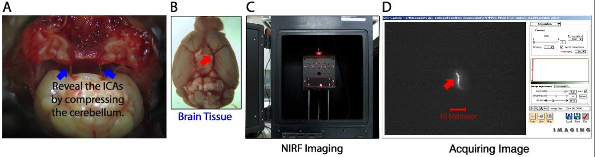
Figure 1: Overview of Ex Vivo Near-Infrared Fluorescent (NIRF) Imaging of Cerebr...
Toegang beperkt. Log in of start een proefperiode om deze inhoud te bekijken.
Materialen
| Naam | Bedrijf | Catalogusnummer | Opmerkingen |
|---|---|---|---|
| Machines | |||
| NIRF imaging system | Roper-scientific,Tucson, AZ | Coolsnap-Ez | |
| Surgical microscope | Leica Microsystems, Seoul, Korea | EZ4HD | |
| Software | |||
| Image J software | Wanye Rasband, NIH, USA | 1.49d | Imaging analysis |
| Micro forceps | Fine Science Tools, Vancouver, Canada | 11253-27 | Dumont L5 |
| Micro scissor | Fine Science Tools, Vancouver, Canada | 15000-03 | Vannas spring |
| Scissor | Fine Science Tools, Vancouver, Canada | 14084-08 | 8.5 cm |
| Black silk suture | Ailee, Busan, Korea | SK6071, SK728 | 6-0 and 7-0 |
| Reagents | |||
| TTC | Amresco, Ohio, USA | 0765-100g | |
| C15 optical agent | Synthesis |
Meer artikelen verkennen
