This video demonstrates the use of high-intensity focused ultrasound (HIFU) to block action potentials from the sciatic nerve to the gastrocnemius muscle in a diabetic rat model. Upon delivering the HIFU beam to the nerve, a decrease in muscle signal confirms HIFU's potential as a non-invasive therapy for diabetic neuropathic pain relief.
Een abonnement op JoVE is vereist om deze inhoud te bekijken. Log in of start vandaag met uw gratis proefperiode.
Methodenartikel
High-Intensity Focused Ultrasound for Blocking Nerve Action Potentials in a Diabetic Rat Model
1K weergaven
⸱
7 augustus 2025
In dit artikel
Samenvatting
Protocol
All procedures involving animal models have been reviewed by the local institutional animal care committee and the JoVE veterinary review board.
1. In Vivo Nerve Conduction Blockage with the High-Intensity Focused Ultrasound (HIFU) Transducer
NOTE: The in vivo experiment starts on week 5 after a 50 mg/kg streptozotocin (STZ) injection.
- Perform animal procedures before blocking the compound muscle action potentials (CMAPs) with HIFU sonication.
- Sterilize the surgical tools (scalpel, scissors, forceps, and glass hook) in an autoclave before the surgery.
- Anesthetize the rats with an intraperitoneal injection of tiletamine/zolazepam mixture (40 mg/kg) and xylazine (10 mg/kg) or via inhalation of 1.75% of isoflurane via isoflurane vaporizer. Place the rats on a heating pad to maintain their body temperature.
- Position the rats for surgery in ventral recumbency. Apply the eye ointment. Extend the leg of the rat and pinch the plantar surface of the foot with fingernails to ascertain the depth of anesthesia. If the rats show withdrawal responses, apply additional anesthesia.
- Remove hair from the thigh and lower back of the rats with an electric clipper. Apply liquid iodine with clean gauze at the surgical site and circularly move the gauze to outward from the surgical site. Use an alcohol pad to wipe the liquid iodine with the same circular movement. This experiment is performed on the left and right surgical sites. Repeat the procedure at the other surgical site when performing the next step of the procedure.
- Use a sterile surgical scissors or scalpel to make an incision of the skin at the dorsal thigh. Use blunt surgical scissors to carefully separate the tissue underneath the skin and secure the skin with skin hooks. The femur can be seen within the muscles.
- Use scissors to carefully separate the muscles parallel to the femur until the mid-thigh sciatic nerve fibers that are embedded in the muscles are visible. Carefully use a glass hook to separate the mid-thigh sciatic nerve from the surrounding connective tissues and muscles.
- Position the sciatic nerve in the HIFU focal zone using a custom-made nerve fixator (Figure 1 and Figure 2).
NOTE: The custom-made nerve fixator consists of 3 components (Figure 2). All the components are made of transparent polymethylmethacrylate (PMMA). The external thread of the top structure of component I is 2.5 mm tall and M10XP0.7 (Figure 3A). The central well is a 4.0 mm-diameter hole through component I. The bottom opening of the well is sealed by a sheet of tape. The diameter of the slot is 1.2 mm, and the distance between the central plane of the slot and the top surface of component I is 3.1 mm. Component II consists of a main body, four legs, and the bottom structure (Figure 3B). The internal thread of the bottom structure is 2.5 mm tall and M10XP0.7 to fit the external thread of component I. The diameter of the central hole is the same as the central well of component I. The dimensions of the main body are 32 mm in diameter and 5.4 mm in thickness. Four legs are symmetrically deployed. Two identical short legs are designed for alignment, and two identical long legs work for hooking on component III. The outer diameter and height of component III are 41 mm and 9.2 mm. The internal thread is M36XP1.0, and the through hole is 27.5 mm in diameter (Figure 3C). The cone is a hollow taper with a top opening of 84 mm in diameter and a bottom opening of 27.5 mm in diameter. The height is 57.5 mm (Figure 4A).- Before the experiment, soak the custom-made acrylic nerve fixator in bleach solution for 30 - 60 min, followed by soaking in sterile water.
- Using a glass hook, lift the nerve carefully and put it in the slot of component I.
- Screw component II to component I. Fill the central well of component I with Ringer's solution for ultrasonic propagation and nerve preservation.
- Screw component III to the HIFU housing cone. Dock component III with component II through the four legs of component II.
NOTE: The geometric center of the three components and the transducer are aligned. The distance between the nerve and the transducer is equal to the focal length, which ensures the nerve is inside the HIFU focal zone.
- Insert one pair of acupuncture needles into the origin of the sciatic nerve and the other pair into the gastrocnemius muscle. Connect each pair of needles to the electrophysiology acquisition system through an electrical coaxial cable (Figure 1).
NOTE: On one end of the cable are two alligator clips to clip two needles separately, and on the other end of the cable is a bayonet Neill–Concelman (BNC) connector to link the system. The pairs of acupuncture needles work as stimulating electrodes at the sciatic nerve and the recording electrodes at the gastrocnemius muscle.- Set the sampling rate and bandwidth of the electrophysiology acquisition system to 50 kHz and 70 Hz-3 kHz, respectively. Apply a supra-maximal stimulus with a pulse width of 0.1 ms to the stimulating electrodes at the origin of the sciatic nerve.
- Record the CMAPs from the recording electrodes and amplify the CMAPs with the built-in amplifier in the electrophysiology acquisition system.
NOTE: Use the built-in amplifier in the electrophysiology acquisition system to amplify the nerve signals and record the CMAPs from the recording electrodes with the electrophysiology acquisition system.
- Use a commercial 2.68 MHz HIFU transducer to suppress the CMAPs in diabetic neuropathic rats.
NOTE: The specifications of the transducer are described as follows: a single-element spherical bowl with an aperture diameter of 6 cm and focal length of 5 cm, and an ellipsoid focal zone of 4 mm in depth and 0.8 mm in width in the free field.- Immerse the spherical cone, the HIFU transducer, and the cone cover in the tank filled with degassed water. Put the HIFU transducer into the spherical cone and fix the cone cover to the top opening of the spherical cone with six head screws (Figure 4B). After bubbles in the spherical cone are expelled naturally due to the low density of bubbles compared with water, seal the front-end opening of the cone with a transparent 0.03 mm-thick tape. Screw component III onto the spherical cone.
- Take out the HIFU transducer with the spherical cone and component III from the degassed water tank.
NOTE: The reverse osmosis (RO) water used in the study is the water purified by the process of reverse osmosis. The RO water is boiled to expel the gas. After cooling, the degassed water is obtained in an individual sealed tank.
- Put component I into the space between the nerve and the muscle carefully, and position the nerve in the gap of component I. Perform the steps 1.2.3 and 1.2.4 to ensure that the nerve is inside the focal zone of the HIFU (Figure 2A).
- Link a function generator and a radiofrequency power amplifier. Connect the power amplifier to the HIFU transducer to generate the HIFU beam. Manually set the voltage output of the function generator to the HIFU transducer via the power amplifier. Manually turn off the function generator once the HIFU exposure time is up. Observe time using a timer.
NOTE: The intensity and energy of the HIFU beam used in this study are 2,810 W/cm2 and 84 J/mm2, respectively. - Simultaneously, deliver the stimulus via the electrophysiology acquisition system (step 1.3) and HIFU beam via the HIFU system (step 1.6) to the sciatic nerve while recording the CMAPs. Gradually increase the HIFU exposure on the sciatic nerve from 3 s, 5 s to 8 s until a decrease or inhibition of the amplitude of CMAPs is observed.
- Record the CMAPs once per second during the delivery of the HIFU beam. After observing the change in the amplitude of CMAPs, turn off the HIFU system and manually click on the record icon on the electrophysiology acquisition software to record CMAPs every 2 min in the early 10 min, every 5 min in the consecutive 30 min, and every 10 min in the last phase until the recording time reaches 2 h.
- Separate components II and III of the nerve fixator (Figure 2) to remove the HIFU transducer from the incision site. Separate components II and I to release the secured sciatic nerve. Suture the surgical site of the diabetic rat with 4-0 chromic catgut sutures after recording the CMAPs. Apply liquid iodine to the surgical site to prevent infection.
- Place the cages on the heating pad and allow the rats to recover in their cages before returning them to the animal facility. Provide the rats with ibuprofen in drinking water for 3 days or an intraperitoneal injection of buprenorphine (0.05 - 0.1 mg/kg).
- Insert stimulating and recording electrodes in the origin of the sciatic nerve and the gastrocnemius muscles of anesthetized diabetic neuropathic rats as described in steps 1.1.2 and 1.3 on days 7, 14, and 28 after the initial HIFU sonication.
Toegang beperkt. Log in of start een proefperiode om deze inhoud te bekijken.
Resultaten
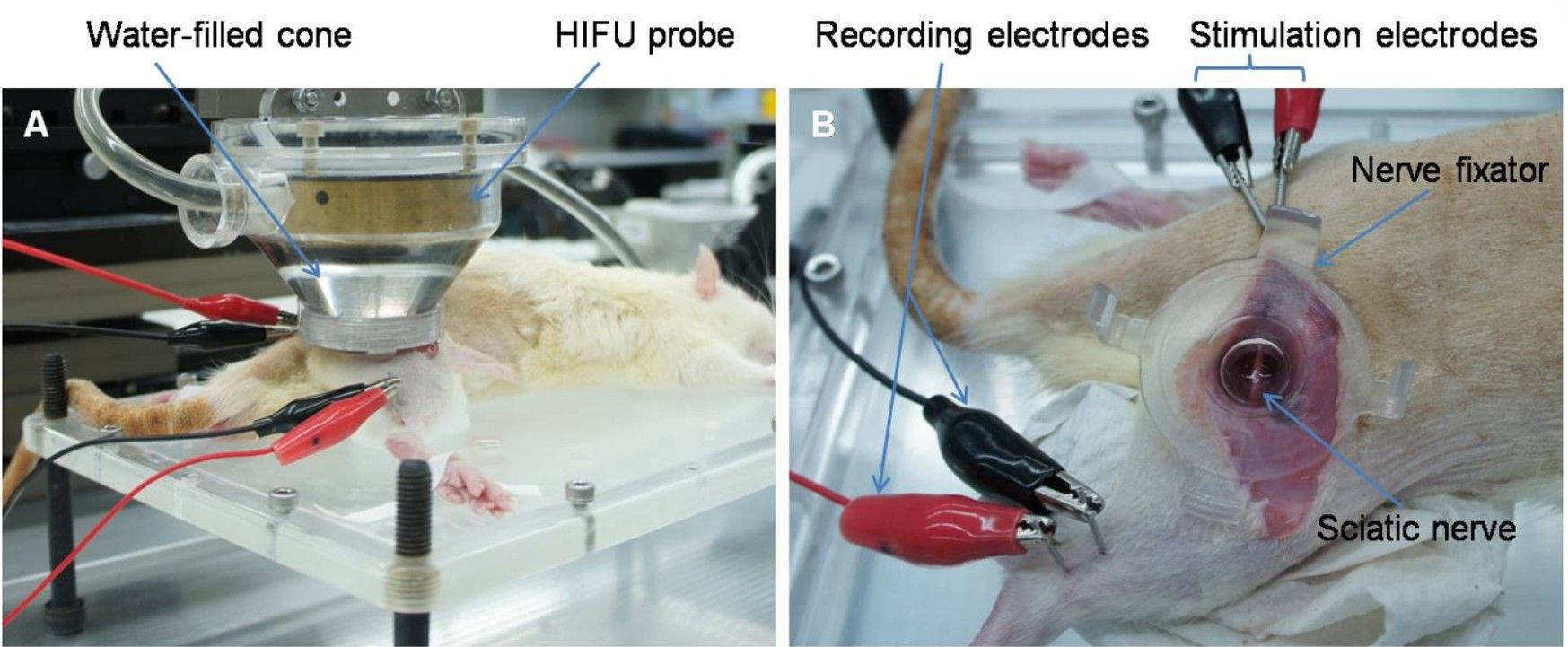
Figure 1: The Experimental Setup for In Vivo Nerve Conduction Block with High...
Toegang beperkt. Log in of start een proefperiode om deze inhoud te bekijken.
Materialen
| Naam | Bedrijf | Catalogusnummer | Opmerkingen |
|---|---|---|---|
| Streptozotocin | Sigma | 85882 | |
| Biopac MP36 acquisition system | Biopac Systems, Inc. | ||
| HIFU transducer | Sonic Concepts | H108 | |
| Function generator | Agilent | 33250A | |
| Power amplifier | Electronics & Innovation | 1040L | |
| Rats | Biolasco taiwan | Sprague-Dawley | |
| Puralube vet ointment | Dechra | ||
| Isoflurane vaporizer | Parkland Scientific | V3000PS | |
| Isoflurane | Attane |
