Het gebruik van varkens in onderzoek is de afgelopen jaren toegenomen. Niettemin worden varkens gekenmerkt door een moeilijke anatomie van de luchtwegen. Door aan te tonen hoe endoscopisch geleide endotracheale intubatie moet worden uitgevoerd, beoogt dit protocol de veiligheid van proefdieren verder te verhogen om dierenleed en onnodige dood te voorkomen.
Een abonnement op JoVE is vereist om deze inhoud te bekijken. Log in of start vandaag met uw gratis proefperiode.
Methodenartikel
Endotracheale intubatie met behulp van een flexibele intubatie-endoscoop als een gestandaardiseerd model voor veilig luchtwegbeheer bij varkens
In dit artikel
Erratum-melding
Important: There has been an erratum issued for this article. View Erratum Notice
Samenvatting
Samenvatting
Endotracheale intubatie is vaak een basisvereiste voor translationeel onderzoek in varkensmodellen voor verschillende interventies die een beveiligde luchtweg of hoge ventilatiedruk vereisen. Endotracheale intubatie is een uitdagende vaardigheid, die een minimum aantal succesvolle endotracheale intubaties vereist om een hoog slagingspercentage onder optimale omstandigheden te bereiken, wat vaak onhaalbaar is voor niet-anesthesiologische onderzoekers. Vanwege de specifieke anatomie van de varkensluchtwegen kan meestal worden uitgegaan van een moeilijke luchtweg. De onmogelijkheid om een veilige luchtweg tot stand te brengen kan leiden tot letsel, bijwerkingen of de dood van het proefdier. Met behulp van een prospectieve, gerandomiseerde, gecontroleerde evaluatiebenadering is aangetoond dat fiberoptic-assisted endotracheale intubatie langer duurt, maar een hoger first-pass succespercentage heeft dan conventionele intubatie zonder klinisch relevante dalingen in zuurstofverzadiging te veroorzaken. Dit model presenteert een gestandaardiseerd regime voor endoscopisch geleide endotracheale intubatie, dat een beveiligde luchtweg biedt, vooral voor onderzoekers die onervaren zijn in de techniek van endotracheale intubatie via directe laryngoscopie. Deze procedure zal naar verwachting dierenleed en onnodige verliezen van dieren minimaliseren.
Inleiding
Endotracheale intubatie is vaak een basisvereiste voor translationeel onderzoek in varkensmodellen voor verschillende interventies die een beveiligde luchtweg of hoge beademingsdruk vereisen (zoals ventilatie tijdens cardiopulmonale reanimatie1 of acuut respiratoir distress-syndroom2) of vereisen dat de cerebrale bloedstroom niet wordt aangetast door interne compressie door supraglottische luchtwegapparaten3 , die soms als alternatief worden vermeerderd in het kader van een verwachte moeilijke luchtweg bij varkens 4,5.
Hoewel de longfysiologie van varkens vergelijkbare kenmerken vertoont als die van mensen6, is het beveiligen van de luchtweg soms aanzienlijk moeilijker7 vanwege specifieke verschillen in de orotracheale anatomie van varkens. De snuit van een varken heeft een smalle opening met een zeer grote tong, het strottenhoofd is extreem mobiel en de epiglottis is relatief groot, met een vrij uiteinde dat zich uitstrekt tot het zachte gehemelte. Caudally, het strottenhoofd vormt een stompe hoek met de luchtpijp. Het arytenoïde kraakbeen is groot8. Het smalste deel van de luchtweg bevindt zich op subglottisch niveau9, vergelijkbaar met de luchtweganatomie van kinderen10. Omdat het strottenhoofd bij varkens zeer mobiel is, bestaat het risico dat het uiteinde van de endotracheale buis door de stembanden gaat, maar het strottenhoofd wordt slechts enkele centimeters caudaal verplaatst, wat kan worden aangezien voor een juiste intubatie 8,11. Bovendien is slokdarmintubatie een veel voorkomend risico bij het omgaan met varkensluchtwegbeheer12.
De percentages van moeilijke of onmogelijke endotracheale intubaties met een overeenkomstige negatieve impact op het experiment of vroege sterfte zijn niet systematisch geregistreerd, maar er zijn verschillende case reports gepubliceerd13,14. Bij mensen is er de mogelijkheid om een flexibele intubatie-endoscoop te gebruiken in de context van een onverwacht moeilijke conventionele intubatie15. Aan deze maatregel gaan vaak verschillende valse intubaties vooraf. Deze herhaalde intubatiepogingen zijn geassocieerd met bijwerkingen bij mensen16,17, vooral luchtwegcomplicaties 18. Dergelijke voorvallen zijn schadelijk bij proefdieren, omdat zij in het eenvoudigste geval een confounder variabele in het experiment vertegenwoordigen; In het ergste geval kunnen ze leiden tot het onnodige verlies van het dier.
De huidige studie heeft een model ontwikkeld op basis van de richtlijnen voor verwacht moeilijk luchtwegbeheer bij mensen 15,19,20,21,22,23,24. Eerder is een vergelijkbare techniek beschreven voor het leren van fiberoptische intubatie in menselijke studies25,26. Het protocol dat in dit rapport wordt gepresenteerd, heeft tot doel een gestandaardiseerd en gemakkelijk aan te passen intubatiemodel te bieden waarmee ook niet-luchtwegspecialisten succesvolle en veilige endotracheale intubatie bij varkens kunnen uitvoeren.
Toegang beperkt. Log in of start een proefperiode om deze inhoud te bekijken.
Protocol
De experimenten in dit protocol zijn goedgekeurd door het Landesuntersuchungsamt Rheinland-Pfalz, Koblenz, Duitsland; goedkeuring nr. G20-1-135). De experimenten werden uitgevoerd volgens de ARRIVE-richtlijnen. In totaal werden 10 verdoofde mannelijke varkens (Sus scrofa domestica) met een gemiddeld gewicht van 30 kg ± 2 kg en een leeftijd van 12-16 weken gebruikt voor dit onderzoek.
1. Voorbereiding van dieren
- Zorg voor een normale omgeving voor de dieren om stress te minimaliseren. Houd voedsel 6 uur voor het geplande experiment achter om het risico op aspiratie te verlagen, maar geef toegang tot water.
- Verdoof de varkens met een gecombineerde injectie van midazolam (0,5 mg/kg) en azapron (2-3 mg/kg) (zie tabel met materialen) in de bilspier of nek met een naald (20 G) voor intramusculaire injectie. Laat de dieren ongemoeid totdat de sedatie optreedt (15-20 min).
OPMERKING: Afhankelijk van de nationale regelgeving kan de toediening van sederende middelen aan een onderzoek worden onderworpen en kan al dan niet het toezicht van een opgeleide dierenarts vereist zijn. Overleg met de lokale autoriteiten voordat u de experimenten plant. - Vervoer de verdoofde dieren van de stallen naar het laboratorium. De transporttijd mag niet langer zijn dan voldoende sedatietijd (hier 30-60 min). Zorg voor voldoende warmteretentie zodat het dier niet onderkoeld raakt (d.w.z. onder de 38 °C), bijvoorbeeld door het lichaam te bedekken met een deken, afhankelijk van de buitentemperatuur.
- Controleer met behulp van een sensor (zie materiaaltabel) die aan het oor of de staart is vastgeklikt de perifere zuurstofverzadiging (SpO2).
- Desinfecteer de huid met een ontsmettingsmiddel (alcoholisch) voordat u een perifere adercanule (22 G) in een oorader inbrengt. Spuit het gebied, veeg één keer af, spuit dan opnieuw en laat het ontsmettingsmiddel drogen. Zet de oorcanule vast met een pleister (zie materiaaltabel).
2. Anesthesie en mechanische beademing
- Dien analgesie toe via een intraveneuze injectie van 4 μg/kg fentanyl. Induceer anesthesie met een intraveneuze injectie van 3 mg/kg propofol (zie tabel met materialen).
OPMERKING: Vanwege de bolustoepassing stroomt het medicijn snel het actieve compartiment binnen, waardoor een snel begin van diepe anesthesie ontstaat. - Plaats het varken op een brancard in rugligging en bevestig het met verbanden. Breng spierverslapper aan via een intraveneuze injectie van 0,5 mg/kg atracurium (zie tabel met materialen).
- Start onmiddellijk niet-invasieve beademing via een hondenventilatiemasker (zie materiaaltabel) of vergelijkbare modellen. Om een strakke pasvorm van het masker te garanderen, plaatst u de thenar eminentie en de duimen van beide handen bovenop het masker terwijl u met de resterende vingers een kaakstoot uitvoert.
OPMERKING: Ventilatieparameters: FiO 2 (inspiratoire zuurstoffractie) = 100%, piekinspiratoire druk = <20 cmH 2 0, ademhalingsfrequentie = 18-20 ademhalingen / min, PEEP (positieve eind-expiratoire druk) = 5 cmH 20. - Handhaaf anesthesie door continue infusie van 0,1-0,2 mg/kg/uur fentanyl en 8-12 mg/kg/uur propofol. Begin continu met 5 ml/kg/h gebalanceerde elektrolytoplossing (zie materiaaltabel). Zorg constant voor een voldoende diepte van anesthesie.
OPMERKING: Surrogaatparameters hiervoor zijn de afwezigheid van beweging, het ontbreken van eigen ademhalingsinspanningen na intubatie en de afwezigheid van een plotselinge toename van de hartslag. Vermijd indien mogelijk permanente spierontspanning om motorische reacties mogelijk te maken als een teken van onvoldoende diepte van anesthesie.
3. Endotracheale intubatie
- Laat een assistent aan de linkerkant van het hoofd staan. Laat de linkerhand van de assistent de mond openen en knijp de tong naar buiten en laat met een kompres links zitten. Vraag de assistent om met de rechter wijsvinger op de rechter bovenlip te drukken om een betere mondopening te bieden.
- Voer directe laryngoscopie uit. Om dit te doen, steekt u de laryngoscoop (zie tabel met materialen) in de rechterkant van de mond en duwt u deze naar voren terwijl u de tong naar links duwt. Verplaats de punt van de laryngoscoop totdat deze in de epiglottische vallecula rust.
OPMERKING: De epiglottis verdoezelt meestal de glottis door aan het zachte gehemelte te kleven. - Duw de epiglottis voorzichtig opzij met een buisgeleidingsdraad (zie Materiaaltabel) met een zachte schepbeweging van de rechter piriforme uitsparing naar links langs het zachte gehemelte.
- Geef het handvat van de laryngoscoop door aan de assistent om deze in de huidige positie te bevestigen.
- Neem nu de flexibele intubatie-endoscoop waarop al een endotracheale buis is gemonteerd en die is aangesloten op een videomonitor. Breng de endoscoop oraal in en beweeg deze over de basis van de tong totdat de glottis is gevisualiseerd.
OPMERKING: Om beslaan van de camera te voorkomen, wordt de voorafgaande toepassing van anti-vernevelingsmiddelen (zie materiaaltabel) aanbevolen. - Verplaats de endoscoop tussen de vocale ligamenten in de luchtpijp. Bevestig de anatomie van de luchtpijp door de kraakbeenachtige ringen en de pars membranacea visueel te identificeren. Beweeg de endoscoop totdat deze boven de carina rust. Probeer het gevoelige slijmvlies niet aan te raken met de punt van de endoscoop om zwelling en bloeding te voorkomen.
- Met behoud van de positie van de endoscoop, vervroegt u de endotracheale buis totdat deze zichtbaar wordt in het camerabeeld.
OPMERKING: Als de endotracheale buis niet door het glottische vlak kan worden gevorderd, is er een mogelijkheid dat deze vast is komen te zitten op het arytenoïde kraakbeen. In dit geval moet de endotracheale buis 1 cm worden teruggetrokken en 90° worden gedraaid voordat deze weer voorzichtig wordt voortbewogen. Indien nodig kan deze manoeuvre worden herhaald. Vergelijkbare kalibers van flexibele intubatie-endoscoop en endotracheale buis kunnen het risico op dit probleem minimaliseren. Als de endotracheale buis ondanks deze manoeuvre niet kan worden gevorderd, is het waarschijnlijk dat de subglottische smalheid - het smalste deel van het strottenhoofd van het varken - niet kan worden gepasseerd. In dit geval moet een kleinere endotracheale buisgrootte worden geselecteerd. Reguliere in de handel verkrijgbare endotracheale buizen in de maten 6,5 cm of 7,0 cm ID moeten de glottis kunnen passeren zolang er geen anatomische afwijkingen aanwezig zijn. De vereisten voor de endotracheale buisgrootte variëren afhankelijk van de biggengrootte en het ras. - Trek de flexibele intubatie-endoscoop terug met behoud van de positie van de endotracheale buis.
- Blaas met een spuit van 10 ml de manchet op met 10 ml lucht. Regel de manchetdruk met een manchetmanager (streefwaarde: 30 cmH2O, zie materiaaltabel).
- Bevestig de juiste plaatsing van de endotracheale buis en adequate ventilatie door periodieke en regelmatige uitademing van koolstofdioxide via capnografie24 en dubbelzijdige ventilatie via auscultatie15.
- Start mechanische ventilatie na het aansluiten van de buis met een ventilator (PEEP = 5 cmH 2O, ademhalingsfrequentie = variabel om een eindgetijde CO 2 van <6 kPa te bereiken, meestal 30-50 min−1, FiO 2 = 0,4, I:E (inspiratie/expiratieverhouding) = 1:2, getijdenvolume = 6-8 ml/kg).
- Breid de monitoring uit (bijvoorbeeld het opzetten van een intra-arteriële bloeddrukmeting, de installatie van een centraal veneuze of pulmonale arteriële katheter27) of ga door met de interventie.
OPMERKING: Afhankelijk van de vraag van de verdere experimenten, grenswaarden definiëren voor de vitale parameters en interventieopties en de monitoring dienovereenkomstig vaststellen in het onderzoeksprotocol.
Toegang beperkt. Log in of start een proefperiode om deze inhoud te bekijken.
Resultaten
Endotracheale intubatie werd uitgevoerd op 10 mannelijke varkens (leeftijd 12-16 weken, gewicht 30 kg ± 3 kg) in een prospectieve, gerandomiseerde, gecontroleerde onderzoekssetting. De varkens werden gerandomiseerd in twee groepen: één was conventioneel laryngoscopisch geïntubeerd (CI-groep) en de andere groep werd geïntubeerd via een flexibele intubatie-endoscoop zoals beschreven in het protocol (FIE-groep). De groepsopdracht werd gedaan door het trekken van verzegelde enveloppen. De onderzoeker werd dagelijks ...
Toegang beperkt. Log in of start een proefperiode om deze inhoud te bekijken.
Discussie
In eerdere studies heeft onze onderzoeksgroep al specifieke details beschreven met betrekking tot de translationele voordelen van het varkensmodel 2,27,32,33. Over het algemeen moet het verminderen van het stressniveau van het dier en onnodige pijn een integraal onderdeel zijn van elk onderzoeksprotocol en is het van het grootste belang voor het genereren van betrouwbaar reproduceerbare gegeven...
Toegang beperkt. Log in of start een proefperiode om deze inhoud te bekijken.
Openbaarmakingen
De flexibele intubatie-endoscoop en zijn accessoires zijn door de fabrikant onvoorwaardelijk alleen voor onderzoeksdoeleinden verstrekt. De auteurs verklaren geen verdere financiële of andere belangenconflicten te hebben.
Dankbetuigingen
De auteurs willen Dagmar Dirvonskis bedanken voor haar uitstekende technische ondersteuning.
Toegang beperkt. Log in of start een proefperiode om deze inhoud te bekijken.
Materialen
| Naam | Bedrijf | Catalogusnummer | Opmerkingen |
|---|---|---|---|
| Ambu aScope Regular | Ambu GmbH, Medizinprodukte, Bad Nauheim, Germany | Disposable fiber optic outer diameter 5 mm | |
| Ambu aView Monitor | Ambu GmbH, Medizinprodukte, Bad Nauheim, Germany | monitor | |
| Atracurium Hikma 50 mg/5mL | Hikma Pharma GmbH, Martinsried | atracurium | |
| Azaperone (Stresnil) 40mg/mL | Lilly Deutschland GmbH, Bad Homburg, Germany | azaperone | |
| BD Discardit II Spritze 2, 5, 10, 20 mL | Becton Dickinson S.A. Carretera, Mequinenza Fraga, Spain | syringe | |
| BD Luer Connecta | Becton Dickinson Infusion Therapy, AB Helsingborg, Sweden | 3-way-stopcock | |
| BD Microlance 3 20 G | Becton Dickinson S.A. Carretera, Mequinenza Fraga, Spain | cannula | |
| Curafix i.v. classics | Lohmann & Rauscher International GmbH & Co. KG, Rengsdorf, Germany | Cannula retention dressing | |
| Engström Carestation | GE Heathcare, Madison USA | ventilator | |
| Fentanyl-Janssen 0.05 mg/mL | Janssen-Cilag GmbH, Neuss | fentanyl | |
| Führungsstab, Durchmesser 4.3 | Rüsch | endotracheal tube introducer | |
| IBM SPSS Statistics for Windows, Version 20 | IBM SPSS Statistics for Windows, Version 20.0. Armonk, NY: IBM Corp.) | Statistical software | |
| Incetomat-line 150 cm | Fresenius, Kabi Deutschland, GmbH | perfusor line | |
| Intrafix Primeline | B. Braun Melsungen AG, Melsungen, Germany | Infusion line | |
| JOZA Einmal Nitril Untersuchungshandschuhe | JOZA, München, Germany | disposable gloves | |
| Laryngoscope, 45.48.50, KL 2000 | Medicon | Laryngoscope handle | |
| Littmann Classic III Stethoscope | 3M Deutschland GmbH, Neuss, Germany | stethoscope | |
| Luer Lock | B.Braun Melsungen AG, Germany | ||
| Maimed Vlieskompresse | Maimed GmbH, Neuenkirchen, Germany | Fleece compress to fix the tongue | |
| Masimo LNCS Adtx SpO2 sensor | Masimo Corporation Irvine, Ca 92618 USA | saturation clip for the tail | |
| Masimo LNCS TC-I SpO2 ear clip sensor | Masimo Corporation Irvine, Ca 92618 USA | Saturation clip for the ear | |
| Masimo Radical 7 | Masimo Corporation Irvine, Ca 92618 USA | periphereal oxygen saturation | |
| Midazolam 15 mg/3 mL | Hameln Pharma GmbH, Hameln, Germany | midazolam | |
| Midmark Canine Mask Small Plastic with Diaphragm FRSCM-0005 | Midmark Corp., Dayton, Ohio, USA | dog ventilation mask | |
| Octeniderm farblos | Schülke & Mayr GmbH, Nordenstedt, Germany | Alcoholic disinfectant | |
| Original Perfusor syringe 50 mL | B.Braun Melsungen AG, Germany | perfusor syringe | |
| Perfusor FM Braun | B.Braun Melsungen AG, Germany | syringe pump | |
| Propofol 2% 20 mg/mL (50 mL flasks) | Fresenius, Kabi Deutschland, GmbH | propofol | |
| RÜSCH Führungsstab für Endotrachealtubus (ID 5.6 mm) | Teleflex Medical Sdn. Bhd, Malaysia | PVC coated tube guiding wire | |
| Rüschelit Super Safety Clear >ID 6/6.5 /7.0 mm | Teleflex Medical Sdn. Bhd, Malaysia | endotracheal tube | |
| Stainless Macintosh Größe 4 | Welch Allyn69604 | blade for laryngoscope | |
| Sterofundin | B.Braun Melsungen AG, Melsungen, Germany | Balanced electrolyte solution | |
| Ultrastop Antibeschlagmittel bottle with dropper 25 mL | Sigmapharm Arzneimittel GmbH, Wien, Austria | Antifog agent | |
| Vasofix Safety 22 G-16 G | B.Braun Melsungen AG, Germany | venous catheter | |
| VBM Cuff Manometer | VBM Medizintechnik GmbH, Sulz a.N., Germany | cuff pressure gauge | |
| Zelette | Lohmann & Rauscher International GmbH & Co. KG, Rengsdorf, Germany | Tissue swab |
Referenties
- Kleinman, M. E., Oh, W., Stonestreet, B. S. Comparison of intravenous and endotracheal epinephrine during cardiopulmonary resuscitation in newborn piglets. Critical Care Medicine. 27 (12), 2748-2754 (1999).
- Rissel, R., et al. Bronchoalveolar lavage and oleic acid-injection in pigs as a double-hit model for acute respiratory distress syndrome (ARDS). Journal of Visualized Experiments. (159), e61358(2020).
- Segal, N., et al. Impairment of carotid artery blood flow by supraglottic airway use in a swine model of cardiac arrest. Resuscitation. 83 (8), 1025-1030 (2012).
- Goldmann, K., Kalinowski, M., Kraft, S. Airway management under general anaesthesia in pigs using the LMA-ProSeal: A pilot study. Veterinary Anaesthesia and Analgesia. 32 (5), 308-313 (2005).
- Wemyss-Holden, S. A., Porter, K. J., Baxter, P., Rudkin, G. E., Maddern, G. J. The laryngeal mask airway in experimental pig anaesthesia. Lab Animal. 33 (1), 30-34 (1999).
- Kobayashi, E., Hishikawa, S., Teratani, T., Lefor, A. T. The pig as a model for translational research: overview of porcine animal models at Jichi Medical University. Transplantation Research. 1 (1), 8(2012).
- Judge, E. P., et al. Anatomy and bronchoscopy of the porcine lung. A model for translational respiratory medicine. American Journal of Respiratory Cell and Molecular Biology. 51 (3), 334-343 (2014).
- Dondelinger, R. F., et al. Relevant radiological anatomy of the pig as a training model in interventional radiology. European Radiology. 8 (7), 1254-1273 (1998).
- Nickel, R., Schummer, A., Seiferle, E. Lehrbuch der Anatomie der Haustiere, Band I: Bewegungsapparat. , Parey. Singhofen, Germany. (2003).
- Wani, T. M., Rafiq, M., Akhter, N., AlGhamdi, F. S., Tobias, J. D. Upper airway in infants-A computed tomography-based analysis. Paediatric Anaesthesia. 27 (5), 501-505 (2017).
- Chum, H., Pacharinsak, C. Endotracheal intubation in swine. Lab Animal. 41 (11), 309-311 (2012).
- Ettrup, K. S., et al. Basic surgical techniques in the Göttingen minipig: Intubation, bladder catheterization, femoral vessel catheterization, and transcardial perfusion. Journal of Visualized Experiments. (52), e2652(2011).
- Steinbacher, R., von Ritgen, S., Moens, Y. P. S. Laryngeal perforation during a standard intubation procedure in a pig. Laboratory Animals. 46 (3), 261-263 (2012).
- Iliff-Sizemore, S. A., Chrisp, C. E., Rush, H. G. Peritracheolaryngeal abscess: An iatrogenic complication of endotracheal intubation in swine. Laboratory Animal Science. 39 (5), 455-458 (1989).
- Piepho, T., et al. S1 guidelines on airway management. Der Anaesthesist. 64 (11), 859-873 (2015).
- Mort, T. C. Emergency tracheal intubation: Complications associated with repeated laryngoscopic attempts. Anesthesia & Analgesia. 99 (2), 607-613 (2004).
- Hasegawa, K., et al. Association between repeated intubation attempts and adverse events in emergency departments: An analysis of a multicenter prospective observational study. Annals of Emergency Medicine. 60 (6), 749-754 (2012).
- Martin, L. D., Mhyre, J. M., Shanks, A. M., Tremper, K. K., Kheterpal, S. 3,423 emergency tracheal intubations at a university hospital: airway outcomes and complications. Anesthesiology. 114 (1), 42-48 (2011).
- Ahmad, I., et al. Difficult Airway Society guidelines for awake tracheal intubation (ATI) in adults. Anaesthesia. 75 (4), 509-528 (2020).
- Frerk, C., et al. Difficult Airway Society 2015 guidelines for management of unanticipated difficult intubation in adults. British Journal of Anaesthesia. 115 (6), 827-848 (2015).
- Cook, T. M., et al. Consensus guidelines for managing the airway in patients with COVID-19: Guidelines from the Difficult Airway Society, the Association of Anaesthetists the Intensive Care Society, the Faculty of Intensive Care Medicine and the Royal College of Anaesthetists. Anaesthesia. 75 (6), 785-799 (2020).
- Kornas, R. L., Owyang, C. G., Sakles, J. C., Foley, L. J., Mosier, J. M. Evaluation and management of the physiologically difficult airway: Consensus recommendations from Society for Airway Management. Anesthesia & Analgesia. 132 (2), 395-405 (2021).
- Higgs, A., et al. Guidelines for the management of tracheal intubation in critically ill adults. British Journal of Anaesthesia. 120 (2), 323-352 (2018).
- Apfelbaum, J. L., et al. American Society of Anesthesiologists Practice Guidelines for Management of the Difficult Airway. Anesthesiology. 136 (1), 31-81 (2022).
- Doyle, D. J. GlideScope-assisted fiberoptic intubation: A new airway teaching method. Anesthesiology. 101 (5), 1252(2004).
- Lenhardt, R., et al. Is video laryngoscope-assisted flexible tracheoscope intubation feasible for patients with predicted difficult airway? A prospective, randomized clinical trial. Anesthesia & Analgesia. 118 (6), 1259-1265 (2014).
- Ruemmler, R., Ziebart, A., Garcia-Bardon, A., Kamuf, J., Hartmann, E. K. Standardized model of ventricular fibrillation and advanced cardiac life support in swine. Journal of Visualized Experiments. (155), e60707(2020).
- Dodge, Y. Kolmogorov-Smirnov Test. The Concise Encyclopedia of Statistics. , Springer. New York, NY. 283-287 (2008).
- Ross, A., Willson, V. L. Independent Samples T-test. Basic and Advanced Statistical Tests: Writing Results Sections and Creating Tables and Figures. , SensePublishers. Rotterdam, The Netherlands. 13-16 (2017).
- Mann, H. B., Whitney, D. R. On a test of whether one of two random variables is stochastically larger than the other. The Annals of Mathematical Statistics. 18 (1), 50-60 (1947).
- Spearman, C. The proof and measurement of association between two things. American Journal of Psychology. 100 (3-4), 441-471 (1987).
- Ziebart, A., et al. Standardized hemorrhagic shock induction guided by cerebral oximetry and extended hemodynamic monitoring in pigs. Journal of Visualized Experiments. (147), e59332(2019).
- Kamuf, J., et al. Oleic acid-injection in pigs as a model for acute respiratory distress syndrome. Journal of Visualized Experiments. (140), e57783(2018).
- Kurita, T., Kawashima, S., Morita, K., Nakajima, Y. Assessment of the benefits of head-up preoxygenation using near-infrared spectroscopy with pulse oximetry in a swine model. Journal of Clinical Monitoring and Computing. 35 (1), 155-163 (2021).
- Ruemmler, R., Ziebart, A., Ott, T., Dirvonskis, D., Hartmann, E. K. Flexible fibreoptic intubation in swine - Improvement for resident training and animal safety alike. BMC Anesthesiology. 20 (1), 206(2020).
- Cook, J. A., Ramsay, C. R., Fayers, P. Using the literature to quantify the learning curve: A case study. International Journal of Technology Assessment in Health Care. 23 (2), 255-260 (2007).
- Buis, M. L., Maissan, I. M., Hoeks, S. E., Klimek, M., Stolker, R. J. Defining the learning curve for endotracheal intubation using direct laryngoscopy: A systematic review. Resuscitation. 99, 63-71 (2016).
- Knapp, S., et al. The assessment of four different methods to verify tracheal tube placement in the critical care setting. Anesthesia & Analgesia. 88 (4), 766-770 (1999).
- Schmidt, R. F. Physiologie des Menschen. 31, Springer. New York, NY. (2010).
- Eberlein, C. M., Luther, I. S., Carpenter, T. A., Ramirez, L. D. First-pass success intubations using video laryngoscopy versus direct laryngoscopy: A retrospective prehospital ambulance service study. Air Medical Journal. 38 (5), 356-358 (2019).
- Lohse, J., Noppens, R. Awake video laryngoscopy - An alternative to awake fiberoptic intubation. Anasthesiologie, Intensivmedizin, Notfallmedizin, Schmerztherapie. 51 (11-12), 656-663 (2016).
- Johnson, C., Roberts, J. T. Clinical competence in the performance of fiberoptic laryngoscopy and endotracheal intubation: A study of resident instruction. Journal of Clinical Anesthesia. 1 (5), 344-349 (1989).
- Geovanini, G. R., Pinna, F. R., Prado, F. A., Tamaki, W. T., Marques, E. Standardization of anesthesia in swine for experimental cardiovascular surgeries. Revista Brasileira de Anestesiologia. 58 (4), 363-370 (2008).
Toegang beperkt. Log in of start een proefperiode om deze inhoud te bekijken.
Herprints en machtigingen
Erratum
Formal Correction: Erratum: Endotracheal Intubation Using a Flexible Intubation Endoscope As a Standardized Model for Safe Airway Management in Swine
Posted by JoVE Editors on 4/03/2023. Citeable Link.
An erratum was issued for: Endotracheal Intubation Using a Flexible Intubation Endoscope As a Standardized Model for Safe Airway Management in Swine. The Protocol, Representative Results, and Discussion sections were updated.
In the Protocol, step 1.5 was updated from:
Disinfect the skin with a disinfectant (alcoholic) before inserting a peripheral vein cannula (22 G) into an ear vein. Spray the area, wipe once, then spray again, and allow the disinfectant to dry.
to:
Disinfect the skin with a disinfectant (alcoholic) before inserting a peripheral vein cannula (22 G) into an ear vein. Spray the area, wipe once, then spray again, and allow the disinfectant to dry. Secure the ear cannula with a band-aid (See Table of Materials).
In the Protocol, step 3.7 was updated from:
While maintaining the position of the endoscope, advance the endotracheal tube until it becomes visible in the camera image.
NOTE: If the endotracheal tube cannot be advanced through the glottic plane, there is a possibility that it has become caught on the arytenoid cartilage. In this case, the endotracheal tube must be withdrawn 1 cm and rotated by 90° before gently advancing again. If necessary, this maneuver can be repeated. Similar calibers of flexible intubation endoscope and endotracheal tube can minimize the risk of this issue occurring. If the endotracheal tube cannot be advanced despite this maneuver, it is likely that the subglottic narrowness-the narrowest part of the porcine larynx-cannot be passed. In this case, a smaller endotracheal tube size needs to be selected. Regular commercially available endotracheal tubes in sizes 6.5 cm or 7.0 cm ID should be able to pass the glottis as long as no anatomic abnormalities are present.
to:
While maintaining the position of the endoscope, advance the endotracheal tube until it becomes visible in the camera image.
NOTE: If the endotracheal tube cannot be advanced through the glottic plane, there is a possibility that it has become caught on the arytenoid cartilage. In this case, the endotracheal tube must be withdrawn 1 cm and rotated by 90° before gently advancing again. If necessary, this maneuver can be repeated. Similar calibers of flexible intubation endoscope and endotracheal tube can minimize the risk of this issue occurring. If the endotracheal tube cannot be advanced despite this maneuver, it is likely that the subglottic narrowness-the narrowest part of the porcine larynx-cannot be passed. In this case, a smaller endotracheal tube size needs to be selected. Regular commercially available endotracheal tubes in sizes 6.5 cm or 7.0 cm ID should be able to pass the glottis as long as no anatomic abnormalities are present. Endotracheal tube size requirements vary depending on the piglet size and breed.
In the Representative Results, the sixth paragraph was updated from:
Statistical analyses were performed using commercially available software (see Table of Materials). Normal distribution was examined using the Kolmogorov-Smirnoff test28. If a normal distribution was determined, group differences were analyzed using t-tests of independent samples29 or the Mann-Whitney U test30 for the non-parametric version. Data are presented as mean (± standard deviation). Correlations of ordinal-scale data were examined using Spearman's correlation coefficient31. A significance level of p < 0.05 was assumed.
to:
Statistical analyses were performed using commercially available software (see Table of Materials). Normal distribution was examined using the Kolmogorov-Smirnoff test28. If a normal distribution was determined, group differences were analyzed using t-tests of independent samples29 or the Mann-Whitney U test30 for the non-parametric version. Data are presented as mean (± standard deviation). Correlations of ordinal-scale data were examined using Spearman's correlation coefficient31. A significance level of p < 0.05 was assumed. All tests were performed with exploratory intention; therefore p-values are descriptive. Nevertheless, p < 0.05 was accepted as indicative of statistical significance.
In the Representative Results, the legend for figure 1 was updated from:
Figure 1: Number of intubation attempts in group comparison. For the group that was intubated using a flexible intubation endoscope, every intubation attempt was successful; in the group that was conventionally intubated, it took an average of 1.4 attempts before the endotracheal tube could be placed correctly. Error bars show the standard deviation. Please click here to view a larger version of this figure.
to:
Figure 1: Number of intubation attempts in group comparison. For the group that was intubated using a flexible intubation endoscope, every intubation attempt was successful; in the group that was conventionally intubated, it took an average of 1.4 attempts before the endotracheal tube could be placed correctly. Error bars show the standard deviation. n = 5 (for each group). Please click here to view a larger version of this figure.
In the Representative Results, figure 2 was updated from:
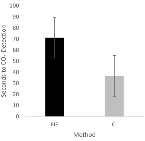
Figure 2: Time until CO2 detection in group comparison. For the group that was intubated using a flexible intubation endoscope, it took significantly longer until end-tidal CO2 could be detected, depicted as mean and standard deviation. Please click here to view a larger version of this figure.
to:
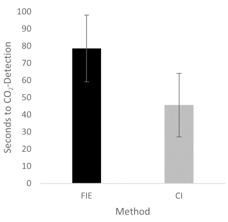
Figure 2: Time until CO2 detection in group comparison. For the group that was intubated using a flexible intubation endoscope, it took significantly longer until end-tidal CO2 could be detected, depicted as mean and standard deviation. n = 5 (for each group). Please click here to view a larger version of this figure.
In the Discussion, the fifth paragraph was updated from:
The increased duration had no clinical significance in this cohort. At no time was the termination criterion-a saturation of less than 93%-reached. This is shown in the results because a procedure change was unnecessary at any time. Prior adequate mask ventilation is a critical step to allow sufficient time for fiberoptic endotracheal tube placement to avoid rapid desaturation34. These results are consistent with previous studies comparing conventional intubation and endoscopically assisted intubations with inexperienced providers35.
to:
The increased duration had no clinical significance in this cohort. At no time was the termination criterion-a saturation of less than 93%-reached. This is shown in the results because a procedure change was unnecessary at any time. Prior adequate mask ventilation is a critical step to allow sufficient time for fiberoptic endotracheal tube placement to avoid rapid desaturation34. These results are consistent with previous studies comparing conventional intubation and endoscopically assisted intubations with inexperienced providers35. We attribute the prolonged duration of fiberoptic intubation to the fact that one must first reorient again after insertion, whereas with conventional intubation, one retains a view of the glottis. It is also important to avoid contact with the mucosa with the flexible intubation endoscope during advancement. This requires occasional corrective maneuvers. Last but not least, after successful placement, retraction of the relatively long endoscope is required, which increases the time to CO2 detection slightly.
