De functie van de ooglens om licht op het netvlies te focussen hangt af van weefseltransparantie, biomechanica en brekingsindex. Deze review benadrukt de driedimensionale architectuur van de lens en bespreekt de complexe mechanismen die de weefselhomeostase in stand houden.
Een abonnement op JoVE is vereist om deze inhoud te bekijken. Log in of start vandaag met uw gratis proefperiode.
Reviewartikel
De ooglens van zoogdieren in focus: Ontwikkeling, Anatomie, Fysiologie, Transparantie, Biomechanica en Leeftijdsgerelateerde Uitdagingen
In dit artikel
Samenvatting
Samenvatting
De lens is een transparant, ellipsoïde orgaan in de voorste kamer van het oog dat licht op het netvlies focust. Dit driedimensionale weefsel bestaat uit epitheelcellen en vezelcellen en is sterk georganiseerd om een helder lichtpad te garanderen en maakt gebruik van meerdere strategieën om een hoge brekingsindex en transparantie te vestigen en te behouden. Bij mensen ondergaat de lens vormveranderingen om licht van verschillende afstanden te focussen in een proces dat accommodatie wordt genoemd, en deze elastische en biomechanische eigenschappen van de lens zijn afhankelijk van weefsel-, cellulaire en moleculaire mechanismen. Staar, gedefinieerd als elke ondoorzichtigheid in de normaal transparante lens, blijft de belangrijkste oorzaak van blindheid in de wereld. Presbyopie, door leeftijdsgebonden veranderingen in de stijfheid van de lenzen, is in verband gebracht met accommodatieve achteruitgang en de noodzaak van leesbrillen. Deze review zal de ontwikkeling en levenslange groei van zoogdierenlenzen, kristallinen en cytoskeletale eiwitten, cel-celcommunicatie en microcirculatie, lensbiomechanica, en de uitdagingen en nieuwe methoden voor het bestuderen van dit unieke weefsel onderzoeken.
Inleiding
De ooglens is op het eerste gezicht een relatief eenvoudig weefsel dat bestaat uit twee celtypen: epitheelcellen en vezelcellen. De bulkmassa van de lens bestaat uit vezelcellen, en er is een monolaag epitheelcellen die de voorste hersenhelft1 bedekt (Figuur 1A). Het gehele weefsel is omhuld door een dun basalmembraan, de zogenaamde lenscapsule, dat voornamelijk bestaat uit collageen IV en laminine, zoals beschreven in een eerdere review2. Omdat er geen bloedtoevoer en zenuwen zijn, hangt de lens in de voorste kamer van het oog door elastische zonulaire vezels (ook wel zonulen genoemd) die de lenscapsule verbinden met het ciliaire lichaam1. Voorste of centrale lens epitheelcellen zijn kuboïdale en gerangschikt in een kasseienpatroon 3,4,5 (Figuur 1A, lichtblauwe cellen, boven). Deze cellen zijn rustend en vermenigvuldigen niet6. Net voor de evenaar van de lens delen epitheelcellen zich in de kiemzone (Figuur 1A, oranje cellen, rechtsboven), en deze equatoriale cellen migreren naar de evenaar waar ze sterk geordend worden in meridionale rijen met hexagonale celvorm 7,8,9,10 (Figuur 1A, groene cellen, rechtsboven). Meridionale rijcellen verankeren hun apicale uiteinden aan het lenspunt of modiolus en beginnen zich te verlengen en te differentiëren tot nieuwe secundaire vezelcellen (Figuur 1A, linksonder). Apicale punten van secundaire vezels migreren naar de voorste pool door te interageren met de apicale zijde van epitheelcellen, waardoor een ongebruikelijke apicale-apikale overgang3 ontstaat (Figuur 1A, rode lijn). De achterste basale punten van nieuw gevormde vezels strekken zich uit naar de achterpool door langs de lenscapsule te bewegen (Figuur 1A, linksonder). Zodra de uiteinden van verlengende vezelcellen de voorste of achterste pool bereiken, losmaken de cellen zich van de voorste epitheelcellen of de achterste capsule en ontmoeten ze de uiteinden van cellen die van de tegenovergestelde zijde van het weefsel uitstrekken om de hechting 11,12,13 te vormen (Figuur 1A, linksboven). Lensvezelcellen zijn een langwerpige zeshoekige vorm in doorsnede met 2 lange/brede zijden, 4 korte zijden en 6 driecellige verbindingen (Figuur 1A, rechtsonder)14. Minimale intercellulaire ruimte tussen naburige vezelcellen vermindert lichtverstrooiingmet 14. De continue toevoeging van corticale vezelcellen aan de rand, overheen op eerdere generaties vezels, bevordert de levenslange groei van de lens14,15. Het midden van de lens wordt de nucleus genoemd, en dit gebied van de lens heeft een verhoogde stijfheid vergeleken met de perifere cortex, zoals beschreven in een eerdere review16.
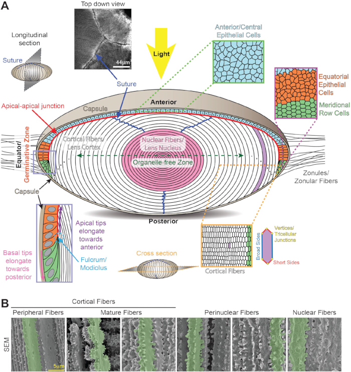
Figuur 1: Lensanatomie en structuur. (A) De lens bestaat uit een monolaag epitheelcellen (lichtblauw, oranje en lichtgroen) die de voorste hersenhelft bedekken, en een grote massa lensvezels (wit en roze). Een dun collageenmembraan, bekend als de lenscapsule (tan), omsluit het weefsel. De lens wordt opgehangen in de voorste kamer van het oog door zonulaire vezels (zonules) die in de capsule rond de evenaar van de lens zijn ingebracht. Voor- en centrale epitheelcellen (lichtblauw, boven) zijn kuboïdaal van vorm en kinderkopjes in doorsnede. Equatoriale epitheelcellen (oranje) in de kiemzone transformeren van willekeurig gepakte cellen (oranje) in georganiseerde meridionale rijen van zeshoekige cellen (groen). Meridionale rijcellen verlengen zich tot nieuwe lagen corticale secundaire vezelcellen (wit) aan de lensperiferie. De apicale uiteinden van nieuw gevormde secundaire vezels verankeren zich aan het draaipunt/modiolus (periwinkle doos, linksonder). De apicale punten van verlengde secundaire vezels migreren naar de voorste pool langs de apicale zijde van de lens epitheelcellen en vormen een ongebruikelijke apicale-apikale overgang (rode lijn). De basale punten van verlengende vezels migreren langs de achterste capsule naar de achterpool. Wanneer secundaire vezels volledig verlengd zijn, loslaten de punten zich van het voorste epitheel of de achterste capsule om de lensnaad te vormen (donkerblauwe lijnen en afbeelding linksboven). Lensvezelcellen zijn langgerekte zeshoeken in doorsnede (rechtsonder) met 2 lange/brede zijden, 4 korte zijden en 6 driecellige verbindingen. Tijdens de rijping van vezelcellen worden alle cellulaire organellen verwijderd om de organellevrije zone (donkergroene dubbelpuntige pijl) te vormen en lichtverstrooiing te minimaliseren. De oudste lensvezelcellen worden samengeperst in het midden van het weefsel om de lenskern (roze) te vormen. Cartoons worden niet op schaal getekend. (B) Scanning-elektronenmicroscoopbeelden van vezelcellen op verschillende dieptes in de lens tonen de veranderende celmorfologie tijdens differentiatie en rijping. Enkele cellen zijn groen gemarkeerd voor nadruk. Corticale perifere vezels hebben kogelvormige interdigitaties langs de lange zijden en kleine uitsteeksels langs de korte zijden. Tijdens de rijping van vezelcellen versieren grote peddels versierd met kleine uitsteeksels en grote uitsteeksels de korte zijkanten van de volwassen cellen. Perinucleaire vezels hebben tong-en-groef interdigitaties over het hele membraan en grote uitsteeksels langs de korte zijden. Kernvezels hebben grote uitsteeksels langs de korte zijden en een bolvormige membraanmorfologie. Schaalbalk, 5 μm. Aangepast van 16.328. Klik hier om een grotere versie van deze figuur te bekijken.
De complexiteit van de lens ligt in de sterk geordende driedimensionale (3D) celopstelling, gespecialiseerde vezelcelrijping die alle cellulaire organellen afbreekt en verwijdert, en moleculaire en cellulaire mechanismen om transparantie en een hoge brekingsindex van het weefsel te vestigen en te behouden. Het ontbreken van cellulaire organellen in de meeste lensvezelcellen vergroot de moeilijkheden om de gezondheid en levensduur van elke lenscel die ooit is gemaakt gedurende het leven van een organisme te behouden. De lens is een uitstekend weefsel voor het bestuderen van veroudering en leeftijdsgerelateerde veranderingen, omdat de meest centrale cellen zijn gevormd tijdens de vroege embryonale ontwikkeling, en de lens blijft gedurende het leven nieuwe cellen aan de rand van het weefsel genereren, waardoor het mogelijk is om cellen te bestuderen die chronologisch oud zijn versus cellen die op hoge leeftijd worden gevormd. De fijne scherpstelfunctie van de lens maakt het mogelijk een helder beeld op het netvlies te vormen van objecten op verschillende afstanden en hangt af van het vermogen van de lens om van vorm te veranderen of zich aan te passen. De lens is een niet-bindend en niet-spierweefsel waarbij biomechanische eigenschappen vereist zijn voor normale functie.
De belangrijkste oorzaak van visuele beperking is17 jaar oud. Leeftijdsgerelateerde veranderingen leiden tot twee grote lenspathologieën: staar en presbyopie18. Staar of welke lens dan ook ondoorzichtigheid blijft wereldwijd de belangrijkste oorzaak van blindheiden wordt veroorzaakt door genetische en omgevingsfactoren, waaronder UV-straling, roken, diabetes en oxidatieve stress, die leiden tot eiwitaggregatie en/of celdefecten, zoals beschreven in eerdere reviews 18,20,21,22,23,24,25. Presbyopie, een leeftijdsgebonden afname in accommodatie, wordt veroorzaakt door een verminderde mogelijkheid van de lens om van vorm te veranderen bij het focussen op nabijgelegen objecten, en daarmee door de noodzaak van leesbrillen 26,27,28. De Wereldgezondheidsorganisatie schat dat er wereldwijd minstens 1 miljard mensen zijn met een niet-behandelde visuele beperking, waaronder 65,2 miljoen mensen met staar en 826 miljoen mensen met presbyopie17. Met een vergrijzende bevolking is er een dringende behoefte om de fysiologie en biologie van de lens beter te begrijpen.
Deze review behandelt weefselarchitectuur, celordening en moleculaire mechanismen die belangrijk zijn voor de functie en homeostase van zoogdierlenzen, met enige informatie over lenzen van andere gewervelden. Het doel is om een algemeen overzicht te geven van lensbiologie en fysiologie, met verwijzingen naar aanvullende gedetailleerde reviews. Onderwerpen zijn onder andere de ontwikkeling van embryonale lenzen, postnatale lensgroei, 3D-cel- en weefselordening, de biomechanische eigenschappen van het weefsel en microcirculatie. Belangrijke lenseiwitten, namelijk de kristallinen, cytoskeletnetwerken en cel-cel communicatie, worden hieronder besproken. De review sluit af met actuele uitdagingen en nieuwe methoden om dit unieke weefsel te bestuderen.
Toegang beperkt. Log in of start een proefperiode om deze inhoud te bekijken.
Review en perspectief
Ontwikkeling van embryonale lenzen en groei van de postnatale lens
Bij gewervelden ontwikkelt de lens zich uit oppervlakte-ectodermcellen, en begint de toekomstige lens met verdikking van het oppervlakte-ectoderm boven het optische blaasje 6,29,30 (Figuur 2A). In combinatie met de vorming van de optische cup dringt het verdikte ectoderm of lensplacode in de lensput 6,29,30 in (Figuur 2B). Actinefilamenten (F-actine) hechten de achterste lensput aan het voorste oppervlak van de zich ontwikkelende optische cup om de beweging en vormveranderingen van deze zich ontwikkelende weefselste coördineren, en apikale vernauwing van lensepitelcellen is vereist voor lensputinvaginatie 35,36,37,38,39,40(Figuur 2C). De opening van de lensput blijft smaller worden totdat het gat wordt gesloten, terwijl lenssteelcellen apoptose 41,42,43 ondergaan. Het lensblaasje wordt gescheiden van het toekomstige hoornvliesectoderm om het lensvesikel te vormen, een holle bol van epitheelcellen 6,29,30 (Figuur 2D). Achterste epitheelcellen in het lensvesikel verlengen zich naar het voorste epitheel en differentiëren tot primaire vezelcellen om de holte te vullen en de embryonale lens te vormen (Figuur 2E). De monolaag van epitheelcellen die de voorste hersenhelft bedekken, blijft behouden terwijl er geen achterste epitheelcellen in de volledig gevormde embryonale lens aanwezig zijn (Figuur 2F). Primaire vezelcellen vormen het middelste deel van de lenskern en worden omringd door nieuwe generaties secundaire lensvezels die gedurendehet leven vanuit de lensrand of cortex worden toegevoegd. Terwijl de meeste lenzen van gewervelde dieren door invaginatie ontstaan, verdikkt bij zebravissen de placode en delamineert vervolgens tot de lens44.
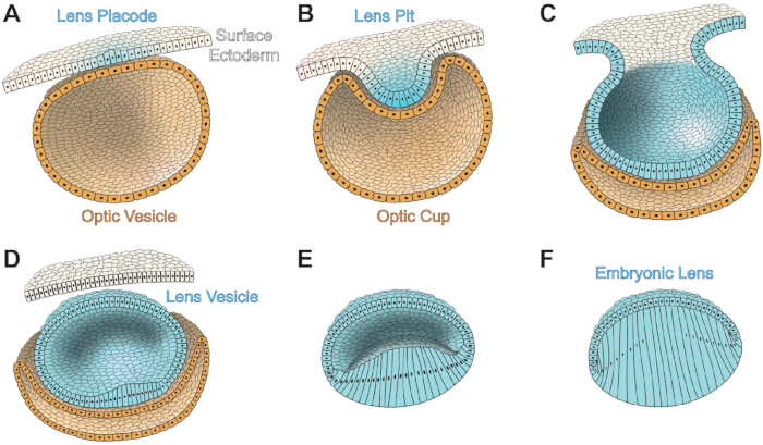
Figuur 2: Ontwikkeling van embryonale lensen in oogdieren van zoogdieren. Oppervlakte-ectodermcellen zijn wit, lenscellen zijn lichtblauw en optische beker/blaascellen zijn zandkleurig. (A) De embryonale lensplacode (lichtblauw) ontwikkelt zich uit het oppervlakte-ectoderm. Achter het oppervlakte-ectoderm en lensplacode bevindt zich de optische blaas. (B) Invaginatie van de lensplacode vormt de lensput, en de invaginatie van het optische blaasje vormt de optische cup. De voorste optische cup ontwikkelt zich tot de iris en het ciliaire lichaam, terwijl de achterste optic cup het netvlies en het retinale gepigmenteerde epitheel vormt. (C) De lens placode-opening versmalt totdat de opening gesloten is. (D) Het lensblaasje scheidt zich af van het oppervlakteectoderm en is een holle bol bekleed met lensepitheelcellen. (E) Achterste epitheelcellen van de lens in het lensblaasje beginnen te verlengen en te differentiëren tot primaire lensvezels. (F) Primaire lensvezels vullen de holte van het lensblaasje om de embryonale lens te vormen. Cartoons worden niet op schaal getekend. Gemodelleerd naar 411. Klik hier om een grotere versie van deze figuur te bekijken.
Tijdens de embryonale ontwikkeling wordt de lens omringd door een mandvormig netwerk van bloedvaten 6,45,46. Deze vaten ontspringen in de hyaloidale slagader van het netvlies en beginnen een netwerk te vormen aan de achterzijde van de lensput en vormen uiteindelijk een geordend netwerk van de tunica vasculosa lentis om de embryonale lens te voeden totdat de voorkamer en de productie van het waterige humor volledig ontwikkeldzijn 6,45. Deze vaten gaan terug en verdwijnen om een helder lichtpad te garanderen vóór de geboorte of voor het openen van de ogen, afhankelijk van de soort 6,45,46,47.
Pax6 is noodzakelijk voor vroege oogontwikkeling 48,49,50,51,52 en is essentieel en voldoende voor lensontwikkeling 53,54,55,56. Fosforylering en SUMOylering van Pax6 reguleren de functie van deze transcriptiefactor tijdens de ontwikkeling van hersenen, centraal zenuwstelsel en ogen 57,58,59,60. Andere belangrijke signaalroutes die nodig zijn voor de ontwikkeling van embryonale ogen en lenzen kruisen Pax6-signalering, waaronder enhancers [Meis, botmorfogene eiwitten (Bmp) en fibroblast growth factor (FGF)] en downstream eiwitten [Foxe3 en retinoïnezuur (RA)], zoals beschreven in een eerdere review61. Meis1 en Meis2 binden aan de enhancer-regio van het Pax6-gen en zijn vereist voor lensinductie bij gewervelden62. Genetische studies tonen aan dat Bmp7 nodig is voor de ontwikkeling van de lens placode door Pax6-expressie63,64 te reguleren. Foxe3 bevindt zich stroomafwaarts van Pax6-signalering en is nodig voor lensontwikkeling 65,66, met name de schone scheiding van de lenssteel van het oppervlakte-ectoderm wanneer het lensblaasje wordt gevormd. Het verlies van Foxe3 leidt tot aangeboren oogafwijkingen die bekend staan als Peters Anomalie67. FGF-receptor (FGFR) signalering werkt samen met Bmp7 om de expressie van Pax6 te reguleren, wat op zijn beurt de Foxe3-niveaus in de ontwikkelende lens68 beïnvloedt. Expressie van een remmende vorm van FGFR leidt tot vertraging in de vorming van lensplacode, onvolledige scheiding van de lenssteel en een klein lensblaasje68. Verstoring van RA-signalering leidt tot oogvormingsdefecten69, en wanneer Pax6 wordt verstoord, is er minder expressie van RA, wat wijst op enige overspraak tussen de twee signaalroutes70.
Nadat de embryonale lens volledig gevormd is, wordt aangenomen dat continue lensgroei door de proliferatie en differentiatie van equatoriale epitheelcellen tot secundaire lensvezels wordt aangedreven door groeifactorsignalering, voornamelijk via FGF in zoogdierlenzen, zoals beschreven in een eerdere review71. FGF's zijn talrijker aanwezig in het glasvocht dan in het waterige humor, wat een gradiënt van toenemende groei- en differentiatiesignalen opzet nabij de evenaar van de lens72,73. De lens drukt 3 van de 4 FGFR's uit: FGFR1, FGFR2 en FGFR3, met toenemende expressie op en achter de evenaar van de lens74,75. Overexpressie van FGF's induceert abnormale verlenging en differentiatie van normaal stil zijnde voorste epitheelcellen tot vezelachtige cellen 76,77,78. Naast FGF-signalering gaven inhibitorassays aan dat de Bmp-route een rol speelt bij de verlenging van vezelcellen79,80, en een abnormale transforming growth factor beta (TGFβ) receptor verstoort de normale rijping van vezelcellen81.
Lenstransparantie en brekingsindex
Lensvezelcellen ondergaan een gespecialiseerd proces om alle cellulaire organellen tijdens de rijping te verwijderen om lichtverstrooiende objecten in het lichtpad te elimineren en om de organellevrije zone (OFZ)82 te creëren (Figuur 1A). Het gecoördineerde verlies van alle cellulaire organellen vindt snel plaats zodra vezelcellen een specifiek stadium van hun rijping bereiken. Abnormale organelafbraak leidt tot staar, en verschillende knockout (KO) zebravis- en muismodellen hebben genen geïdentificeerd voor chromatine- en DNA-afbraak 89,90,91,92,93, cycline-afhankelijke kinasen 94,95, hitteschokfactor 4 91,96,97,98 en PLATT fosfolipase 99 die belangrijk zijn voor de vorming van het OFZ. Abnormale expressie of mutaties in celmembraan en cytoskeleteiwitten 100,101,102,103,104, kristallinen 105,106,107 en oncogenen108,109 kunnen ook invloed hebben op het verwijderen van organelen en de transparantie van de lens. Het exacte signaal dat de organeldegradatie in vezelcellen veroorzaakt, blijft echter onduidelijk. Recent onderzoek heeft het belang van RNA-bindende eiwitten in lensontwikkeling en cataractogenese aan het licht gebracht, zoals beschreven in een eerdere review110. Verstoring van RNA-bindende eiwitten, waaronder Tdrd7111, caprin2112, Rbm24113 en Celf1114, 115, 116, leidt tot abnormale lensontwikkeling, verstoorde organelafbraak en staar.
In vergelijking met de meeste celtypen in het lichaam hebben zoogdierlensvezelcellen een extreem hoge eiwitconcentratie (200–450 mg/ml) 117.118.119.120. Eiwitten vormen 30%–35% van het totale gewicht van de lens, en daardoor heeft de lens een relatief laag watergehalte, ongeveer 65%–70%121.122. Negentig procent van de lenseiwitten naar massa bestaat uit crystallins123, en er zijn 3 families crystallineproteïnen in zoogdierlenzen, α, β en γ, zoals beschreven in eerdere reviews124.125. Een hoog eiwitgehalte zou normaal gesproken het risico verhogen op aggregaten die groter zijn dan de helft van de golflengte van licht, wat leidt tot lichtverstrooiingvan 126, maar kristallineproteïnen zijn georganiseerd in het cytoplasma van de vezelcel met een korteafstandsvolgorde om een hoge brekingsindex en transparantievan 117.127 te bevorderen. Eiwitaggregatie wordt verzwakt door de chaperonachtige functies van α-kristallinen, die hieronder worden besproken. De gradiëntbrekingsindex (GRIN) over de lens bevordert lichtfocussen, zoals beschreven in eerdere reviews128.129, en de maximale brekingsindex in het midden van de lens correleert met de verdichting van kernvezelcellen 130.131.132. In vislenzen suggereren gegevens dat een hoge brekingsindex in de kern het gevolg is van eiwittransport naar het centrum van hetweefsel 133. De uniforme verdeling van kristalline-eiwitten over cellen en tussen vezelschalen wordt verondersteld mogelijk te worden gemaakt door een macromoleculaire transportroute via membraanfusies langs vezelcelmembranen134.135. Membraanfusies zijn echter niet duidelijk waargenomen in muislenzen 136,137,138, en impedantiestudies in lenzen van meerdere soorten detecteren geen membraanfusies 139,140,141,142. Gegevens van muismodellen met verstoring van de communicatie tussen de gap junctions suggereren dat cel-tot-cel communicatie en vezelcelverlenging ook bijdragen aan het macromoleculaire transportpad143.
Kristallinen hebben een molecuulgewicht van ~20–30 kDa en spelen een structurele en brekende rol in de lens. Alfa-kristallinen bestaan uit αA- en αB-subeenheden, die 55%–60% homologie delen (144.145.146), en hoewel αB in de meeste cellen alomtegenwoordig tot expressie komt, wordt αA-kristalline voornamelijk in de lens tot expressie gebracht en is het op lage niveaus in andere weefsels aangetroffen, waaronder de milt en thymus147.148. Recent onderzoek heeft ook aangetoond dat retinale gepigmenteerde epitheelcellen en retinale ganglioncellen αA-kristalline kunnen expresseren tijdens stress of ziekte, vermoedelijk om een neuroprotectiefeffect te verkrijgen. Alfa-kristallinen associëren zich typisch in grote heteromultimeren van 30–35 gemengde αA- en αB-subeenheden123. In het epiteel van muis, runderen en menselijke lenzen wordt alleen αB gedetecteerd in de voorste cellen, terwijl αA en αB in een verhouding van 1:3 voorkomen in equatoriale cellen; Deze verhouding keert zich tijdens differentiëring en elongatie van secundaire vezelcellen naar 3:1 123.152.153. Alfa-kristallinen behoren tot de familie van kleine hitteshock-eiwitten en hebben chaperonachtige functies154, waardoor deze eiwitten kunnen binden en abnormale of ongevouwen eiwitten kunnen binden en vastleggen, waardoor verdere eiwitaggregatie wordt voorkomen. Kleine α-crystallinepeptiden zijn onderzocht ter preventie van door eiwitaggregatie veroorzaakte ziekten, waaronder staar, zoals beschreven in een eerdere review155. Echter, verhoogde of verminderde chaperonne-achtige activiteit van α-kristallinen in muismodellen toont aan dat beide scenario's leiden tot een verhoogde ernst van staar en suggereren dat de juiste chaperonachtige activiteit van α-kristallines cruciaal is voor lenstransparantie156. Bèta- en γ-kristallines behoren tot hun eigen familie van β/γ-kristallline-eiwitten en zijn vezelcelspecifiek; Bij zoogdieren zijn er 7 isoformen van β-kristallijnen die bestaan als dimeren, tetrameren of hogere-orde oligomeren, en er zijn 7 isovormen van γ-kristallijnen die monomeren zijn, zoals beschreven in een eerdere review125. Naast structurele functies binden β en γ-kristallijnen calciumionen en reguleren ze de beschikbaarheid en reserves van calciumionen in lensvezels 157,158,159,160,161.
Lens cytoskeletale en membraaneiwitten
Lensepitel- en vezelcellen hebben netwerken van F-actine, keratine of vimentine tussenfilamenten, gespecialiseerde parel-intermediaire filamenten en microtubuli, zoals beschreven in eerdere reviews. 162.163.164.165.166. Naast functies in oog- en lensontwikkeling 31,32,33,34, zijn diverse F-actinenetwerken in lensepitel- en vezelcellen belangrijk voor lenstransparantie 167,168,169,170,171,172. Recent onderzoek heeft aangetoond dat verandering van het F-actinenetwerk de lensbiomechanicabeïnvloedt. Intermediaire filamenten bestaande uit vimentine of keratine zijn alleen aanwezig in de vroege stadia van de differentiatie van lensvezelcellen of tijdens embryonale ontwikkeling, respectievelijk 177,178,179,180,181; daarentegen hebben alle lensvezels gezaagde tussenfilamenten bestaande uit heterodimeren van filensine (ook bekend als CP95/CP115 en gecodeerd door Bsfp1) en CP49 (ook wel phakinine genoemd en gecodeerd door Bsfp2)182,183,184. Het kralenachtige uiterlijk van deze heterodimeren is te danken aan oligomeren die bestaan uit CP49 en filensine die de filamentkern185,186 decoreren, en de binding van α-kristallijnen langs deze gespecialiseerde tussenliggende filamenten187,188,189,190,191. Het verlies van filensin of CP49 leidt tot het volledige verlies van dit netwerk 192.193.194.195. Verstoring van vimentine tussenliggende filamenten of kralen-intermediaire filamenten veroorzaakt staar bij mensen en diermodellen 192,193,196,197,198,199,200,201,202,203,204, wat het belang van deze cytoskeletnetwerken voor lenstransparantie aantoont. Recent onderzoek suggereert dat microtubuli belangrijk zijn voor lensvezelcelverlenging205.206 en de vorming van de organellevrije zone177.
Adherens-verbindingen die interageren met het actine-cytoskelet zijn belangrijk voor lensvorming207 en de differentiatie van vezelcellen. E-cadherine en N-cadherine worden beide tot expressie gebracht in lensepitelcellen 208,209,210,211, en E-cadherine wordt uitgeschakeld aan het begin van vezelceldifferentiatie 212. N-cadherine wordt sterk tot expressie gebracht in lensvezelcellen en is verrijkt op de middenpunten van de basale hexagonaal voet van verlengende vezelcellen 8.209. Verlies van N-cadherine na vorming van primaire vezelcellen veroorzaakt verstoring van de verlenging van secundaire vezelcellen door abnormale migratie van de apicale toppen van differentiërende vezels langs het apicale oppervlak van anterieure epitheelcellen213. Verstoring van Eph-efrine bidirectionele signalering in de lens veroorzaakt defecten in adherens-verbindingen 4,7,214,215,216.
Andere belangrijke eiwitten van het lensmembraan zijn aquaporinen; connexines; MP20, zoals beschreven in een eerdere review217; galectine 3; galectine-gerelateerd intervezeleiwit (GRIFIN); integrines, zoals beschreven in een eerdere review218; en het afscheiden van zuur en rijk aan cysteïne (SPARC). Aquaporines en connexines worden besproken in de volgende sectie. MP20 (ook wel MP17 of Lim2 genoemd) is een lens-specifiek membraaneiwit met cel-celadhesiefuncties 85.219 dat overvloedig voorkomt in vezelcellen220.221; mutaties in MP20 leiden tot staar 222,223,224,225,226,227,228,229,230. Recent onderzoek heeft interacties aangetoond tussen MP20 en galectine 3, een ander belangrijk cel-cel adhesiemolecuul 231,232,233. GRIFIN, een lensspecifieke isoform van galectine, is gelokaliseerd op vezelcelmembranen en interageert met Pax6, kristallinen en cadherines 234,235,236,237. Integrines zijn belangrijke verbinders tussen de extracellulaire matrix (ECM) en het cytoskelet die nodig zijn voor normale lensontwikkeling. Integrine-isoformen die in de lens tot expressie komen, dienen als receptoren voor capsulecomponenten zoals laminine, collageen of fibronectine. 8.238.239.240.241.242.243.244.245. Elke matrixcomponent kan interageren met meerdere integrines, wat leidt tot diverse signalering via de ruimtelijke expressie van integrines tijdens lensontwikkeling en groei218. Er is bijzondere interesse in het begrijpen van veranderingen in TGFβ, integrines en ECM-componenten tijdens de epitheel- naar mesenchymale overgang in resterende lensepitheelcellen na een staaroperatie, wat leidt tot posterieure capsule-opacificatie, zoals beschreven in een eerdere review218. SPARC, een cel-cel adhesie- en proliferatiemolecuul, is gelokaliseerd in het lensepitheel en ontbreekt in differentiërende vezels246. Verlies van SPARC leidt tot staar 246.247.248, en recente studies hebben aangetoond dat SPARC mogelijk chaperonachtige functies heeft, integrines en ECM-eiwitten beïnvloedt(250.251.252), en de microcirculatieroutebeïnvloedt.
Het microcirculatiepad van een avasculair weefsel
Door een gebrek aan bloedtoevoer genereert de volledig gevormde lens een eigen microcirculatiepad om voedingsstoffen af te voeren en afvalstoffen af te voeren, zoals beschreven in eerdere reviews 254,255,256,257. Een ionische stroom komt de lens binnen bij de voorste en achterste polen en verlaat de lens bij de evenaar258. De inwaartse stroom berust op de beweging van natrium naar de extracellulaire ruimtes tussen lenscellen bij elke pool via een concentratiegradiënt gegenereerd door actief transport, en de uitwaartse stroom wordt gefaciliteerd door gap junction-kanalen op vezelcelmembranen die geconcentreerd zijn nabij de lensevenaar om natriumionen te laten ontsnappen bij de lensevenaar259. Equatoriale epitheliale en perifere vezelcellen hebben een hoge expressie van Na+/K+ ATPase-pompen die actief 3 natriumionen uit de cellen uitwisselen voor 2 kaliumionen uit de extralenticulaire omgeving 260.261.262. De lens behoudt een hoge kaliumconcentratie (135mM) en een lage natriumconcentratie (16mM) om de omstandigheden te creëren zodat natrium bij de polen, langs de concentratiegradiënt259.263, weer in de lens kan binnenkomen. Naast Na+/K+ ATPase helpt de ionencotransporter Na-K-2Cl cotransporter 1 (NKCC1) ook bij het reguleren van de ionenconcentraties van de lens door activatie door mechanosensitive kanalen, zoals beschreven in een eerdere review264.
Gap junctions in de lens bestaan uit drie connexines: connexine 43 (Cx43 of α1), Cx46 (of α3) en Cx50 (of α8)265.266. Cx43 wordt voornamelijk tot expressie gebracht in lensepitheelcellen267, en hoewel de initiatie van vezelcelvorming normaal is in Cx43 KO-lenzen, heeft de interface tussen epitheel- en vezelcellen een vergrote extracellulaire ruimte en intracellulaire vacuolen, wat wijst op een functie van Cx43 in osmotische balans268. Cx46 is geconcentreerd in lensvezelcellen, en Cx50 is aanwezig in zowel epitheel- als vezelcellen269. Cx23 wordt ook tot expressie gebracht in vezelcellen van muislenzen, maar niet in menselijke lenzen 270.271.272.273. Een dominante mutatie in Cx23 veroorzaakt vezelceldefecten bij muizen270, maar Cx23 KO-lenzen hebben geen noemenswaardige defecten274. Zes connexine-subunits vormen een connexon of hemikanaal, en een connexon op één cel koppelt zich aan een connexon op de naburige cel om een gap junction te vormen, die een ~10 angström porie tussen de naburige cellen creëert, waardoor de uitwisseling van kleine moleculen, ionen en watermogelijk is (275.276). Connexine-hemikanalen maken het mogelijk om materialen tussen het intracellulaire cytoplasma en de extracellulaire omgeving te passeren, zoals beschreven in eerdere reviews277.278, en hemikanalen zijn belangrijk voor de microcirculatie279, mechano-sensitieve transport van voedingsstoffen en antioxidanten280.281.282.283, en de oxidatieve stressrespons284.285,286.287.288. Gap-verbindingen komen samen tot grote plaques op het vezelcelmembraan; In de lens kunnen de plaques micron-groot zijn in diameter en zijn ze uitgelijnd langs de lange zijden van aangrenzende vezelcellen289. Verlies van Cx46 in muislenzen leidt tot dichte nucleaire staar door blokkade van het uitstroompad van de lensmicrocirculatie 290.291. De assemblage en/of stabiliteit van grote Cx46-plaques op het vezelcelmembraan vereist het F-actine en de geparelde tussenliggende filamentnetwerken289. Cx50 KO leidt tot microftalmie, milde nucleaire staar en kleine lenzen door abnormale proliferatie van epitheelcellen292.293. Verlies van Cx50 beïnvloedt de gating van de resterende Cx46 gap junction plaques in lensvezelcellen139. Vervanging van Cx50 door knock-in Cx46, uitgedrukt onder het endogene Cx50-locus (4 kopieën van Cx46), redt staar door verlies van Cx50 en verhoogt de koppeling tussen vezels in de gap junction, maar deze transparante knock-in lenzen blijven klein142.294. Cx46 kan dus de lensgroeifunctie van Cx50 niet vervangen. De knock-in-strategie is ook gebruikt om nucleaire staar veroorzaakt door γB-crystalline-mutante eiwitten te redden, wat suggereert dat verhoogde gap-junctionale koppeling een strategie kan zijn voor verbeterde lenshomeostase en staarpreventie295.
De ionische flux die door natriumionen wordt gegenereerd, gaat gepaard met waterstroom naar lenscellen via aquaporinekanalen259. Aquaporinekanalen bestaan uit Aqp1 in epitheelcellen, terwijl in vezelcellen Aqp0 (ook MIP genoemd) en Aqp5 waterkanalen vormen, afhankelijk van de rijpheid van de cellen, zoals beschreven in eerdere reviews 264.296.297. Beweging van water door gap junction-kanalen genereert een hydrostatische drukgradiënt over de lens298.299, en deze gradiënt handhaaft de lensosmotische druk via transient receptorpotentiaal vanilloïde 1 (TRPV1) en TRPV4 mechanosensitive kanalen die respectievelijk de lens hydrostatische druk299.300.301.302 verhogen en verlagen. Verminderde zonulaire spanning op de lens verlaagt de hydrostatische druk, waardoor TRPV1 en vervolgens NKCC1 worden geactiveerd om de ionenconcentratie in de lens te verhogen en de waterstroom in de lens te stimuleren om de normale hydrostatische druk300.301 te herstellen. Omgekeerd activeren verhoogde zonulaire spanning en lensvolume TRPV4 en vervolgens Na+/K+ ATPase om de ionenconcentratie van de lens te verlagen en daarmee de hydrostatische drukmet 300.301.302 te verlagen.
Essentiële voedingsstoffen en aminozuren worden via het microcirculatiepad303 in de lens getransporteerd, en dit transport is vooral belangrijk voor OFZ-vezels. Facilitatieve glucosetransporters (GLUT) worden gevonden in lensepitel- en vezelcellen; GLUT1 wordt in beide celtypen tot expressie gebracht, en GLUT3 wordt tot expressie gebracht in epitheelcellen 304,305,306,307. GLUT1 KO-muizen ontwikkelen staar, wat suggereert dat glucoseopname vereist is voor lenstransparantie308. Glutathion (GSH), een belangrijke antioxidant voor lenstransparantie309,310, wordt in de lens gemaakt en kan vanuit het waterige humor 311,312,313,314,315 in de lens worden getransporteerd. Cysteïne, glutamaat en glycine zijn essentiële aminozuren voor de synthese van GSH, en de ophoping van deze aminozuren is aanwezig in de cortex en nucleaire gebieden van de lens314.315. Leeftijdsgebonden afname van GSH-synthese of verhoogd GSH-gebruik kan leiden tot een verminderde beschikbaarheid van GSH in de lens, vooral in de kern 316,317,318,319,320,321. Glutathionperoxidasen (GPX) gebruiken GSH als co-enzym om hydroperoxide te neutraliseren, en recent onderzoek toont aan dat ferroptose door giftige lipideperoxiden kan leiden tot staar door verstoring van GPX4322,323.
3D-lensarchitectuur en biomechanica
Het patroon van lensvezels in georganiseerde rijen zeshoekige cellen vindt zijn oorsprong in het patroon van equatoriale epitheelcellen naar meridionale rijen (Figuur 1A). De mechanismen die de organisatie van willekeurig gepakte en keienvormige equatoriale epitheelcellen in hexagonische meridionale rijcellen aansturen, draaien rond het actinecytoskelet. Signaaloverdracht van de receptor tyrosine kinase EphA2 is nodig om de ophoping van F-actine aan de hoekpunten van hexagonale meridionale rijcellen te stimuleren en zo celorganisatie 7.324 te bevorderen. Myosine IIA, een contractiel eiwit dat bindt aan F-actine, is ook belangrijk voor het bevorderen van de zeshoekige celvorm 8.325.326. Er werd al lang aangenomen dat deze sterk geordende verpakking van vezelcellen noodzakelijk is voor lenstransparantie, maar verkeerde verpakking door verlies van EphA2 7.324, mutaties in myosine IIA325.326, of misvormde cellen gemaakt op ouderdom130 suggereren dat hexagonaal celoppakken niet nodig is voor transparantie.
Vezelcellen die zich uitstrekken van de voorste tot de achterpool kunnen enkele millimeters lang zijn, afhankelijk van de diersoort en de grootte van delens 327. Typisch zijn vezelcellen slechts 4–7 μm in doorsnede327, en deze lange en dunne cellen worden bij elkaar gehouden door dynamische cel-cel interdigitaties die een 3D-rits vormen tussen aangrenzende vezelcellen, zoals beschreven in een eerdere review328. Interdigitaties van lensvezelcellen hebben complexe patronen die veranderen naarmate de cellen differentiëren en rijpen van 1) perifere, nieuw gevormde vezels met kleine uitsteeksels langs de korte zijden en kogelvormige uitsteeksels langs de lange zijden, 2) volwassen vezelcellen die organelverlies ondergaan en grote paddels hebben met kleine in elkaar grijpende uitsteeksels langs de korte zijden, 3) perinucleaire vezels met grote, in elkaar grijpende uitsteeksels langs de korte zijden en tong- en groefinterdigitaties over het gehele membraan, en 4) kernvezels met grote uitstulpingen langs de korte zijden en bolvormige membraanmorfologie over het gehele membraan 137.328.329.330.331.332.333.334.335, 336.337.338.339 (Figuur 1B). Recent onderzoek heeft direct bewijs geleverd dat interdigitaties tussen lensvezelcellen die afhankelijk zijn van F-actinenetwerken belangrijk zijn voor weefselstijfheid174, wat een lang gekoesterde hypothese ondersteunt. Verlies van ARVCF, een onderdeel van adherens-junctions, leidt tot abnormale interdigitatie van vezelcellen, een vergrote extracellulaire ruimte en vroegtijdig optredende corticale cataracten340. Defecten in stijfheid en grootte van de lenskern worden gecorreleerd met veranderingen in de interdigitaties van de tong en groef en vertraagde overgang naar de morfologie van het globulaire membraan131.
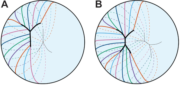
Figuur 3: Lenshechtingenpatroon. Elke gekleurde lijn vertegenwoordigt een enkele vezelcel. (A) Vezelcellen in de embryonale menselijke lens vormen een Y-hechting. De Y-naad is gespiegeld aan de voorste en achterste polen. (B) Naarmate de menselijke lens zich verder ontwikkelt en groeit, zal de hechting meer vertakt worden. Hier wordt een viervertakkige hechting afgebeeld voor de eenvoud. Het hechtpatroon is verschoven tussen de twee polen. Cartoons worden niet op schaal getekend. Gemodelleerd naar 45. Klik hier om een grotere versie van deze figuur te bekijken.
De algehele vorm van de lens verschilt afhankelijk van de soort, en er worden theorieën gesteld dat de kromming en het type hechting van de vezelcel de lensvorm341.342 bepalen. De hechting wordt gevormd door het samenkomen van de uiteinden van verlengende vezelcellen aan de voorste en achterste pool, en de hechting wordt gespiegeld tussen de twee polen11, 12, 13, 343 (Figuur 3). Bij muislenzen is de hechting Y-vormig, en bij objectieven en mensen begint de hechting in een Y-vorm en wordt het meer vertakt rondde leeftijd van 11, 12 en 45 jaar. Interessant genoeg beperkt de Y-hechting in muislenzen de elasticiteit of veerkracht van de lens nadat een mechanische belasting is verwijderd, en vertakte hechtingen in KO-lenzen met verstoring van Eph-efrine-signalering correleren met verhoogde veerkracht344. De normale vorming van de lenshechting vereist communicatie tussen de gap junction345, en leeftijdsgebonden voorste staar in muislenzen is het resultaat van onvolledige hechtingsvorming130.
Biomechanische eigenschappen van de lens kunnen worden beïnvloed door diverse moleculaire, cellulaire en weefselfactoren, zoals beschreven in een eerdere review16. Leeftijdsgebonden toename van lensstijfheid wordt beschouwd als een belangrijke oorzaak van presbyopie346. Bij mensen, wanneer ze naar verre objecten kijken, rekt ontspanning van de ciliaire spieren de zonulaire vezels die aan de capsule vastzitten uit, wat resulteert in het afvlakken van de lens1. Bij het focussen op iets van dichtbij trekt de ciliaire spier samen waardoor de zonules ontspannen, en de lens rondt af om het licht van het nabije object1 te focussen. Dit proces van accommodatie vereist biomechanische eigenschappen van de lens, contractie van de ciliaire spier en zonulaire spanning. Met de leeftijd hebben studies weinig tot geen verandering aangetoond in ciliaire spiercontractiliteit en zonulaire flexibiliteit en spanning 347.348.349.350. Verschillende studies suggereren dat een grotere lensgrootte en kerngrootte en stijfheid bijdragen aan de algehele versteviging van de hele lens bijleeftijd 26.351.352.353.354. Studies naar lensstijfheid met de leeftijd bij muizen suggereren echter dat er weinig correlatie is tussen lens- en kerngrootte met de totale lensstijfheidvan 130.132, wat erop wijst dat andere factoren de leeftijdsgebonden lensstijfheid beïnvloeden. Recent onderzoek heeft aangetoond dat veranderingen in cytoskeletnetwerken de stijfheid van de lens beïnvloeden; verlies van tropomyosine 3.5, een F-actine stabiliserend eiwit, beïnvloedt de samenstelling en ordening van F-actinenetwerken in het membraan van vezelcellen en vermindert de stijfheid van de lens173. Evenzo leiden spontane mutaties in verschillende inteelt wildtype-muislijnen tot spontane KO van CP49192.355 en zachtere lenzen356.357. Membranen van lensvezelcels hebben een ongewoon hoog cholesterolgehaltevan 358.359.360.361.362.363, wat de elasticiteit van het membraan kan reguleren. Veranderingen in het membraancholesterolgehalte en de binding van α-kristallinen aan het vezelcelmembraan met de leeftijd worden verondersteld de stijfheid van de lens te verhogen365.366.367.368.369.370.371.372.
Uitdagingen en nieuwe methoden voor het bestuderen van de lens
De 3D-structuur van de lens vormt uitdagingen voor het begrijpen van cel-cel ordening en voor het creëren van geschikte in vitro modellen die weefselarchitectuur reproduceren. Hoewel waardevolle inzichten over signalering en gemuteerde eiwitlokalisatie zijn opgehelderd met behulp van geïmmortaliseerde of primaire kweekcellen, hebben de geïsoleerde lensepitheelcellen niet de karakteristieke kuboïdale morfologie of dezelfde genexpressie of cytoskeletstructuren als native lensepitheelcellen373. In vitrolentoïde, zoals beschreven in een eerdere review374 en lensmodellen375, creëren vezelachtige cellen en celballen die respectievelijk lijken op een mini-lens. Deze modellen creëren een transparante, lensachtige structuur die nuttig kan zijn voor snelle drugsscreening, maar geen van beide modellen reproduceert de normale structuur van vezelcellen of de 3D-structuur van de lens. Er zijn veel protocollen ontwikkeld voor het lokaliseren van eiwitten in lensweefselsecties en immunokleuring, en deze methode blijft de meest efficiënte manier om te begrijpen of eiwitten tot expressie komen in specifieke celcompartimenten. Weefselsectie voor histologie en immunokleuring kan echter moeilijk zijn, vooral bij monsters van oudere zoogdieren, vanwege onvolledige fixatie van lens137; en het bestuderen van 3D-structuur in 2D-secties verwijdert details die belangrijk zijn voor het begrijpen van de vorm van cellen, interacties tussen cellen en lokalisatie van eiwitten. Hoewel elektronenmicroscopie (EM) de gouden standaard blijft voor het bestuderen van vezelcelvorm, blijft het lokaliseren van eiwitten in EM-monsters een uitdaging 376.377.378. Recent werk met vlakke mounts van de epitheelcelmonolaag 4,7,324,373, enkelvezelcelkleuring 85,173,174,379 en whole lensbeeldvorming 4,7,324,344,380,381 hebben belangrijke informatie onthuld over cel-cel adhesies, lensbiomechanica en veranderende cytoskeletnetwerken in lensvezels.
Lensepitheel- en vezelcellen hebben verschillende functies, transcriptomen en proteomen, en eerdere studies die de verschillende celcompartimenten vergeleken waren moeilijk omdat er slechts een monolaag epitheelcellen is die de voorste hemisfeer bedekt, waardoor de hoeveelheid RNA of eiwit die kan worden verzameld zonder veel monsters te verzamelen beperkt wordt. Vooruitgang in RNA-extractietechnieken, realtime PCR en geautomatiseerde capillaire elektroforese Westerns hebben gen- en eiwitanalyse mogelijk gemaakt van lensepitheelcelmonsters verzameld uit één paar muisogen 130.132.373.382 of microdissecte gebieden van het lensepitheel, corticale vezels en kernvezels383. Single-cell transcriptomics maken diepgaande profilering van lenscellen van zebravisembryo's en larve- en kuikenembryo'smogelijk. Deze nieuwe methoden openen de deur om de moleculaire mechanismen te onthullen die elk lenscelcompartiment in stand houden tijdens normale veroudering en pathologie.
Naarmate het vakgebied vordert, is het belangrijk te onthouden dat de genetische achtergrond van muizen een belangrijke rol kan spelen in lenspathologieen biomechanica355.356.357, en daarom zijn de controles van nestgenoten voor experimenten cruciaal voor data-analyse. Mutant- en KO-muismodellen bieden een systeem om leeftijdsgebonden staar te bestuderen en een toename van lensstijfheid 130.356.357.388.389.390.391.392.393.394.395.396.397, en vanwege de relatief korte levensduur van muizen zouden verouderingsstudies op oude dieren moeten worden uitgevoerd in plaats van gegevens van jonge dieren te extrapoleren.
Toegang beperkt. Log in of start een proefperiode om deze inhoud te bekijken.
Conclusies
Technologische vooruitgang voor transcriptomics, proteomics, beeldvorming en biomechanische tests heeft ons begrip van lensbiologie, fysiologie en pathologie verbeterd. Onze groeiende kennis over de verschillen tussen verschillende populaties van lensepitel- en vezelcellen zal belangrijk zijn om veranderingen tijdens veroudering te begrijpen en de mechanismen van de levensduur van eiwitten en cellen. Er is meer belangstelling voor het ontwikkelen van farmaceutische interventies om staar ...
Toegang beperkt. Log in of start een proefperiode om deze inhoud te bekijken.
Openbaarmakingen
De auteurs hebben niets te onthullen.
Dankbetuigingen
Dit werk werd gefinancierd door subsidie R01 EY032056 (naar CC) van het National Eye Institute. De auteurs bedanken Isaiah J. Innis en Gryffin M. Flowers voor hun behulpzame opmerkingen.
Toegang beperkt. Log in of start een proefperiode om deze inhoud te bekijken.
Referenties
- Freddo, T. F., Chaum, E. Anatomy of the Eye and Orbit: The Clinical Essentials, 1e. , Lippincott Williams & Wilkins, a Wolters Kluwer business. (2018).
- Danysh, B. P., Duncan, M. K. The lens capsule. Exp Eye Res. 88 (2), 151-164 (2009).
- Zampighi, G. A., Eskandari, S., Kreman, M. Epithelial organization of the mammalian lens. Exp Eye Res. 71 (4), 415-435 (2000).
- Cheng, C., Gong, X. Diverse roles of Eph/ephrin signaling in the mouse lens. PLoS One. 6 (11), e28147(2011).
- Parreno, J., Cheng, C., Nowak, R. B., Fowler, V. M. The effects of mechanical strain on mouse eye lens capsule and cellular microstructure. Mol Biol Cell. 29 (16), 1963-1974 (2018).
- Lovicu, F. J., Robinson, M. L. Development of the Ocular Lens. , Cambridge University Press. (2011).
- Cheng, C., Ansari, M. M., Cooper, J. A., Gong, X. EphA2 and Src regulate equatorial cell morphogenesis during lens development. Development. 140 (20), 4237-4245 (2013).
- Bassnett, S., Missey, H., Vucemilo, I. Molecular architecture of the lens fiber cell basal membrane complex. J Cell Sci. 112 (Pt 13), 2155-2165 (1999).
- Bassnett, S., Sikic, H. The lens growth process. Prog Retin Eye Res. 60, 181-200 (2017).
- Sikic, H., Shi, Y., Lubura, S., Bassnett, S. A full lifespan model of vertebrate lens growth. R Soc Open Sci. 4 (1), 160695(2017).
- Kuszak, J. R. The development of lens sutures. Progress in Retinal and Eye Research. 14 (2), 567-591 (1995).
- Kuszak, J. R., Mazurkiewicz, M., Zoltoski, R. Computer modeling of secondary fiber development and growth: I. Nonprimate lenses. Mol Vis. 12, 251-270 (2006).
- Kuszak, J. R., Zoltoski, R. K., Tiedemann, C. E. Development of lens sutures. Int J Dev Biol. 48 (8-9), 889-902 (2004).
- Kuszak, J. R. The ultrastructure of epithelial and fiber cells in the crystalline lens. Int Rev Cytol. 163, 305-350 (1995).
- Kuszak, J. R., Macsai, M. S., Rae, J. L. Stereo scanning electron microscopy of the crystalline lens. Scan Electron Microsc. (Pt 3), 1415-1426 (1983).
- Cheng, C. Tissue, cellular, and molecular level determinants for eye lens stiffness and elasticity. Front Ophthalmol (Lausanne). 4, 1456474(2024).
- World Health Organization. World report on vision. , (2019).
- Michael, R., Bron, A. J. The ageing lens and cataract: a model of normal and pathological ageing. Philos Trans R Soc Lond B Biol Sci. 366 (1568), 1278-1292 (2011).
- Asbell, P. A., et al. Age-related cataract. Lancet. 365 (9459), 599-609 (2005).
- Hejtmancik, J. F. Oxidative Stress in Genetic Cataract Formation. Antioxidants (Basel). 13 (11), (2024).
- Shiels, A., Hejtmancik, J. F. Biology of Inherited Cataracts and Opportunities for Treatment. Annu Rev Vis Sci. 5, 123-149 (2019).
- Truscott, R. J. W., Friedrich, M. G. Molecular Processes Implicated in Human Age-related Nuclear Cataract. Invest Ophthalmol Vis Sci. 60 (15), 5007-5021 (2019).
- Fan, X., Monnier, V. M. Protein posttranslational modification (PTM) by glycation: Role in lens aging and age-related cataractogenesis. Exp Eye Res. 210, 108705(2021).
- Budnar, P., Tangirala, R., Bakthisaran, R., Rao, C. M. Protein Aggregation and Cataract: Role of Age-related Modifications and Mutations in alpha Crystallins. Biochemistry (Mosc). 87 (3), 225-241 (2022).
- Shiels, A., Hejtmancik, J. F. Inherited cataracts: Genetic mechanisms and pathways new and old. Exp Eye Res. 209, 108662(2021).
- Heys, K. R., Cram, S. L., Truscott, R. J. Massive increase in the stiffness of the human lens nucleus with age: the basis for presbyopia? Mol Vis. 10, 956-963 (2004).
- Glasser, A., Campbell, M. C. Biometric, optical and physical changes in the isolated human crystalline lens with age in relation to presbyopia. Vision Res. 39 (11), 1991-2015 (1999).
- Weeber, H. A., et al. Dynamic mechanical properties of human lenses. Exp Eye Res. 80 (3), 425-434 (2005).
- Piatigorsky, J. Lens differentiation in vertebrates. A review of cellular and molecular features. Differentiation. 19 (3), 134-153 (1981).
- Gunhaga, L. The lens: a classical model of embryonic induction providing new insights into cell determination in early development. Philos Trans R Soc Lond B Biol Sci. 366 (1568), 1193-1203 (2011).
- Chauhan, B. K., et al. Cdc42 and IRSp53 dependent contractile filopodia tether presumptive lens and retina to coordinate epithelial invagination. Development. 136 (21), 3657-3667 (2009).
- McAvoy, J. W. Cytoplasmic processes interconnect lens placode and optic vesicle during eye morphogenesis. Exp Eye Res. 31 (5), 527-534 (1980).
- Mann, I. The development of the human eye. , 3d, Grune & Stratton. (1964).
- Eiraku, M., et al. Self organizing optic cup morphogenesis in three dimensional culture. Nature. 472 (7341), 51-56 (2011).
- Lang, R. A., Herman, K., Reynolds, A. B., Hildebrand, J. D., Plageman, T. F. Jr p120 catenin dependent junctional recruitment of Shroom3 is required for apical constriction during lens pit morphogenesis. Development. 141 (16), 3177-3187 (2014).
- Plageman, T. F. Jr, et al. A Trio RhoA Shroom3 pathway is required for apical constriction and epithelial invagination. Development. 138 (23), 5177-5188 (2011).
- Borges, R. M., Lamers, M. L., Forti, F. L., Santos, M. F., Yan, C. Y. Rho signaling pathway and apical constriction in the early lens placode. Genesis. 49 (5), 368-379 (2011).
- Quintin, S., Gally, C., Labouesse, M. Epithelial morphogenesis in embryos: asymmetries, motors and brakes. Trends Genet. 24 (5), 221-230 (2008).
- Muccioli, M., Qaisi, D., Herman, K., Plageman, T. F. Jr Lens placode planar cell polarity is dependent on Cdc42 mediated junctional contraction inhibition. Dev Biol. 412 (1), 32-43 (2016).
- Houssin, N. S., Martin, J. B., Coppola, V., Yoon, S. O., Plageman, T. F. Jr Formation and contraction of multicellular actomyosin cables facilitate lens placode invagination. Dev Biol. 462 (1), 36-49 (2020).
- Glucksmann, A. Cell deaths in normal vertebrate ontogeny. Biol Rev Camb Philos Soc. 26 (1), 59-86 (1951).
- Ozeki, H., Ogura, Y., Hirabayashi, Y., Shimada, S. Suppression of lens stalk cell apoptosis by hyaluronic acid leads to faulty separation of the lens vesicle. Exp Eye Res. 72 (1), 63-70 (2001).
- Yan, Q., Liu, J. P., Li, D. W. Apoptosis in lens development and pathology. Differentiation. 74 (5), 195-211 (2006).
- Easter, S. S. Jr, Nicola, G. N. The development of vision in the zebrafish (Danio rerio). Dev Biol. 180 (2), 646-663 (1996).
- Mann, I. The development of the human eye. , The University Press. (1928).
- Darche, M., et al. Three dimensional characterization of developing and adult ocular vasculature in mice using in toto clearing. Communications Biology. 5 (1), 1135(2022).
- Wang, Z., Liu, C. H., Huang, S., Chen, J. Assessment and Characterization of Hyaloid Vessels in Mice. J Vis Exp. (147), e59222(2019).
- Hill, R. E., et al. Mouse small eye results from mutations in a paired like homeobox containing gene. Nature. 354 (6354), 522-525 (1991).
- Jordan, T., et al. The human PAX6 gene is mutated in two patients with aniridia. Nat Genet. 1 (5), 328-332 (1992).
- Hogan, B. L., et al. Small eyes (Sey): a homozygous lethal mutation on chromosome 2 which affects the differentiation of both lens and nasal placodes in the mouse. J Embryol Exp Morphol. 97, 95-110 (1986).
- Hanson, I. M., et al. Mutations at the PAX6 locus are found in heterogeneous anterior segment malformations including Peters' anomaly. Nat Genet. 6 (2), 168-173 (1994).
- Grindley, J. C., Davidson, D. R., Hill, R. E. The role of Pax 6 in eye and nasal development. Development. 121 (5), 1433-1442 (1995).
- Fujiwara, M., Uchida, T., Osumi Yamashita, N., Eto, K. Uchida rat (rSey): a new mutant rat with craniofacial abnormalities resembling those of the mouse Sey mutant. Differentiation. 57 (1), 31-38 (1994).
- Collinson, J. M., Hill, R. E., West, J. D. Different roles for Pax6 in the optic vesicle and facial epithelium mediate early morphogenesis of the murine eye. Development. 127 (5), 945-956 (2000).
- Quinn, J. C., West, J. D., Hill, R. E. Multiple functions for Pax6 in mouse eye and nasal development. Genes Dev. 10 (4), 435-446 (1996).
- Ashery Padan, R., Marquardt, T., Zhou, X., Gruss, P. Pax6 activity in the lens primordium is required for lens formation and for correct placement of a single retina in the eye. Genes Dev. 14 (21), 2701-2711 (2000).
- Kim, E. A., et al. Phosphorylation and transactivation of Pax6 by homeodomain interacting protein kinase 2. J Biol Chem. 281 (11), 7489-7497 (2006).
- Mikkola, I., Bruun, J. A., Bjorkoy, G., Holm, T., Johansen, T. Phosphorylation of the transactivation domain of Pax6 by extracellular signal regulated kinase and p38 mitogen activated protein kinase. J Biol Chem. 274 (21), 15115-15126 (1999).
- Yan, Q., et al. Sumoylation activates the transcriptional activity of Pax 6, an important transcription factor for eye and brain development. Proc Natl Acad Sci U S A. 107 (49), 21034-21039 (2010).
- Yan, Q., et al. Protein phosphatase 1 modulates the function of Pax 6, a transcription factor controlling brain and eye development. J Biol Chem. 282 (19), 13954-13965 (2007).
- Cvekl, A., Zhang, X. Signaling and Gene Regulatory Networks in Mammalian Lens Development. Trends Genet. 33 (10), 677-702 (2017).
- Zhang, X., Friedman, A., Heaney, S., Purcell, P., Maas, R. L. Meis homeoproteins directly regulate Pax6 during vertebrate lens morphogenesis. Genes Dev. 16 (16), 2097-2107 (2002).
- Dudley, A. T., Lyons, K. M., Robertson, E. J. A requirement for bone morphogenetic protein 7 during development of the mammalian kidney and eye. Genes Dev. 9 (22), 2795-2807 (1995).
- Wawersik, S., et al. BMP7 acts in murine lens placode development. Dev Biol. 207 (1), 176-188 (1999).
- Blixt, A., et al. A forkhead gene, FoxE3, is essential for lens epithelial proliferation and closure of the lens vesicle. Genes Dev. 14 (2), 245-254 (2000).
- Brownell, I., Dirksen, M., Jamrich, M. Forkhead Foxe3 maps to the dysgenetic lens locus and is critical in lens development and differentiation. Genesis. 27 (2), 81-93 (2000).
- Dimanlig, P. V., Faber, S. C., Auerbach, W., Makarenkova, H. P., Lang, R. A. The upstream ectoderm enhancer in Pax6 has an important role in lens induction. Development. 128 (22), 4415-4424 (2001).
- Faber, S. C., et al. Fgf receptor signaling plays a role in lens induction. Development. 128 (22), 4425-4438 (2001).
- Bavik, C., Ward, S. J., Chambon, P. Developmental abnormalities in cultured mouse embryos deprived of retinoic by inhibition of yolk sac retinol binding protein synthesis. Proc Natl Acad Sci U S A. 93 (7), 3110-3114 (1996).
- Enwright, J. F. 3rd, Grainger, R. M. Altered retinoid signaling in the heads of small eye mouse embryos. Dev Biol. 221 (1), 10-22 (2000).
- Lovicu, F. J., McAvoy, J. W. Growth factor regulation of lens development. Dev Biol. 280 (1), 1-14 (2005).
- Schulz, M. W., Chamberlain, C. G., de Iongh, R. U., McAvoy, J. W. Acidic and basic FGF in ocular media and lens: implications for lens polarity and growth patterns. Development. 118 (1), 117-126 (1993).
- Lovicu, F. J., Chamberlain, C. G., McAvoy, J. W. Differential effects of aqueous and vitreous on fiber differentiation and extracellular matrix accumulation in lens epithelial explants. Invest Ophthalmol Vis Sci. 36 (7), 1459-1469 (1995).
- de Iongh, R. U., Lovicu, F. J., Chamberlain, C. G., McAvoy, J. W. Differential expression of fibroblast growth factor receptors during rat lens morphogenesis and growth. Invest Ophthalmol Vis Sci. 38 (9), 1688-1699 (1997).
- de Iongh, R. U., Lovicu, F. J., Hanneken, A., Baird, A., McAvoy, J. W. FGF receptor 1 (flg) expression is correlated with fibre differentiation during rat lens morphogenesis and growth. Dev Dyn. 206 (4), 412-426 (1996).
- Lovicu, F. J., Overbeek, P. A. Overlapping effects of different members of the FGF family on lens fiber differentiation in transgenic mice. Development. 125 (17), 3365-3377 (1998).
- Robinson, M. L., et al. Extracellular FGF 1 acts as a lens differentiation factor in transgenic mice. Development. 121 (2), 505-514 (1995).
- Robinson, M. L., et al. Disregulation of ocular morphogenesis by lens specific expression of FGF 3/int 2 in transgenic mice. Dev Biol. 198 (1), 13-31 (1998).
- Faber, S. C., Robinson, M. L., Makarenkova, H. P., Lang, R. A. Bmp signaling is required for development of primary lens fiber cells. Development. 129 (15), 3727-3737 (2002).
- Belecky Adams, T., et al. Pax 6, Prox 1, and Chx10 homeobox gene expression correlates with phenotypic fate of retinal precursor cells. Invest Ophthalmol Vis Sci. 38 (7), 1293-1303 (1997).
- de Iongh, R. U., et al. Requirement for TGFbeta receptor signaling during terminal lens fiber differentiation. Development. 128 (20), 3995-4010 (2001).
- Bassnett, S. Lens organelle degradation. Exp Eye Res. 74 (1), 1-6 (2002).
- Bassnett, S. The fate of the Golgi apparatus and the endoplasmic reticulum during lens fiber cell differentiation. Invest Ophthalmol Vis Sci. 36 (9), 1793-1803 (1995).
- Bassnett, S., Beebe, D. C. Coincident loss of mitochondria and nuclei during lens fiber cell differentiation. Dev Dyn. 194 (2), 85-93 (1992).
- Bassnett, S., Shi, Y., Vrensen, G. F. Biological glass: structural determinants of eye lens transparency. Philos Trans R Soc Lond B Biol Sci. 366 (1568), 1250-1264 (2011).
- Counis, M. F., et al. Analysis of nuclear degradation during lens cell differentiation. Cell Death Differ. 5 (4), 251-261 (1998).
- Vrensen, G. F., Graw, J., De Wolf, A. Nuclear breakdown during terminal differentiation of primary lens fibres in mice: a transmission electron microscopic study. Exp Eye Res. 52 (6), 647-659 (1991).
- Costello, M. J., et al. Autophagy and mitophagy participate in ocular lens organelle degradation. Exp Eye Res. 116, 141-150 (2013).
- De Maria, A., Bassnett, S. DNase IIbeta distribution and activity in the mouse lens. Invest Ophthalmol Vis Sci. 48 (12), 5638-5646 (2007).
- He, S., et al. Chromatin remodeling enzyme Brg1 is required for mouse lens fiber cell terminal differentiation and its denucleation. Epigenetics Chromatin. 3 (1), 21(2010).
- Cui, X., et al. HSF4 regulates DLAD expression and promotes lens de nucleation. Biochim Biophys Acta. 1832 (8), 1167-1172 (2013).
- Nakahara, M., et al. Degradation of nuclear DNA by DNase II like acid DNase in cortical fiber cells of mouse eye lens. FEBS J. 274 (12), 3055-3064 (2007).
- Nishimoto, S., et al. Nuclear cataract caused by a lack of DNA degradation in the mouse eye lens. Nature. 424 (6952), 1071-1074 (2003).
- Chaffee, B. R., et al. Nuclear removal during terminal lens fiber cell differentiation requires CDK1 activity: appropriating mitosis related nuclear disassembly. Development. 141 (17), 3388-3398 (2014).
- Lyu, L., et al. Unfolded protein response associated stabilization of p27(Cdkn1b) interferes with lens fiber cell denucleation, leading to cataract. FASEB J. 30 (3), 1087-1095 (2016).
- Gao, M., et al. HSF4 regulates lens fiber cell differentiation by activating p53 and its downstream regulators. Cell Death Dis. 8 (10), e3082(2017).
- Fujimoto, M., et al. HSF4 is required for normal cell growth and differentiation during mouse lens development. EMBO J. 23 (21), 4297-4306 (2004).
- Min, J. N., Zhang, Y., Moskophidis, D., Mivechi, N. F. Unique contribution of heat shock transcription factor 4 in ocular lens development and fiber cell differentiation. Genesis. 40 (4), 205-217 (2004).
- Morishita, H., et al. Organelle degradation in the lens by PLAAT phospholipases. Nature. 592 (7855), 634-638 (2021).
- Capetanaki, Y., Smith, S., Heath, J. P. Overexpression of the vimentin gene in transgenic mice inhibits normal lens cell differentiation. J Cell Biol. 109 (4 Pt 1), 1653-1664 (1989).
- Hamai, Y., Kuwabara, T. Early cytologic changes of Fraser cataract. An electron microscopic study. Invest Ophthalmol. 14 (7), 517-527 (1975).
- Muggleton Harris, A. L., Festing, M. F., Hall, M. A gene location for the inheritance of the cataract Fraser (CatFr) mouse congenital cataract. Genet Res. 49 (3), 235-238 (1987).
- Hamai, Y., Fukui, H. N., Kuwabara, T. Morphology of hereditary mouse cataract. Exp Eye Res. 18 (6), 537-546 (1974).
- Takehana, M. Hereditary cataract of the Nakano mouse. Exp Eye Res. 50 (6), 671-676 (1990).
- Uga, S., Kador, P. F., Kuwabara, T. Cytological study of Philly mouse cataract. Exp Eye Res. 30 (1), 79-92 (1980).
- Nakamura, M., Russell, P., Carper, D. A., Inana, G., Kinoshita, J. H. Alteration of a developmentally regulated, heat stable polypeptide in the lens of the Philly mouse. Implications for cataract formation. J Biol Chem. 263 (35), 19218-19221 (1988).
- Wang, K., et al. GammaD crystallin associated protein aggregation and lens fiber cell denucleation. Invest Ophthalmol Vis Sci. 48 (8), 3719-3728 (2007).
- Cavalheiro, G. R., et al. N myc regulates growth and fiber cell differentiation in lens development. Dev Biol. 429 (1), 105-117 (2017).
- Khillan, J. S., et al. Defects in lens fiber differentiation are linked to c mos overexpression in transgenic mice. Genes Dev. 1 (10), 1327-1335 (1987).
- Lachke, S. A. RNA binding proteins and post transcriptional regulation in lens biology and cataract: Mediating spatiotemporal expression of key factors that control the cell cycle, transcription, cytoskeleton and transparency. Exp Eye Res. 214, 108889(2022).
- Lachke, S. A., et al. Mutations in the RNA granule component TDRD7 cause cataract and glaucoma. Science. 331 (6024), 1571-1576 (2011).
- Dash, S., Dang, C. A., Beebe, D. C., Lachke, S. A. Deficiency of the RNA binding protein caprin2 causes lens defects and features of Peters anomaly. Dev Dyn. 244 (10), 1313-1327 (2015).
- Dash, S., et al. The master transcription factor SOX2, mutated in anophthalmia/microphthalmia, is post transcriptionally regulated by the conserved RNA binding protein RBM24 in vertebrate eye development. Hum Mol Genet. 29 (4), 591-604 (2020).
- Siddam, A. D., et al. The RNA binding protein Celf1 post transcriptionally regulates p27Kip1 and Dnase2b to control fiber cell nuclear degradation in lens development. PLoS Genet. 14 (3), e1007278(2018).
- Siddam, A. D., et al. High Throughput Transcriptomics of Celf1 Conditional Knockout Lens Identifies Downstream Networks Linked to Cataract Pathology. Cells. 12 (7), (2023).
- Aryal, S., et al. The cataract linked RNA binding protein Celf1 post transcriptionally controls the spatiotemporal expression of the key homeodomain transcription factors Pax6 and Prox1 in lens development. Hum Genet. 139 (12), 1541-1554 (2020).
- Takemoto, L., Sorensen, C. M. Protein protein interactions and lens transparency. Exp Eye Res. 87 (6), 496-501 (2008).
- Oyster, C. W. The human eye : structure and function. , Sinauer Associates, Inc.. (1999).
- Flaugh, S. L., Kosinski Collins, M. S., King, J. Contributions of hydrophobic domain interface interactions to the folding and stability of human gammaD crystallin. Protein Sci. 14 (3), 569-581 (2005).
- Papanikolopoulou, K., et al. Formation of amyloid fibrils in vitro by human gammaD crystallin and its isolated domains. Mol Vis. 14, 81-89 (2008).
- Graw, J. Genetics of crystallins: cataract and beyond. Exp Eye Res. 88 (2), 173-189 (2009).
- Kosinski Collins, M. S., King, J. In vitro unfolding, refolding, and polymerization of human gammaD crystallin, a protein involved in cataract formation. Protein Sci. 12 (3), 480-490 (2003).
- Horwitz, J., Bova, M. P., Ding, L. L., Haley, D. A., Stewart, P. L. Lens alpha crystallin: function and structure. Eye (Lond). 13 (Pt 3b), 403-408 (1999).
- Hejtmancik, J. F., et al. Progress in Molecular Biology and Translational Science. Hejtmancik, J. F., Nickerson, J. M. 134, Academic Press. 169-201 (2015).
- Bloemendal, H., et al. Ageing and vision: structure, stability and function of lens crystallins. Prog Biophys Mol Biol. 86 (3), 407-485 (2004).
- Benedek, G. B. Theory of transparency of the eye. Appl Opt. 10 (3), 459-473 (1971).
- Delaye, M., Tardieu, A. Short range order of crystallin proteins accounts for eye lens transparency. Nature. 302 (5907), 415-417 (1983).
- Bassnett, S., Costello, M. J. The cause and consequence of fiber cell compaction in the vertebrate lens. Exp Eye Res. 156, 50-57 (2017).
- Pierscionek, B. K., Regini, J. W. The gradient index lens of the eye: an opto biological synchrony. Prog Retin Eye Res. 31 (4), 332-349 (2012).
- Cheng, C., et al. Age-related changes in eye lens biomechanics, morphology, refractive index and transparency. Aging (Albany NY). 11 (24), 12497-12531 (2019).
- Cheng, C., et al. EphA2 Affects Development of the Eye Lens Nucleus and the Gradient of Refractive Index. Invest Ophthalmol Vis Sci. 63 (1), 2(2022).
- Flowers, G. M., et al. Eph ephrin signaling affects lens growth and shape, nucleus size, and gradient refractive index in adult mice. Frontiers in Ophthalmology. 5, (2025).
- Kozlowski, T. M., Kroger, R. H. H. Constant lens fiber cell thickness in fish suggests crystallin transport to denucleated cells. Vision Res. 162, 29-34 (2019).
- Shestopalov, V. I., Bassnett, S. Expression of autofluorescent proteins reveals a novel protein permeable pathway between cells in the lens core. J Cell Sci. 113 (Pt 11), 1913-1921 (2000).
- Shestopalov, V. I., Bassnett, S. Development of a macromolecular diffusion pathway in the lens. J Cell Sci. 116 (Pt 20), 4191-4199 (2003).
- Dunia, I., et al. Structural and immunocytochemical alterations in eye lens fiber cells from Cx46 and Cx50 knockout mice. Eur J Cell Biol. 85 (8), 729-752 (2006).
- Blankenship, T., Bradshaw, L., Shibata, B., Fitzgerald, P. Structural specializations emerging late in mouse lens fiber cell differentiation. Invest Ophthalmol Vis Sci. 48 (7), 3269-3276 (2007).
- Lo, W. K., Reese, T. S. Multiple structural types of gap junctions in mouse lens. J Cell Sci. 106 (Pt 1), 227-235 (1993).
- Baldo, G. J., et al. Gap junctional coupling in lenses from alpha(8) connexin knockout mice. J Gen Physiol. 118 (5), 447-456 (2001).
- Baldo, G. J., Mathias, R. T. Spatial variations in membrane properties in the intact rat lens. Biophys J. 63 (2), 518-529 (1992).
- Duncan, G., Patmore, L., Pynsent, P. B. Impedance of the amphibian lens. J Physiol. 312, 17-27 (1981).
- Martinez Wittinghan, F. J., et al. Lens gap junctional coupling is modulated by connexin identity and the locus of gene expression. Invest Ophthalmol Vis Sci. 45 (10), 3629-3637 (2004).
- Cheng, C., et al. Gap junction communication influences intercellular protein distribution in the lens. Exp Eye Res. 86 (6), 966-974 (2008).
- Bloemendal, H. The vertebrate eye lens. Science. 197 (4299), 127-138 (1977).
- van der Ouderaa, F. J., de Jong, W. W., Bloemendal, H. The amino acid sequence of the alphaA2 chain of bovine alpha crystallin. Eur J Biochem. 39 (1), 207-222 (1973).
- Bloemendal, H., de Jong, W. W. Lens proteins and their genes. Prog Nucleic Acid Res Mol Biol. 41, 259-281 (1991).
- Kato, K., et al. Immunoreactive alpha A crystallin in rat non lenticular tissues detected with a sensitive immunoassay method. Biochim Biophys Acta. 1080 (2), 173-180 (1991).
- Srinivasan, A. N., Nagineni, C. N., Bhat, S. P. alpha A crystallin is expressed in non ocular tissues. J Biol Chem. 267 (32), 23337-23341 (1992).
- Nath, M., Fort, P. E. alphaA Crystallin Mediated Neuroprotection in the Retinal Neurons Is Independent of Protein Kinase B. Front Neurosci. 16, 912757(2022).
- Skeie, J. M., Mahajan, V. B. Proteomic interactions in the mouse vitreous retina complex. PLoS One. 8 (11), e82140(2013).
- Ruebsam, A., et al. A specific phosphorylation regulates the protective role of alphaA crystallin in diabetes. JCI Insight. 3 (4), (2018).
- Vermorken, A. J., Hilderink, J. M., van de Ven, W. J., Bloemendal, H. Lens differentiation. Crystallin synthesis in isolated epithelia from calf lenses. J Cell Biol. 76 (1), 175-183 (1978).
- Boyle, D. L., Takemoto, L., Brady, J. P., Wawrousek, E. F. Morphological characterization of the Alpha A and Alpha B crystallin double knockout mouse lens. BMC Ophthalmol. 3, 3(2003).
- Horwitz, J. Alpha crystallin. Exp Eye Res. 76 (2), 145-153 (2003).
- Raju, M., Santhoshkumar, P., Krishna Sharma, K. Alpha crystallin derived peptides as therapeutic chaperones. Biochim Biophys Acta. 1860 (1 Pt B), 246-251 (2016).
- Cheng, C., et al. Altered chaperone like activity of alpha crystallins promotes cataractogenesis. J Biol Chem. 285 (52), 41187-41193 (2010).
- Aravind, P., et al. The betagamma crystallin superfamily contains a universal motif for binding calcium. Biochemistry. 48 (51), 12180-12190 (2009).
- Srivastava, S. S., Mishra, A., Krishnan, B., Sharma, Y. Ca2+ binding motif of betagamma crystallins. J Biol Chem. 289 (16), 10958-10966 (2014).
- Vanmarle, J., Jonges, R., Vrensen, G. F., Dewolf, A. Calcium and its localization in human lens fibres: an electron tomographic study. Exp Eye Res. 65 (1), 83-88 (1997).
- Jobby, M. K., Sharma, Y. Calcium binding to lens betaB2 and betaA3 crystallins suggests that all beta crystallins are calcium binding proteins. FEBS J. 274 (16), 4135-4147 (2007).
- Bergman, M. R., Deravi, L. F. Manipulating polydispersity of lens beta crystallins using divalent cations demonstrates evidence of calcium regulation. Proc Natl Acad Sci U S A. 119 (48), e2212051119(2022).
- Cheng, C., Nowak, R. B., Fowler, V. M. The lens actin filament cytoskeleton: Diverse structures for complex functions. Exp Eye Res. 156, 58-71 (2017).
- Quinlan, R. A., Clark, J. I. Insights into the biochemical and biophysical mechanisms mediating the longevity of the transparent optics of the eye lens. J Biol Chem. 298 (11), 102537(2022).
- Song, S., et al. Functions of the intermediate filament cytoskeleton in the eye lens. J Clin Invest. 119 (7), 1837-1848 (2009).
- FitzGerald, P. G. Lens intermediate filaments. Exp Eye Res. 88 (2), 165-172 (2009).
- Logan, C. M., Menko, A. S. Microtubules: Evolving roles and critical cellular interactions. Exp Biol Med (Maywood). 244 (15), 1240-1254 (2019).
- More, M. I., Kirsch, F. P., Rathjen, F. G. Targeted ablation of NrCAM or ankyrin B results in disorganized lens fibers leading to cataract formation. J Cell Biol. 154 (1), 187-196 (2001).
- Lin, Q., et al. Genetic variations and polymorphisms in the ezrin gene are associated with age-related cataract. Mol Vis. 19, 1572-1579 (2013).
- Hao, J., Kunishima, S., Guo, X., Hu, R., Gao, W. A large family with MYH9 disorder caused by E1841K mutation suffering from serious kidney and hearing impairment and cataracts. Ann Hematol. 91 (7), 1147-1148 (2012).
- De Rocco, D., et al. MYH9 related disease: five novel mutations expanding the spectrum of causative mutations and confirming genotype/phenotype correlations. Eur J Med Genet. 56 (1), 7-12 (2013).
- Economou, M., et al. MYH9 related disorders: report on a patient of Greek origin presenting with macroscopic hematuria and presenile cataract, caused by an R1165C mutation. J Pediatr Hematol Oncol. 34 (6), 412-415 (2012).
- Saposnik, B., et al. Mutation spectrum and genotype phenotype correlations in a large French cohort of MYH9 Related Disorders. Mol Genet Genomic Med. 2 (4), 297-312 (2014).
- Cheng, C., et al. Tropomyosin 3.5 protects the F-actin networks required for tissue biomechanical properties. J Cell Sci. 131 (23), (2018).
- Cheng, C., et al. Tropomodulin 1 Regulation oF-actin Is Required for the Formation of Large Paddle Protrusions Between Mature Lens Fiber Cells. Invest Ophthalmol Vis Sci. 57 (10), 4084-4099 (2016).
- Maddala, R., Walters, M., Brophy, P. J., Bennett, V., Rao, P. V. Ankyrin B directs membrane tethering of periaxin and is required for maintenance of lens fiber cell hexagonal shape and mechanics. Am J Physiol Cell Physiol. 310 (2), C115-C126 (2016).
- Won, G. J., Fudge, D. S., Choh, V. The effects of actomyosin disruptors on the mechanical integrity of the avian crystalline lens. Mol Vis. 21, 98-109 (2015).
- Kasper, M., Moll, R., Stosiek, P., Karsten, U. Patterns of cytokeratin and vimentin expression in the human eye. Histochemistry. 89 (4), 369-377 (1988).
- Kasper, M., Viebahn, C. Cytokeratin expression and early lens development. Anat Embryol (Berl). 186 (3), 285-290 (1992).
- Ellis, M., Alousi, S., Lawniczak, J., Maisel, H., Welsh, M. Studies on lens vimentin. Exp Eye Res. 38 (2), 195-202 (1984).
- Ramaekers, F. C., Dunia, I., Dodemont, H. J., Benedetti, E. L., Bloemendal, H. Lenticular intermediate sized filaments: biosynthesis and interaction with plasma membrane. Proc Natl Acad Sci U S A. 79 (10), 3208-3212 (1982).
- Ramaekers, F. C., et al. Identification of the cytoskeletal proteins in lens forming cells, a special epitheloid cell type. Exp Cell Res. 127 (2), 309-327 (1980).
- FitzGerald, P. G. Immunochemical characterization of a Mr 115 lens fiber cell specific extrinsic membrane protein. Curr Eye Res. 7 (12), 1243-1253 (1988).
- Ireland, M. E., et al. Up regulation of novel intermediate filament proteins in primary fiber cells: an indicator of all vertebrate lens fiber differentiation? Anat Rec. 258 (1), 25-33 (2000).
- Quinlan, R. A., Stewart, M. Molecular interactions in intermediate filaments. Bioessays. 13 (11), 597-600 (1991).
- Goulielmos, G., et al. Filensin and phakinin form a novel type of beaded intermediate filaments and coassemble de novo in cultured cells. J Cell Biol. 132 (4), 643-655 (1996).
- Georgatos, S. D., Gounari, F., Goulielmos, G., Aebi, U. To bead or not to bead? Lens specific intermediate filaments revisited. J Cell Sci. 110 (Pt 21), 2629-2634 (1997).
- Ireland, M., Maisel, H. A cytoskeletal protein unique to lens fiber cell differentiation. Exp Eye Res. 38 (6), 637-645 (1984).
- Bloemendal, H., et al. Interaction of crystallins with the cytoskeletal plasma membrane complex of the bovine lens. Ciba Found Symp. 106, 177-190 (1984).
- Carter, J. M., Hutcheson, A. M., Quinlan, R. A. In vitro studies on the assembly properties of the lens proteins CP49, CP115: coassembly with alpha crystallin but not with vimentin. Exp Eye Res. 60 (2), 181-192 (1995).
- Chaves, J. M., Gupta, R., Srivastava, K., Srivastava, O. Human alpha A crystallin missing N terminal domain poorly complexes with filensin and phakinin. Biochem Biophys Res Commun. 494 (1-2), 402-408 (2017).
- FitzGerald, P. G., Graham, D. Ultrastructural localization of alpha A crystallin to the bovine lens fiber cell cytoskeleton. Curr Eye Res. 10 (5), 417-436 (1991).
- Sandilands, A., et al. Bfsp2 mutation found in mouse 129 strains causes the loss of CP49' and induces vimentin dependent changes in the lens fibre cell cytoskeleton. Exp Eye Res. 78 (4), 875-889 (2004).
- Sandilands, A., et al. Knockout of the intermediate filament protein CP49 destabilises the lens fibre cell cytoskeleton and decreases lens optical quality, but does not induce cataract. Exp Eye Res. 76 (3), 385-391 (2003).
- Alizadeh, A., et al. Targeted deletion of the lens fiber cell specific intermediate filament protein filensin. Invest Ophthalmol Vis Sci. 44 (12), 5252-5258 (2003).
- Alizadeh, A., et al. Targeted genomic deletion of the lens specific intermediate filament protein CP49. Invest Ophthalmol Vis Sci. 43 (12), 3722-3727 (2002).
- Perng, M. D., Zhang, Q., Quinlan, R. A. Insights into the beaded filament of the eye lens. Exp Cell Res. 313 (10), 2180-2188 (2007).
- Muller, M., et al. Dominant cataract formation in association with a vimentin assembly disrupting mutation. Hum Mol Genet. 18 (6), 1052-1057 (2009).
- Bornheim, R., et al. A dominant vimentin mutant upregulates Hsp70 and the activity of the ubiquitin proteasome system, and causes posterior cataracts in transgenic mice. J Cell Sci. 121 (Pt 22), 3737-3746 (2008).
- Jakobs, P. M., et al. Autosomal dominant congenital cataract associated with a deletion mutation in the human beaded filament protein gene BFSP2. Am J Hum Genet. 66 (4), 1432-1436 (2000).
- Conley, Y. P., et al. A juvenile onset, progressive cataract locus on chromosome 3q21 q22 is associated with a missense mutation in the beaded filament structural protein 2. Am J Hum Genet. 66 (4), 1426-1431 (2000).
- Zhang, Q., et al. Clinical description and genome wide linkage study of Y sutural cataract and myopia in a Chinese family. Mol Vis. 10, 890-900 (2004).
- Cui, X., et al. The E233del mutation in BFSP2 causes a progressive autosomal dominant congenital cataract in a Chinese family. Mol Vis. 13, 2023-2029 (2007).
- Ma, X., et al. A new mutation in BFSP2 (G1091A) causes autosomal dominant congenital lamellar cataracts. Mol Vis. 14, 1906-1911 (2008).
- Ramachandran, R. D., Perumalsamy, V., Hejtmancik, J. F. Autosomal recessive juvenile onset cataract associated with mutation in BFSP1. Hum Genet. 121 (3-4), 475-482 (2007).
- Logan, C. M., Bowen, C. J., Menko, A. S. Functional role for stable microtubules in lens fiber cell elongation. Exp Cell Res. 362 (2), 477-488 (2018).
- Basu, S., Rajakaruna, S., Reyes, B., Van Bockstaele, E., Menko, A. S. Suppression of MAPK/JNK MTORC1 signaling leads to premature loss of organelles and nuclei by autophagy during terminal differentiation of lens fiber cells. Autophagy. 10 (7), 1193-1211 (2014).
- Pontoriero, G. F., et al. Co operative roles for E-cadherin and N-cadherin during lens vesicle separation and lens epithelial cell survival. Dev Biol. 326 (2), 403-417 (2009).
- Leonard, M., Zhang, L., Bleaken, B. M., Menko, A. S. Distinct roles for N-cadherin linked c Src and fyn kinases in lens development. Dev Dyn. 242 (5), 469-484 (2013).
- Leonard, M., et al. Modulation of N-cadherin junctions and their role as epicenters of differentiation specific actin regulation in the developing lens. Dev Biol. 349 (2), 363-377 (2011).
- Maddala, R., Nagendran, T., Lang, R. A., Morozov, A., Rao, P. V. Rap1 GTPase is required for mouse lens epithelial maintenance and morphogenesis. Dev Biol. 406 (1), 74-91 (2015).
- Wigle, J. T., Chowdhury, K., Gruss, P., Oliver, G. Prox1 function is crucial for mouse lens fibre elongation. Nat Genet. 21 (3), 318-322 (1999).
- Xu, L., Overbeek, P. A., Reneker, L. W. Systematic analysis of E , N and P cadherin expression in mouse eye development. Exp Eye Res. 74 (6), 753-760 (2002).
- Logan, C. M., et al. N-cadherin regulates signaling mechanisms required for lens fiber cell elongation and lens morphogenesis. Dev Biol. 428 (1), 118-134 (2017).
- Cooper, M. A., et al. Loss of ephrin A5 function disrupts lens fiber cell packing and leads to cataract. Proc Natl Acad Sci U S A. 105 (43), 16620-16625 (2008).
- Son, A. I., et al. Further analysis of the lens of ephrin A5 / mice: development of postnatal defects. Mol Vis. 19, 254-266 (2013).
- Biswas, S., Son, A. I., Yu, Q., Zhou, R., Lo, W. K. Breakdown of interlocking domains may contribute to formation of membranous globules and lens opacity in ephrin A5( / ) mice. Exp Eye Res. 145, 130-139 (2016).
- Maher, G. J., Black, G. C., Manson, F. D. Focus on molecules: lens intrinsic membrane protein (LIM2/MP20). Exp Eye Res. 103, 115-116 (2012).
- Walker, J., Menko, A. S. Integrins in lens development and disease. Exp Eye Res. 88 (2), 216-225 (2009).
- Shi, Y., et al. Further analysis of the lens phenotype in Lim2 deficient mice. Invest Ophthalmol Vis Sci. 52 (10), 7332-7339 (2011).
- Mulders, J. W., et al. MP17, a fiber specific intrinsic membrane protein from mammalian eye lens. Curr Eye Res. 7 (2), 207-219 (1988).
- Voorter, C. E., et al. Distribution of MP17 in isolated lens fibre membranes. Curr Eye Res. 8 (7), 697-706 (1989).
- Steele, E. C. Jr, et al. Identification of a mutation in the MP19 gene, Lim2, in the cataractous mouse mutant To3. Mol Vis. 3, 5(1997).
- Irum, B., et al. Mutation in LIM2 Is Responsible for Autosomal Recessive Congenital Cataracts. PLoS One. 11 (11), e0162620(2016).
- Wang, X., et al. Elongated axial length and myopia related fundus changes associated with the Arg130Cys mutation in the LIM2 gene in four Chinese families with congenital cataracts. Ann Transl Med. 9 (3), 235(2021).
- Berry, V., et al. A recurrent variant in LIM2 causes an isolated congenital sutural/lamellar cataract in a Japanese family. Ophthalmic Genet. 43 (5), 622-626 (2022).
- Pei, R., et al. A novel mutation of LIM2 causes autosomal dominant membranous cataract in a Chinese family. Int J Ophthalmol. 13 (10), 1512-1520 (2020).
- Ponnam, S. P., Ramesha, K., Tejwani, S., Matalia, J., Kannabiran, C. A missense mutation in LIM2 causes autosomal recessive congenital cataract. Mol Vis. 14, 1204-1208 (2008).
- Pras, E., et al. A missense mutation in the LIM2 gene is associated with autosomal recessive presenile cataract in an inbred Iraqi Jewish family. Am J Hum Genet. 70 (5), 1363-1367 (2002).
- Goyal, S., Singh, R., Singh, J. R., Vanita, V. Novel and known variants in GJA3 and LIM2 in congenital cataract families from North India. BMC Genomics. 25 (1), 31(2024).
- Puk, O., Ahmad, N., Wagner, S., de Angelis, M. H., Graw, J. Microphakia and congenital cataract formation in a novel Lim2(C51R) mutant mouse. Mol Vis. 17, 1164-1171 (2011).
- Dahm, R., Bramke, S., Dawczynski, J., Nagaraj, R. H., Kasper, M. Developmental aspects of galectin 3 expression in the lens. Histochem Cell Biol. 119 (3), 219-226 (2003).
- Gonen, T., Donaldson, P., Kistler, J. Galectin 3 is associated with the plasma membrane of lens fiber cells. Invest Ophthalmol Vis Sci. 41 (1), 199-203 (2000).
- Gonen, T., Grey, A. C., Jacobs, M. D., Donaldson, P. J., Kistler, J. MP20, the second most abundant lens membrane protein and member of the tetraspanin superfamily, joins the list of ligands of galectin 3. BMC Cell Biol. 2, 17(2001).
- Barton, K. A., Hsu, C. D., Petrash, J. M. Interactions between small heat shock protein alpha crystallin and galectin related interfiber protein (GRIFIN) in the ocular lens. Biochemistry. 48 (18), 3956-3966 (2009).
- Garcia Caballero, G., et al. Chicken lens development: complete signature of expression of galectins during embryogenesis and evidence for their complex formation with alpha , beta , delta , and tau crystallins, N CAM, and N-cadherin obtained by affinity chromatography. Cell Tissue Res. 379 (1), 13-35 (2020).
- Garcia Caballero, G., et al. Chicken GRIFIN: binding partners, developmental course of localization and activation of its lens specific gene expression by L Maf/Pax6. Cell Tissue Res. 375 (3), 665-683 (2019).
- Ogden, A. T., et al. GRIFIN, a novel lens specific protein related to the galectin family. J Biol Chem. 273 (44), 28889-28896 (1998).
- Menko, A. S., Philip, N. J. Beta 1 integrins in epithelial tissues: a unique distribution in the lens. Exp Cell Res. 218 (2), 516-521 (1995).
- Menko, S., Philp, N., Veneziale, B., Walker, J. Integrins and development: how might these receptors regulate differentiation of the lens. Ann N Y Acad Sci. 842, 36-41 (1998).
- Walker, J. L., Menko, A. S. alpha6 Integrin is regulated with lens cell differentiation by linkage to the cytoskeleton and isoform switching. Dev Biol. 210 (2), 497-511 (1999).
- Walker, J. L., Zhang, L., Zhou, J., Woolkalis, M. J., Menko, A. S. Role for alpha 6 integrin during lens development: Evidence for signaling through IGF 1R and ERK. Dev Dyn. 223 (2), 273-284 (2002).
- Wederell, E. D., et al. Laminin binding integrins in rat lens morphogenesis and their regulation during fibre differentiation. Exp Eye Res. 81 (3), 326-339 (2005).
- Barbour, W., et al. Expression patterns of beta1 related alpha integrin subunits in murine lens during embryonic development and wound healing. Curr Eye Res. 29 (1), 1-10 (2004).
- Wu, J. E., Santoro, S. A. Complex patterns of expression suggest extensive roles for the alpha 2 beta 1 integrin in murine development. Dev Dyn. 199 (4), 292-314 (1994).
- Duncan, M. K., Kozmik, Z., Cveklova, K., Piatigorsky, J., Cvekl, A. Overexpression of PAX6(5a) in lens fiber cells results in cataract and upregulation of (alpha)5(beta)1 integrin expression. J Cell Sci. 113 (Pt 18), 3173-3185 (2000).
- Bassuk, J. A., et al. Disruption of the Sparc locus in mice alters the differentiation of lenticular epithelial cells and leads to cataract formation. Exp Eye Res. 68 (3), 321-331 (1999).
- Gilmour, D. T., et al. Mice deficient for the secreted glycoprotein SPARC/osteonectin/BM40 develop normally but show severe age onset cataract formation and disruption of the lens. EMBO J. 17 (7), 1860-1870 (1998).
- Kantorow, M., Horwitz, J., Carper, D. Up regulation of osteonectin/SPARC in age-related cataractous human lens epithelia. Mol Vis. 4, 17(1998).
- Emerson, R. O., Sage, E. H., Ghosh, J. G., Clark, J. I. Chaperone like activity revealed in the matricellular protein SPARC. J Cell Biochem. 98 (4), 701-705 (2006).
- Weaver, M. S., Sage, E. H., Yan, Q. Absence of SPARC in lens epithelial cells results in altered adhesion and extracellular matrix production in vitro. J Cell Biochem. 97 (2), 423-432 (2006).
- Weaver, M. S., Workman, G., Sage, E. H. The copper binding domain of SPARC mediates cell survival in vitro via interaction with integrin beta1 and activation of integrin linked kinase. J Biol Chem. 283 (33), 22826-22837 (2008).
- Yan, Q., Perdue, N., Blake, D., Sage, E. H. Absence of SPARC in murine lens epithelium leads to increased deposition of laminin 1 in lens capsule. Invest Ophthalmol Vis Sci. 46 (12), 4652-4660 (2005).
- Greiling, T. M., Stone, B., Clark, J. I. Absence of SPARC leads to impaired lens circulation. Exp Eye Res. 89 (3), 416-425 (2009).
- Giannone, A. A., Li, L., Sellitto, C., White, T. W. Physiological Mechanisms Regulating Lens Transport. Front Physiol. 12, 818649(2021).
- Donaldson, P. J., Chen, Y., Petrova, R. S., Grey, A. C., Lim, J. C. Regulation of lens water content: Effects on the physiological optics of the lens. Prog Retin Eye Res. 95, 101152(2023).
- Donaldson, P. J., Grey, A. C., Maceo Heilman, B., Lim, J. C., Vaghefi, E. The physiological optics of the lens. Prog Retin Eye Res. 56, e1-e24 (2017).
- Mathias, R. T., Kistler, J., Donaldson, P. The lens circulation. J Membr Biol. 216 (1), 1-16 (2007).
- Robinson, K. R., Patterson, J. W. Localization of steady currents in the lens. Curr Eye Res. 2 (12), 843-847 (1982).
- Mathias, R. T., Rae, J. L., Baldo, G. J. Physiological properties of the normal lens. Physiol Rev. 77 (1), 21-50 (1997).
- Gao, J., Sun, X., Yatsula, V., Wymore, R. S., Mathias, R. T. Isoform specific function and distribution of Na/K pumps in the frog lens epithelium. J Membr Biol. 178 (2), 89-101 (2000).
- Alvarez, L. J., Candia, O. A., Grillone, L. R. Na+ K+ ATPase distribution in frog and bovine lenses. Curr Eye Res. 4 (2), 143-152 (1985).
- Delamere, N. A., Dean, W. L. Distribution of lens sodium potassium adenosine triphosphatase. Invest Ophthalmol Vis Sci. 34 (7), 2159-2163 (1993).
- Paterson, C. A. Distribution and movement of ions in the ocular lens. Doc Ophthalmol. 31 (1), 1-28 (1972).
- Donaldson, P. J., Petrova, R. S., Nair, N., Chen, Y., Schey, K. L. Regulation of water flow in the ocular lens: new roles for aquaporins. J Physiol. 602 (13), 3041-3056 (2024).
- Goodenough, D. A. The crystalline lens. A system networked by gap junctional intercellular communication. Semin Cell Biol. 3 (1), 49-58 (1992).
- Mathias, R. T., White, T. W., Gong, X. Lens gap junctions in growth, differentiation, and homeostasis. Physiol Rev. 90 (1), 179-206 (2010).
- Beyer, E. C., Kistler, J., Paul, D. L., Goodenough, D. A. Antisera directed against connexin43 peptides react with a 43 kD protein localized to gap junctions in myocardium and other tissues. J Cell Biol. 108 (2), 595-605 (1989).
- Gao, Y., Spray, D. C. Structural changes in lenses of mice lacking the gap junction protein connexin43. Invest Ophthalmol Vis Sci. 39 (7), 1198-1209 (1998).
- Rong, P., et al. Disruption of Gja8 (alpha8 connexin) in mice leads to microphthalmia associated with retardation of lens growth and lens fiber maturation. Development. 129 (1), 167-174 (2002).
- Puk, O., et al. Mutation in a novel connexin like gene (Gjf1) in the mouse affects early lens development and causes a variable small eye phenotype. Invest Ophthalmol Vis Sci. 49 (4), 1525-1532 (2008).
- Sonntag, S., et al. Mouse lens connexin23 (Gje1) does not form functional gap junction channels but causes enhanced ATP release from HeLa cells. Eur J Cell Biol. 88 (2), 65-77 (2009).
- Bassnett, S., Wilmarth, P. A., David, L. L. The membrane proteome of the mouse lens fiber cell. Mol Vis. 15, 2448-2463 (2009).
- Wang, Z., Han, J., David, L. L., Schey, K. L. Proteomics and phosphoproteomics analysis of human lens fiber cell membranes. Invest Ophthalmol Vis Sci. 54 (2), 1135-1143 (2013).
- Berthoud, V. M., et al. Connexin23 deletion does not affect lens transparency. Exp Eye Res. 146, 283-288 (2016).
- Fleishman, S. J., Unger, V. M., Yeager, M., Ben Tal, N. A Calpha model for the transmembrane alpha helices of gap junction intercellular channels. Mol Cell. 15 (6), 879-888 (2004).
- Unger, V. M., Kumar, N. M., Gilula, N. B., Yeager, M. Three dimensional structure of a recombinant gap junction membrane channel. Science. 283 (5405), 1176-1180 (1999).
- Hu, Z., Riquelme, M. A., Gu, S., Jiang, J. X. Regulation of Connexin Gap Junctions and Hemichannels by Calcium and Calcium Binding Protein Calmodulin. Int J Mol Sci. 21 (21), (2020).
- Quan, Y., Du, Y., Tong, Y., Gu, S., Jiang, J. X. Connexin Gap Junctions and Hemichannels in Modulating Lens Redox Homeostasis and Oxidative Stress in Cataractogenesis. Antioxidants (Basel). 10 (9), (2021).
- Ebihara, L., Korzyukov, Y., Kothari, S., Tong, J. J. Cx46 hemichannels contribute to the sodium leak conductance in lens fiber cells. Am J Physiol Cell Physiol. 306 (5), C506-C513 (2014).
- Liu, J., et al. Mechanosensitive collaboration between integrins and connexins allows nutrient and antioxidant transport into the lens. J Cell Biol. 219 (12), (2020).
- Tong, Y., et al. Mechano activated connexin hemichannels and glutathione transport protect lens fiber cells against oxidative insults. Redox Biol. 73, 103216(2024).
- Wang, G., Quan, Y., Ma, B., Gu, S., Jiang, J. X. Mechano activated connexin hemichannels mediate intercellular glutathione transport and support lens redox homeostasis. Redox Biol. 85, 103767(2025).
- Shi, W., Riquelme, M. A., Gu, S., Jiang, J. X. Connexin hemichannels mediate glutathione transport and protect lens fiber cells from oxidative stress. J Cell Sci. 131 (6), (2018).
- Ramachandran, S., Xie, L. H., John, S. A., Subramaniam, S., Lal, R. A novel role for connexin hemichannel in oxidative stress and smoking induced cell injury. PLoS One. 2 (8), e712(2007).
- Quan, Y., Du, Y., Wu, C., Gu, S., Jiang, J. X. Connexin hemichannels regulate redox potential via metabolite exchange and protect lens against cellular oxidative damage. Redox Biol. 46, 102102(2021).
- Quan, Y., Du, Y., Wu, C., Gu, S., Jiang, J. X. Data on Connexin 43 hemichannels regulation of cellular redox in lens. Data Brief. 39, 107572(2021).
- Ren, Q., et al. Cataract causing mutation of human connexin 46 impairs gap junction, but increases hemichannel function and cell death. PLoS One. 8 (9), e74732(2013).
- Siller Jackson, A. J., et al. Adaptation of connexin 43 hemichannel prostaglandin release to mechanical loading. J Biol Chem. 283 (39), 26374-26382 (2008).
- Cheng, C., et al. Lens ion homeostasis relies on the assembly and/or stability of large connexin 46 gap junction plaques on the broad sides of differentiating fiber cells. Am J Physiol Cell Physiol. 308 (10), C835-C847 (2015).
- Gong, X., et al. Disruption of alpha3 connexin gene leads to proteolysis and cataractogenesis in mice. Cell. 91 (6), 833-843 (1997).
- Gong, X., Baldo, G. J., Kumar, N. M., Gilula, N. B., Mathias, R. T. Gap junctional coupling in lenses lacking alpha3 connexin. Proc Natl Acad Sci U S A. 95 (26), 15303-15308 (1998).
- White, T. W., Goodenough, D. A., Paul, D. L. Targeted ablation of connexin50 in mice results in microphthalmia and zonular pulverulent cataracts. J Cell Biol. 143 (3), 815-825 (1998).
- Sellitto, C., Li, L., White, T. W. Connexin50 is essential for normal postnatal lens cell proliferation. Invest Ophthalmol Vis Sci. 45 (9), 3196-3202 (2004).
- White, T. W. Unique and redundant connexin contributions to lens development. Science. 295 (5553), 319-320 (2002).
- Li, L., et al. Connexin mediated cataract prevention in mice. PLoS One. 5 (9), (2010).
- Schey, K. L., et al. Lens Aquaporins in Health and Disease: Location is Everything! Front Physiol. 13, 882550(2022).
- Schey, K. L., Petrova, R. S., Gletten, R. B., Donaldson, P. J. The Role of Aquaporins in Ocular Lens Homeostasis. Int J Mol Sci. 18 (12), (2017).
- Gao, J., et al. Lens intracellular hydrostatic pressure is generated by the circulation of sodium and modulated by gap junction coupling. J Gen Physiol. 137 (6), 507-520 (2011).
- Gao, J., Sun, X., White, T. W., Delamere, N. A., Mathias, R. T. Feedback Regulation of Intracellular Hydrostatic Pressure in Surface Cells of the Lens. Biophys J. 109 (9), 1830-1839 (2015).
- Shahidullah, M., et al. TRPV1 activation stimulates NKCC1 and increases hydrostatic pressure in the mouse lens. Am J Physiol Cell Physiol. 318 (5), C969-C980 (2020).
- Petrova, R. S., et al. Modulation of Membrane Trafficking of AQP5 in the Lens in Response to Changes in Zonular Tension Is Mediated by the Mechanosensitive Channel TRPV1. Int J Mol Sci. 24 (10), (2023).
- Chen, Y., Petrova, R. S., Qiu, C., Donaldson, P. J. Intracellular hydrostatic pressure regulation in the bovine lens: a role in the regulation of lens optics? Am J Physiol Regul Integr Comp Physiol. 322 (3), R263-R279 (2022).
- Donaldson, P. J., Musil, L. S., Mathias, R. T. Point: A critical appraisal of the lens circulation model an experimental paradigm for understanding the maintenance of lens transparency? Invest Ophthalmol Vis Sci. 51 (5), 2303-2306 (2010).
- Merriman Smith, B. R., Krushinsky, A., Kistler, J., Donaldson, P. J. Expression patterns for glucose transporters GLUT1 and GLUT3 in the normal rat lens and in models of diabetic cataract. Invest Ophthalmol Vis Sci. 44 (8), 3458-3466 (2003).
- Merriman Smith, R., Donaldson, P., Kistler, J. Differential expression of facilitative glucose transporters GLUT1 and GLUT3 in the lens. Invest Ophthalmol Vis Sci. 40 (13), 3224-3230 (1999).
- Zahraei, A., et al. Mapping Glucose Uptake, Transport and Metabolism in the Bovine Lens Cortex. Front Physiol. 13, 901407(2022).
- Lim, J. C., Perwick, R. D., Li, B., Donaldson, P. J. Comparison of the expression and spatial localization of glucose transporters in the rat, bovine and human lens. Exp Eye Res. 161, 193-204 (2017).
- Swarup, A., et al. Deletion of GLUT1 in mouse lens epithelium leads to cataract formation. Exp Eye Res. 172, 45-53 (2018).
- Reddy, V. N. Metabolism of glutathione in the lens. Exp Eye Res. 11 (3), 310-328 (1971).
- Truscott, R. J. Age-related nuclear cataract oxidation is the key. Exp Eye Res. 80 (5), 709-725 (2005).
- Kannan, R., Yi, J. R., Zlokovic, B. V., Kaplowitz, N. Molecular characterization of a reduced glutathione transporter in the lens. Invest Ophthalmol Vis Sci. 36 (9), 1785-1792 (1995).
- Mackic, J. B., et al. Transport of circulating reduced glutathione at the basolateral side of the anterior lens epithelium: physiologic importance and manipulations. Exp Eye Res. 62 (1), 29-37 (1996).
- Li, B., Li, L., Donaldson, P. J., Lim, J. C. Dynamic regulation of GSH synthesis and uptake pathways in the rat lens epithelium. Exp Eye Res. 90 (2), 300-307 (2010).
- Lim, J., Li, L., Jacobs, M. D., Kistler, J., Donaldson, P. J. Mapping of glutathione and its precursor amino acids reveals a role for GLYT2 in glycine uptake in the lens core. Invest Ophthalmol Vis Sci. 48 (11), 5142-5151 (2007).
- Li, L., Lim, J., Jacobs, M. D., Kistler, J., Donaldson, P. J. Regional differences in cystine accumulation point to a sutural delivery pathway to the lens core. Invest Ophthalmol Vis Sci. 48 (3), 1253-1260 (2007).
- Rathbun, W. B. Activity of glutathione synthesis enzymes in the rhesus monkey lens related to age: a model for the human lens. Curr Eye Res. 5 (2), 161-166 (1986).
- Sethna, S. S., Holleschau, A. M., Rathbun, W. B. Activity of glutathione synthesis enzymes in human lens related to age. Curr Eye Res. 2 (11), 735-742 (1982).
- Giblin, F. J., et al. Nuclear light scattering, disulfide formation and membrane damage in lenses of older guinea pigs treated with hyperbaric oxygen. Exp Eye Res. 60 (3), 219-235 (1995).
- Padgaonkar, V. A., et al. Hyperbaric oxygen in vivo accelerates the loss of cytoskeletal proteins and MIP26 in guinea pig lens nucleus. Exp Eye Res. 68 (4), 493-504 (1999).
- Padgaonkar, V., Giblin, F. J., Reddy, V. N. Disulfide cross linking of urea insoluble proteins in rabbit lenses treated with hyperbaric oxygen. Exp Eye Res. 49 (5), 887-899 (1989).
- Lou, M. F., Dickerson, J. E. Jr Protein thiol mixed disulfides in human lens. Exp Eye Res. 55 (6), 889-896 (1992).
- Wei, Z., et al. Aging lens epithelium is susceptible to ferroptosis. Free Radic Biol Med. 167, 94-108 (2021).
- Wei, Z., et al. Deficiency in glutathione peroxidase 4 (GPX4) results in abnormal lens development and newborn cataract. Proc Natl Acad Sci U S A. 121 (48), e2407842121(2024).
- Cheng, C., Fowler, V. M., Gong, X. EphA2 and ephrin A5 are not a receptor ligand pair in the ocular lens. Exp Eye Res. 162, 9-17 (2017).
- Islam, S. T., Cheheltani, S., Cheng, C., Fowler, V. M. Disease related non muscle myosin IIA D1424N rod domain mutation, but not R702C motor domain mutation, disrupts mouse ocular lens fiber cell alignment and hexagonal packing. Cytoskeleton (Hoboken). 81 (12), 789-805 (2024).
- Islam, S. T., Cheng, C., Parreno, J., Fowler, V. M. Nonmuscle Myosin IIA Regulates the Precise Alignment of Hexagonal Eye Lens Epithelial Cells During Fiber Cell Formation and Differentiation. Invest Ophthalmol Vis Sci. 64 (4), 20(2023).
- Forrester, J. V., Dick, A. D., McMenamin, P. G., Roberts, F., Pearlman, E. The Eye (Fourth Edition). Forrester, J. V. , W.B. Saunders. 1-102.e102 (2016).
- D’Amore, P. A. Encyclopedia of the Eye (Second Edition). , Elsevier. 514-520 (2025).
- Vrensen, G. F. Aging of the human eye lens a morphological point of view. Comp Biochem Physiol A Physiol. 111 (4), 519-532 (1995).
- Vrensen, G. F., Duindam, H. J. Maturation of fiber membranes in the human eye lens. Ultrastructural and Raman microspectroscopic observations. Ophthalmic Res. 27 Suppl 1, 78-85 (1995).
- Willekens, B., Vrensen, G. The three dimensional organization of lens fibers in the rabbit. A scanning electron microscopic reinvestigation. Albrecht Von Graefes Arch Klin Exp Ophthalmol. 216 (4), 275-289 (1981).
- Willekens, B., Vrensen, G. The three dimensional organization of lens fibers in the rhesus monkey. Graefes Arch Clin Exp Ophthalmol. 219 (3), 112-120 (1982).
- Willekens, B., Vrensen, G. Lens fiber organization in four avian species: a scanning electron microscopic study. Tissue Cell. 17 (3), 359-377 (1985).
- Lo, W. K., et al. Aquaporin 0 targets interlocking domains to control the integrity and transparency of the eye lens. Invest Ophthalmol Vis Sci. 55 (3), 1202-1212 (2014).
- Lo, W. K., Harding, C. V. Square arrays and their role in ridge formation in human lens fibers. J Ultrastruct Res. 86 (3), 228-245 (1984).
- Kistler, J., et al. Membrane interlocking domains in the lens. Invest Ophthalmol Vis Sci. 27 (10), 1527-1534 (1986).
- Dickson, D. H., Crock, G. W. Interlocking patterns on primate lens fibers. Invest Ophthalmol. 11 (10), 809-815 (1972).
- Kuszak, J., Alcala, J., Maisel, H. The surface morphology of embryonic and adult chick lens fiber cells. Am J Anat. 159 (4), 395-410 (1980).
- Kuwabara, T. The maturation of the lens cell: a morphologic study. Exp Eye Res. 20 (5), 427-443 (1975).
- Martin, J. B., et al. Arvcf Dependent Adherens Junction Stability is Required to Prevent Age-related Cortical Cataracts. Front Cell Dev Biol. 10, 840129(2022).
- Chen, Y., Stump, R. J., Lovicu, F. J., Shimono, A., McAvoy, J. W. Wnt signaling is required for organization of the lens fiber cell cytoskeleton and development of lens three dimensional architecture. Dev Biol. 324 (1), 161-176 (2008).
- Sugiyama, Y., et al. Concave to convex curve conversion of fiber cells correlates with Y shaped suture formation at the poles of the rodent lens. Exp Eye Res. 248, 110066(2024).
- Kuszak, J. R., Sivak, J. G., Weerheim, J. A. Lens optical quality is a direct function of lens sutural architecture. Invest Ophthalmol Vis Sci. 32 (7), 2119-2129 (1991).
- Cheng, C. EphA2 and Ephrin A5 Guide Eye Lens Suture Alignment and Influence Whole Lens Resilience. Invest Ophthalmol Vis Sci. 62 (15), 3(2021).
- Xia, C. H., et al. Absence of alpha3 (Cx46) and alpha8 (Cx50) connexins leads to cataracts by affecting lens inner fiber cells. Exp Eye Res. 83 (3), 688-696 (2006).
- BEERS, A. P. A., van der HEIJDE, G. L. Age-related Changes in the Accommodation Mechanism. Optometry and Vision Science. 73 (4), 235-242 (1996).
- van Alphen, G. W., Graebel, W. P. Elasticity of tissues involved in accommodation. Vision Res. 31 (7-8), 1417-1438 (1991).
- Fisher, R. F. The ciliary body in accommodation. Trans Ophthalmol Soc U K (1962). 105 (Pt 2), 208-219 (1986).
- Graebel, W. P., van Alphen, G. W. H. M. The Elasticity of Sclera and Choroid of the Human Eye, and Its Implications on Scleral Rigidity and Accommodation. Journal of Biomechanical Engineering. 99 (4), 203-208 (1977).
- Tamm, S., Tamm, E., Rohen, J. W. Age-related changes of the human ciliary muscle. A quantitative morphometric study. Mech Ageing Dev. 62 (2), 209-221 (1992).
- Scarcelli, G., Kim, P., Yun, S. H. In vivo measurement of age-related stiffening in the crystalline lens by Brillouin optical microscopy. Biophys J. 101 (6), 1539-1545 (2011).
- Zhou, H., Yan, H., Yan, W., Wang, X., Li, Q. In vivo ultraosound elastographic evaluation of the age-related change of human lens nuclear stiffness. BMC Ophthalmol. 20 (1), 135(2020).
- Hermans, E., Dubbelman, M., van der Heijde, R., Heethaar, R. The shape of the human lens nucleus with accommodation. J Vis. 7 (10), 16.1-10 (2007).
- Fisher, R. F., Pettet, B. E. Presbyopia and the water content of the human crystalline lens. J Physiol. 234 (2), 443-447 (1973).
- Alizadeh, A., et al. Characterization of a mutation in the lens specific CP49 in the 129 strain of mouse. Invest Ophthalmol Vis Sci. 45 (3), 884-891 (2004).
- Fudge, D. S., et al. Intermediate filaments regulate tissue size and stiffness in the murine lens. Invest Ophthalmol Vis Sci. 52 (6), 3860-3867 (2011).
- Gokhin, D. S., et al. Tmod1 and CP49 synergize to control the fiber cell geometry, transparency, and mechanical stiffness of the mouse lens. PLoS One. 7 (11), e48734(2012).
- Duindam, J. J., Vrensen, G. F., Otto, C., Greve, J. Aging affects the conformation of cholesterol in the human eye lens. Ophthalmic Res. 28 Suppl 1, 86-91 (1996).
- Li, L. K., So, L., Spector, A. Age dependent changes in the distribution and concentration of human lens cholesterol and phospholipids. Biochim Biophys Acta. 917 (1), 112-120 (1987).
- Mainali, L., Raguz, M., O'Brien, W. J., Subczynski, W. K. Changes in the Properties and Organization of Human Lens Lipid Membranes Occurring with Age. Curr Eye Res. 42 (5), 721-731 (2017).
- Truscott, R. J. Age-related nuclear cataract: a lens transport problem. Ophthalmic Res. 32 (5), 185-194 (2000).
- Subczynski, W. K., Raguz, M., Widomska, J., Mainali, L., Konovalov, A. Functions of cholesterol and the cholesterol bilayer domain specific to the fiber cell plasma membrane of the eye lens. J Membr Biol. 245 (1), 51-68 (2012).
- Widomska, J., Subczynski, W. K. Why Is Very High Cholesterol Content Beneficial for the Eye Lens but Negative for Other Organs? Nutrients. 11 (5), (2019).
- Khadka, N. K., Mortimer, M. F., Marosvari, M., Timsina, R., Mainali, L. Membrane elasticity modulated by cholesterol in model of porcine eye lens lipid membrane. Exp Eye Res. 220, 109131(2022).
- Bracchi, P. G., Carta, F., Fasella, P., Maraini, G. Selective binding of aged alpha crystallin to lens fibre ghosts. Exp Eye Res. 12 (1), 151-154 (1971).
- Maraini, G., Fasella, P. Reversible binding of soluble lens proteins to lens fibre ghosts. Exp Eye Res. 10 (1), 133-139 (1970).
- Broekhuyse, R. M., Kuhlmann, E. D. Lens membranes. IV. Preparative isolation and characterization of membranes and various membrane proteins from calf lens. Exp Eye Res. 26 (3), 305-320 (1978).
- Friedrich, M. G., Truscott, R. J. Large scale binding of alpha crystallin to cell membranes of aged normal human lenses: a phenomenon that can be induced by mild thermal stress. Invest Ophthalmol Vis Sci. 51 (10), 5145-5152 (2010).
- Khadka, N. K., Timsina, R., Mainali, L. An AFM Approach Applied in a Study of alpha Crystallin Membrane Association: New Insights into Lens Hardening and Presbyopia Development. Membranes (Basel). 12 (5), (2022).
- Timsina, R., Khadka, N. K., Maldonado, D., Mainali, L. Interaction of alpha crystallin with four major phospholipids of eye lens membranes. Exp Eye Res. 202, 108337(2021).
- Timsina, R., Mainali, L. Association of Alpha Crystallin with Fiber Cell Plasma Membrane of the Eye Lens Accompanied by Light Scattering and Cataract Formation. Membranes (Basel). 11 (6), (2021).
- Timsina, R., Trossi Torres, G., O'Dell, M., Khadka, N. K., Mainali, L. Cholesterol and cholesterol bilayer domains inhibit binding of alpha crystallin to the membranes made of the major phospholipids of eye lens fiber cell plasma membranes. Exp Eye Res. 206, 108544(2021).
- Parreno, J., et al. Methodologies to unlock the molecular expression and cellular structure of ocular lens epithelial cells. Front Cell Dev Biol. 10, 983178(2022).
- Cvekl, A., Camerino, M. J. Generation of Lens Progenitor Cells and Lentoid Bodies from Pluripotent Stem Cells: Novel Tools for Human Lens Development and Ocular Disease Etiology. Cells. 11 (21), (2022).
- Murphy, P., et al. Light focusing human micro lenses generated from pluripotent stem cells model lens development and drug induced cataract in vitro. Development. 145 (1), (2018).
- Goldberg, M. W. High Resolution Scanning Electron Microscopy and Immuno Gold Labeling of the Nuclear Lamina and Nuclear Pore Complex. Methods Mol Biol. 1411, 441-459 (2016).
- Goldberg, M. W., Fiserova, J. Immunogold Labeling for Scanning Electron Microscopy. Methods Mol Biol. 1474, 309-325 (2016).
- Hermann, R., Walther, P., Muller, M. Immunogold labeling in scanning electron microscopy. Histochem Cell Biol. 106 (1), 31-39 (1996).
- Vu, M. P., Cheng, C. Preparation and Immunofluorescence Staining of Bundles and Single Fiber Cells from the Cortex and Nucleus of the Eye Lens. J Vis Exp. (196), e65638(2023).
- Emin, G., et al. Whole Mount Imaging to Visualize and Quantify Peripheral Lens Structure, Cell Morphology, and Organization. J Vis Exp. (203), e66017(2024).
- Zhou, Y., Shiels, A. Epha2 and Efna5 participate in lens cell pattern formation. Differentiation. 102, 1-9 (2018).
- Huynh, P. N., Cheng, C. Spatial temporal comparison of Eph/Ephrin gene expression in ocular lenses from aging and knockout mice. Front Ophthalmol (Lausanne). 4, 1410860(2024).
- Disatham, J., et al. Lens differentiation is characterized by stage specific changes in chromatin accessibility correlating with differentiation state specific gene expression. Dev Biol. 453 (1), 86-104 (2019).
- Farnsworth, D. R., Posner, M., Miller, A. C. Single cell transcriptomics of the developing zebrafish lens and identification of putative controllers of lens development. Exp Eye Res. 206, 108535(2021).
- Tangeman, J. A., et al. Integrated single cell multiomics uncovers foundational regulatory mechanisms of lens development and pathology. Development. 151 (1), (2024).
- Xia, C. H., et al. Periaxin gene variants are linked to age-related cataracts in Cx46 deficient lenses. Commun Biol. 8 (1), 1356(2025).
- Gong, X., Agopian, K., Kumar, N. M., Gilula, N. B. Genetic factors influence cataract formation in alpha 3 connexin knockout mice. Dev Genet. 24 (1-2), 27-32 (1999).
- Baradia, H., Nikahd, N., Glasser, A. Mouse lens stiffness measurements. Exp Eye Res. 91 (2), 300-307 (2010).
- Sindhu Kumari, S., et al. Role of Aquaporin 0 in lens biomechanics. Biochem Biophys Res Commun. 462 (4), 339-345 (2015).
- Cheng, C., Gokhin, D. S., Nowak, R. B., Fowler, V. M. Sequential Application of Glass Coverslips to Assess the Compressive Stiffness of the Mouse Lens: Strain and Morphometric Analyses. J Vis Exp. (111), e53986(2016).
- Wolf, N. S., Li, Y., Pendergrass, W., Schmeider, C., Turturro, A. Normal mouse and rat strains as models for age-related cataract and the effect of caloric restriction on its development. Exp Eye Res. 70 (5), 683-692 (2000).
- Martin Montalvo, A., et al. Metformin improves healthspan and lifespan in mice. Nat Commun. 4, 2192(2013).
- Pearson, K. J., et al. Resveratrol delays age-related deterioration and mimics transcriptional aspects of dietary restriction without extending life span. Cell Metab. 8 (2), 157-168 (2008).
- Pendergrass, W., Penn, P., Possin, D., Wolf, N. Accumulation of DNA, nuclear and mitochondrial debris, and ROS at sites of age-related cortical cataract in mice. Invest Ophthalmol Vis Sci. 46 (12), 4661-4670 (2005).
- Pendergrass, W. R., Penn, P. E., Li, J., Wolf, N. S. Age-related telomere shortening occurs in lens epithelium from old rats and is slowed by caloric restriction. Exp Eye Res. 73 (2), 221-228 (2001).
- Schriner, S. E., et al. Extension of murine life span by overexpression of catalase targeted to mitochondria. Science. 308 (5730), 1909-1911 (2005).
- Wolf, N., et al. Age-related cataract progression in five mouse models for anti oxidant protection or hormonal influence. Exp Eye Res. 81 (3), 276-285 (2005).
- Panja, S., et al. Aggrelyte 2 promotes protein solubility and decreases lens stiffness through lysine acetylation and disulfide reduction: Implications for treating presbyopia. Aging Cell. 22 (4), e13797(2023).
- Panja, S., Nam, M. H., Gaikwad, H., Rankenberg, J., Nagaraj, R. H. Topical ocular application of aggrelyte 2A reduces lens stiffness in mice. Frontiers in Ophthalmology. 3, (2023).
- Kador, P. F., Guo, C., Kawada, H., Randazzo, J., Blessing, K. Topical nutraceutical Optixcare EH ameliorates experimental ocular oxidative stress in rats. J Ocul Pharmacol Ther. 30 (7), 593-602 (2014).
- Kador, P. F., Webb, T. R., Bras, D., Ketring, K., Wyman, M. Topical KINOSTAT ameliorates the clinical development and progression of cataracts in dogs with diabetes mellitus. Vet Ophthalmol. 13 (6), 363-368 (2010).
- Kador, P. F., Wyman, M., Oates, P. J. Aldose reductase, ocular diabetic complications and the development of topical Kinostat((R)). Prog Retin Eye Res. 54, 1-29 (2016).
- Maden, M. Salamanders as Key Models for Development and Regeneration Research. Methods Mol Biol. 2562, 1-23 (2023).
- Grogg, M. W., Call, M. K., Tsonis, P. A. Signaling during lens regeneration. Semin Cell Dev Biol. 17 (6), 753-758 (2006).
- Tsonis, P. A., Makarev, E. On dorsal/ventral specific genes in the iris during lens regeneration. Cell Mol Life Sci. 65 (1), 41-44 (2008).
- Kha, C. X., Son, P. H., Lauper, J., Tseng, K. A. A model for investigating developmental eye repair in Xenopus laevis. Exp Eye Res. 169, 38-47 (2018).
- Gwon, A. E., Gruber, L. J., Mundwiler, K. E. A histologic study of lens regeneration in aphakic rabbits. Invest Ophthalmol Vis Sci. 31 (3), 540-547 (1990).
- Gwon, A., Gruber, L. J., Mantras, C. Restoring lens capsule integrity enhances lens regeneration in New Zealand albino rabbits and cats. J Cataract Refract Surg. 19 (6), 735-746 (1993).
- Medvedovic, M., Tomlinson, C. R., Call, M. K., Grogg, M., Tsonis, P. A. Gene expression and discovery during lens regeneration in mouse: regulation of epithelial to mesenchymal transition and lens differentiation. Mol Vis. 12, 422-440 (2006).
- Call, M. K., Grogg, M. W., Del Rio Tsonis, K., Tsonis, P. A. Lens regeneration in mice: implications in cataracts. Exp Eye Res. 78 (2), 297-299 (2004).
- Kuszak, J. R., Brown, H. G. Embryology and anatomy of the crystallin lens. , W.B. Saunders. 82-96 (1994).
Toegang beperkt. Log in of start een proefperiode om deze inhoud te bekijken.