Two patients with partial thickness rotator cuff tears (PTRCTs) underwent arthroscopic repair and perinatal tissue membrane allograft augmentation (PTAMA). Arthroscopic augmentation was successful; the allograft was secured as an onlay covering the tears, the patients had minimal pain, and recovered strength and a full range of motion.
Casusrapport
Arthroscopic Matrix Augmentation of Partial-Thickness Rotator Cuff Tear Repairs Using a Full-Thickness Human Perinatal Tissue Allograft
In dit artikel
Samenvatting
Samenvatting
Partial-thickness rotator cuff tears (PTRCTs) are common and many progress to full-thickness tears. A persistently symptomatic PTRCT can result in pain, weakness, sleep disturbances, and functional loss. The high incidence, reported progression rates, limited intrinsic healing capability, and functional impairments motivate investigation into effective surgical strategies when conservative care fails. Human perinatal tissue allografts have been previously used for full-thickness rotator cuff tear repair in limited instances, however their fragility makes arthroscopic handling challenging. Herein, we describe two patients who were successfully treated with perinatal tissue membrane allograft matrix augmentation (PTAMA) using an arthroscopic onlay technique after failed conservative care. The perinatal tissue allograft (MLG–COMPLETE; Samaritan Biologics) was found to have handling characteristics that could withstand arthroscopic manipulation and suturing. Following PTAMA, both patients had a significant reduction in pain levels (VAS) at 3 months and improved function (ASES and SANE) at final follow-up (13–15 months). No complications were seen in either patient. At final follow-up, strength and range of motion had returned to normal in both patients. PTRCTs are common and can be challenging to treat. Incorporation of an arthroscopic perinatal tissue membrane allograft onlay is feasible in symptomatic PTRCT after failed conservative treatments.
Inleiding
The estimated prevalence of rotator cuff tears in the US is between 10.9 million and 36 million1, and of these, more than 250,000 require surgical intervention every year2,3. Much attention of the scientific and clinical community has focused on the development of techniques and therapies for the repair of chronic, full-thickness, large to massive rotator cuff (RC) tears, as the incidence of retear and reoperation rate in these cases has been shown to be 27%–40%4,5, and 3%–16%6,7, respectively. However, partial–thickness rotator cuff tears (PTRCTs) affect between 13%–32% of the population8 and may be nearly twice as common as full-thickness RC tears9. PTRCTs can manifest intratendinously or at the articular and/or bursal surfaces and are positively associated with increasing age10,11,12. While PTRCTs often have an asymptomatic, indiscriminate onset, they can progress to causing pain, crepitus, decreased range of motion, and ultimately the development of full-thickness tears13,14. More specifically, Yamanaka and Matsumoto reported that 28% of PTRCTs progressed to full-thickness tears and 80% of tears increased in size over a mean of 1.1 years postoperatively15. Multiple clinical imaging and histological studies reviewed by Matthewson et al. demonstrated that healing of PTRCTs over time is rare14. Taken together, the prevalence and clinical impact of PTRCTs motivate the development of clinical approaches that can reduce pain, disability, and help to attenuate tear propagation in symptomatic patients. The etiology of PTRCTs, propagation, and lack of healing is hypothesized to be due to multiple factors, including both extrinsic (i.e., mechanical) and intrinsic (i.e., tendon biology) influences11. Extrinsic factors can include subacromial impingement and glenohumeral joint instability16,17, as well as repetitive traumatic (i.e., tendon overloading) events18. Intrinsic factors include the relative hypocellularity and avascular nature of the RC tendons19, and the presence of inflammation, which can decrease tendon extracellular matrix (ECM) production concomitant with an increase in the presence of proteases (i.e., matrix metalloproteinases [MMPs]) produced by resident tenocytes that lead to tissue breakdown20. Ultimately, tendon degeneration/tendinopathy can progress, resulting in tissue that does not have the capacity for endogenous repair, eventuating in inferior tendon tissue quality and the potential for tear propagation.
While arthroscopic operative techniques, including debridement, subacromial decompression/acromioplasty, and repair, are used to address extrinsic factors contributing to PTRCTs14, these approaches do not address compromised tendon biology. Thus, augmentation of RC repairs has been of significant clinical interest. The efficacy of autologous blood products, including platelet-rich plasma and bone marrow aspirate concentrate, growth factors, and/or autologous point-of-care cell-based approaches, has been explored to aid in the repair of chronic, large-to-massive RC tears21,22; however, these adjuncts have not been widely investigated for PTRCTs. Reports suggest that the use of augmentation can result in both positive and inconsequential clinical impacts. These conflicting findings may result from several factors, including that autologous product composition and potency depend on the general health and age of the patient from whom they are derived, as well as the inability to target or sequester these therapies at the tendon tear repair site. Taken together, alternative augmentation strategies should be explored that may positively impact tendon biology and healing at the site of PTRCTs, with the goal of improving patient outcomes.
An alternative and underexplored approach to augmenting RC tears is the use of dehydrated human perinatal tissue membrane allografts. Derived from donated placental tissue membranes (i.e., the amnion and chorion) following the birth of full–term healthy babies, these membranes are aseptically cleaned, dehydrated, and cut into sheets of various sizes prior to packaging and undergoing terminal sterilization to achieve a sterility assurance level of 10-6. Perinatal tissue membranes have been used clinically for over 100 years as protective coverings/onlays for a variety of tissue wounds. These membranes, although often devoid of viable cells, contain ECM proteins, growth factors, and cytokines that have been deposited by cells throughout the 9-month gestation period. Basic science research has shown that the composition of these membranes endows the tissue with antiinflammatory, antifibrotic, antimicrobial, proangiogenic, and pro–regenerative properties, while also possessing immune-privileged characteristics23. These membranes and their derivatives have been explored for use in a variety of orthopedic applications24,25. In the context of tendon repair, preclinical studies have demonstrated that perinatal membranes can promote tenocyte migration, proliferation, and ECM production while attenuating the impacts of inflammation and enhanced protease activity26,27,28,29,30. Clinically, perinatal membranes have been used to wrap tendon repair sites, including the Achilles and peroneus tendons31,32; however, clinical reports evaluating the use of these membranes in arthroscopic rotator cuff repair are limited33. The lack of utilization of the perinatal membranes as a matrix to augment arthroscopic RC repair is due in part to the challenges associated with the delivery and securement of these allografts. Despite their therapeutic potential, dehydrated perinatal tissue membrane allografts can be thin and fragile, may not hold suture, and once hydrated can be exceedingly difficult to manipulate and maneuver within the shoulder, particularly during arthroscopic procedures in the presence of fluid.
MLG-COMPLETE is a dehydrated, full-thickness perinatal tissue membrane allograft that contains a continuous and intact amnion, intermediate layer, and chorion that has been designed for use in challenging surgical applications, including robotic, arthroscopic, and minimally invasive procedures34. Its use as a covering/onlay matrix augment for arthroscopic repair has yet to be explored. Thus, the primary objective of this case study was to evaluate the feasibility of arthroscopically delivering and securing the perinatal tissue allograft in patients with PTRCTs.
Case Presentation:
Between April and June 2024, two patients underwent RCR with perinatal tissue membrane allograft matrix augmentation (PTAMA) for bursal-sided, high-grade partial thickness rotator cuff tears (Ellman classification Grade 3 with an estimated involvement between 50%–75% tendon thickness) (Table 1). No near full-thickness or full-thickness tear areas were observed, and thus a full take-down of the partially torn tendons and conventional repair with suture anchors was not deemed necessary by the operating surgeon. Both patients had intact rotator cuff cable, and the tears involved the crescentic portion of the posterior supraspinatus and anterior infraspinatus. This segmental tendon involvement measured approximately 2 cm medial to lateral as well as anterior to posterior.
Surgical intervention was considered reasonable for both patients, as they had attempted a minimum of 6 months of conservative care, including a minimum of two corticosteroid injections in the subacromial space and a supervised physical therapy program for at least 6 weeks. The patients were considered for PTAMA due to persistent symptoms and the inability to return to work (which required occupational overhead reaching and lifting), the presence of a high-grade, bursal-sided partial thickness tear of the supraspinatus and infraspinatus on preoperative MRI (Figure 1), and confirmation of the partial rotator cuff tearing at the time of arthroscopy (Figure 2). Preoperative and postoperative range of motion (ROM), American shoulder and elbow surgeons (ASES) score, visual analogue score (VAS) for pain, and single assessment numeric evaluation (SANE) scores were obtained.
Diagnosis, Assessment, and Plan:
Patient 1 was a 60-year-old male who presented with a high-grade partial tear of the supraspinatus and infraspinatus tendons. Patient 1’s past medical history was notable for hypertension and hypercholesterolemia. The patient was a nonsmoker and was employed as a lineman, a job that required extensive work in an overhead position. The patient’s exam revealed normal active and passive range of motion in the involved shoulder; pain and weakness were noted in an overhead position. More specifically, the preoperative range of motion included forward elevation of 100°, limited by pain, 40° external rotation, and internal rotation to the sacrum. While a normal passive motion was observed, positive impingement signs were noted. Patient 1 underwent 8 months of conservative care, which included two subacromial corticosteroid injections that relieved the pain for approximately 1 month, and 8 weeks of formal physical therapy. Due to recurring pain and inability to perform his job duties, he elected to undergo surgical intervention.
Patient 2 was a 39-year-old otherwise healthy male who presented with a worker’s compensation injury of high-grade partial tears of the supraspinatus and infraspinatus tendons related to repetitive work as an electrician. The patient had pain and weakness with empty can testing but preserved range of motion. More specifically, preoperative range of motion included forward elevation of 120°, limited by pain, 50° external rotation, and internal rotation to the lumbosacral junction. While a normal passive motion was observed, positive impingement signs were noted. The patient underwent 6 months of nonsurgical treatments and light-duty work. Specifically, he had two rounds of subacromial corticosteroid injection with formal physical therapy. The physical therapist reported very little progress in therapy, with continued pain in the overhead position and decreased strength compared to the contralateral side. He elected to have surgery due to an inability to achieve his prior level of work duty.
Protocol
The outcomes reported herein were assessed retrospectively under an IRB-approved protocol (IRB#: 2410781–1 “Improving patient outcomes following shoulder surgery”).
1.Surgical procedure
- The procedure was performed arthroscopically with the patients in the lateral decubitus position.
- A single interscalene regional anesthesia was used for both patients.
- Viewing from a high posterolateral portal, the bursal surfaces of the supraspinatus and infraspinatus were visualized and confirmed to have segmental partial-thickness tearing (Figure 2).
- The frayed bursal surfaces of the tendons were lightly debrided with an arthroscopic shaver.
- No full-thickness tendon tearing was evident before or after debriding.
- Fraying of the coracoacromial (CA) ligament was seen in both cases (Figure 2), suggesting external impingement on the bursal surface of the superior rotator cuff tendons.
- A subacromial decompression was done in both cases, which included a release of the CA ligament and acromioplasty to create a flat undersurface of the anterior and lateral acromion.
- An 8 mm × 4 cm silicone cannula was placed in the anterior and lateral portals.
- Two high-strength sutures were passed into the medial aspect of the rotator cuff tendons using a suture lasso (Figure 3). This created four limbs of suture within the rotator cuff tendon to arthroscopically tie the perinatal tissue membrane allograft medially.
- After passing the suture limbs through the rotator cuff tendon, the allograft was removed from its sterile packaging and prehydrated on the back table in sterile saline for at least 30 s.
- Next, the middle two suture limbs were retrieved through the lateral portal and were placed through the prehydrated perinatal tissue allograft (2 cm × 3 cm).
- The limb from the anterior suture is placed through the anteromedial corner of the allograft, and the posterior suture is placed through the posteromedial corner of the allograft.
- Both limbs are then tied with a bulky knot outside the cannula prior to cutting the extra suture limb length.
- A luggage tag suture was also placed in the remaining corners of the allograft for eventual lateral fixation of the allograft.
- The most posterior limb of the suture passing through the rotator cuff was retrieved through the portal just off the acromial edge, and the most anterior limb of the suture was retrieved from the anterior portal.
- The allograft was then folded in half twice so that it could be grasped by a non–penetrating (i.e., no perforating teeth) Allis clamp.
- The clamp was used to gently insert the folded graft into the cannula while the free suture limbs were gently tensioned from the other portals to avoid entanglement of the suture during allograft advancement to the bursal surface of the rotator cuff. The advancement technique was termed a double-pulley technique.
- Once the allograft was secured onto the bursal surface of the rotator cuff (Figure 3), the focus shifted to lateral fixation of the allograft.
- Lateral fixation was accomplished with two 3.5 mm knotless anchors using the luggage tag stitch on the lateral aspect of the allograft with one medial limb of the suture each (Figure 4).
2.Rehabilitation protocol
- The postoperative rehabilitation protocol consisted of sling immobilization for the first 2 weeks, with initiation of pendulum exercises and range of motion exercises of the elbow, wrist, and hand on postoperative day 1.
- Beginning at 2 weeks, a gentle active range of motion up to 90° elevation was allowed. At 6 weeks, gradual progression of the overhead range of motion was permitted, and strengthening was initiated.
- Both patients received formal physical therapy to ensure compliance and appropriate progression. At 12 weeks, return to work was allowed if a sufficient range of motion and strength were achieved through physical therapy.
Resultaten
Both procedures were done using standard arthroscopic portals. The augmentation portion of the procedure took approximately 15 min. No adverse reactions to the allograft materials occurred.
Patient 1
Preoperatively, the patient's VAS score was 7, ASES score was 58, and SANE score was 60 (Table 2). At 7 days postoperatively, the patient had very little discomfort and was sleeping through the night. At 3 months postoperatively, the patient had no pain (VAS 0), an ASES score of 85, and a SANE score of 96. At his final follow-up (15 months), he continued to have no pain (VAS 0), an ASES score of 92, and a SANE score of 98. Following physical therapy, the patient had normal active and passive ROM and had returned to all activities, including weightlifting, and had no difficulties with work duties.
Patient 2
The baseline scores were VAS 8, ASES 52, and SANE 50 (Table 2). The patient followed the same postoperative course and had no pain in his first 7 days postoperatively. He was off all medications within 24 h after surgery. He remained on light duty for 8 weeks, then transitioned to 10-pound lifting at 8weeks, and was released to full work, including overhead motion, at 12 weeks. At 3 months postoperatively, his shoulder range of motion matched his nonoperative shoulder, and his strength had returned to normal. At 3 months, his VAS was 2, ASES score was 88, and SANE score was 98. At his final follow-up (13 months), he had no pain (VAS 0), an ASES score of 94, and a SANE score of 100. His active and passive range of motion returned to normal, and he was back to full work duties and reporting no difficulties with his overhead work as an electrician following the course of physical therapy.
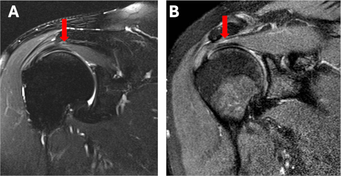
Figure 1: Representative preoperative MRI of the patients. MRI of the shoulder of (A) patient 1 and (B) patient 2 illustrating high-grade partial tears of the supraspinatus tendons (arrows). Please click here to view a larger version of this figure.
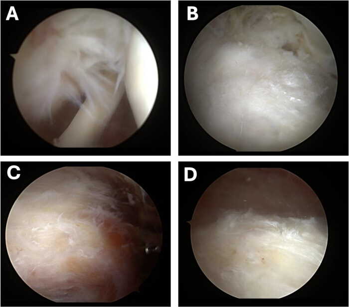
Figure 2: Representative arthroscopic images confirming the presence of segmental partial-thickness rotator cuff tears prior to debridement. Intraoperative images of patient 1 illustrating fraying of the bursal surface of the (A) infraspinatus and (B) supraspinatus tendons. Intraoperative images illustrating fraying of the coracoacromial ligament in (C) patient 1 and (D) patient 2. Please click here to view a larger version of this figure.
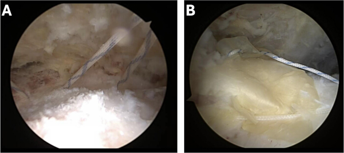
Figure 3: Representative intraoperative arthroscopic images. Representative intraoperative arthroscopic images illustrating (A) placement of suture into the medial aspect of the rotator cuff tendon and (B) securement of the MLG-COMPLETE allograft on the bursal surface of the rotator cuff. Please click here to view a larger version of this figure.
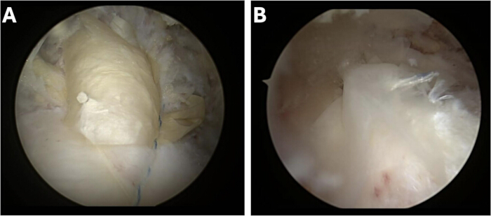
Figure 4: Representative intraoperative arthroscopic images. Representative intraoperative arthroscopic images illustrating final securement of the allograft using a luggage tag stitch from the lateral aspect of the allograft in (A) patient 1 and (B) patient 2. Please click here to view a larger version of this figure.
Table 1: Patient demographic and preoperative partial thickness rotator cuff tear classification.Please click here to download this Table.
Table 2: Summary of combined (averaged) clinical outcomes. Please click here to download this Table.
Discussie
The objective of this observational case study was to demonstrate the feasibility of arthroscopically delivering and securing a full-thickness perinatal tissue membrane allograft as a matrix augment in patients with PTRCTs. The primary findings from this work were that perinatal tissue membrane allograft matrix augmentation (PTAMA) was successfully performed on two patients with PTRCTs. The allograft was successfully delivered arthroscopically utilizing a double-pulley suture technique in conjunction with atraumatic graspers to advance and guide the allograft into the shoulder, where it was secured as an onlay directly over the PTRCTs. Additionally, patients reported minimal to no pain or discomfort at 7 days postoperatively, and continuous improvements in functional outcomes were observed at 3 months and at final follow-ups of 15 and 13 months for patients 1 and 2, respectively. Taken together, these initial findings establish the feasibility of arthroscopically delivering perinatal tissue allografts as a matrix augment for PTRCTs.
The surgical treatments for partial-thickness rotator cuff tears can include tear completion with repair, isolated bioinductive onlay augmentation, or combined completion/repair with onlay augmentation. Factors that can influence treatment choice are tear depth (Ellman grade), tissue quality, and patient risk factors for retear. In general, low-grade partial tears (generally <50% thickness, particularly bursal-sided lesions) with reasonable residual tendon integrity are well suited to isolated onlay augmentation, such as a bioinductive collagen implant or the perinatal tissue allograft used in this study. This strategy aims to promote new tissue formation and increase tendon thickness rather than provide mechanical support. Furthermore, the onlay approach preserves the native footprint, avoids the morbidity of formal fixation, and has shown clinical and MRI outcomes comparable to tear completion and repair in recent prospective and randomized data, including improved early functional recovery in some series of high-grade tears treated this way35. High-grade partial tears (>50% thickness) with substantial fiber disruption, delamination, or poor-quality residual tissue are generally managed with traditional completion of the tear and formal transosseous-equivalent repair to a well-vascularized footprint, since an isolated onlay graft cannot reliably restore mechanical strength across a compromised tendon, and its benefit is unproven once a tear approaches full thickness. However, recent literature has challenged this as even full-thickness small to medium tears with an intact rotator cuff cable may be successfully managed with bioinductive grafts alone36. The combined approach of completion and repair augmented with a bioinductive graft is reasonable for higher-risk scenarios, such as larger tears, revision surgery, poor native tissue quality, or patient factors (age, diabetes, smoking). These high-risk scenarios are associated with elevated retear rates, and augmentation has shown improved structural healing and lower retear rates versus repair alone37. However, supporting literature is still limited by relatively small cohorts, heterogeneous grading criteria, and short-to-midterm follow-up.
The first major finding from this work was that matrix augmentation of PTRCTs with MLG–COMPLETE perinatal tissue membrane allografts could feasibly and safely be introduced arthroscopically via the use of a double-pulley suture technique in conjunction with an atraumatic grasper and secured as an onlay covering the PTRCT using luggage tag sutures. Prior case studies report either injecting micronized (i.e., particulated) perinatal tissue allografts into the shoulder joint of patients with PTRCTs38, or implanting intact amnion-only containing membrane allografts at the interface of the greater tuberosity and supraspinatus tendon during the repair of full-thickness tears33. Notable differences between the present case study and previous work include being able to keep the arthroscopic saline source on, not having to dry the surgical site prior to perinatal allograft implantation, and the avoidance of having to use a spinal needle or other instrumentation to keep the graft in place prior to its securement. This was enabled in part due to the use of a full-thickness perinatal tissue membrane allograft that exhibited handling characteristics which allowed it to: 1) pass through an 8 mm diameter cannula with the aid of an atraumatic grasper/clamp, 2) withstand the shear forces applied to it while utilizing the double – pully suture passing mechanism to ‘parachute/zip line’ the grafts into place over the PTRCT, and 3) be secured in place with sutures. It should be noted that although some fraying of the graft was observed following passage through the cannula, the graft remained intact and could be guided and secured directly over the PTRCT as an onlay.
The second major finding from this study was that patients reported no pain or discomfort as early as 7 days postoperatively. In our experience, this is typically not the case for patients receiving debridement and decompression alone, in which patients typically experience soreness and have difficulties sleeping for the first 3–4 weeks after surgery. Furthermore, the patients experienced an average improvement in VAS, ASES, and SANE scores of 86.7%, 57.3%, and 76.4% at the 3-month follow-up, and 100%, 69.1%, and 80% at final follow–up, respectively. The patients ultimately experienced a return to the normal range of motion. The patients in this study achieved near full strength and were allowed to resume normal work, recreation, and sports by 3 months, whereas 5–6 months is often required for standard rotator cuff repair in our experience. The findings herein are in agreement with prior clinical reports. Ackley et al. demonstrated that intra-articular injection of micronized, human cryopreserved amnion/umbilical cord resulted in significant improvements in pain, function, patient satisfaction, and Penn shoulder scores at 6 months following injection in patients with MRI-confirmed partial tears of the supraspinatus tendon compared to preoperative values38. The patients noted overall improvement in quality of life, and follow-up MRI indicated no change in depth of rotator cuff tear concomitant with decreases in fluid intensity and effusion in some patients38. In regard to intact (i.e., non-micronized) perinatal tissue membrane allografts, Mirzayan et al arthroscopically repaired full-thickness tears in rotator cuff tendons having poor tissue quality via augmentation with human amniotic membrane allografts placed between the torn tendon and the humeral insertion33. They found that the interposition grafts did not prevent tendon-to-bone healing, as demonstrated by radiographic evidence, and that the repair resulted in significant improvements in ASES, Oxford, VAS, and SANE scores compared to preoperative values33. Taken together, despite the limited number of reports on the use of perinatal tissue allografts as a matrix augment in rotator cuff repair, early findings suggest the feasibility and safety of using these grafts.
Regarding the mechanisms by which perinatal tissue allografts may impact tendon biology and healing, preclinical studies are beginning to uncover potential mechanisms beyond their utility as a covering. McQuilling et al. demonstrated that perinatal tissue allografts contain growth factors known to positively influence tendon healing, including insulin-like growth factor (IGF), basic fibroblast growth factor (bFGF), and platelet-derived growth factor (PDGF)27. Moreover, the extracellular matrix (ECM) of the membranes supports tenocyte attachment, and the soluble factors released from the ECM promote tenocyte proliferation and migration27. Similarly, Kimmerling and colleagues found that a micronized amniotic membrane suspension allograft promoted increased tenocyte proliferation and migration while also increasing tenocyte production of tendon ECM components, including collagen, elastin, and glycosaminoglycan26. Moreno et al. demonstrated that soluble factors released from dehydrated amniotic membrane allografts may protect tenocytes from the detrimental effects of the proinflammatory cytokine interleukin-1β, which is often found at the site of tendon injury, while also exhibiting the potential to differentially influence new blood vessel formation in the presence of vascular endothelial cells30. Preclinical, in vivo models of tendon injury and repair augmentation with perinatal tissue allografts have also illustrated therapeutic potential. Injection of micronized human amnion/chorion allograft at the time of reattachment of a full-thickness chronic supraspinatus injury in rats resulted in a heightened inflammatory response and evidence of increased regenerating muscle fibers compared to saline injection controls39. Smith et al. demonstrated that bursal-sided amnion matrix augmentation of articular-sided partial thickness tears of the supraspinatus tendon in canines resulted in improved pain and functional assessments, enhanced histological appearance of the tendon, and reduced intra-articular pathology at 6 months postoperatively compared to debridement alone40.
Of note, conventional tendon-to-bone repair employing suture anchors was not utilized in the cases presented herein. Previous studies have demonstrated that take-down repair of PTRCTs with and without allografts demonstrated improved results with graft-only repairs35. Therefore, conventional repair would only be considered for full-thickness tears observed intraoperatively. Additionally, onlay augmentation with extracellular matrix-containing products has demonstrated success, as they are hypothesized to ‘biologically augment’ the tendon/repair microenvironment rather than mechanically reinforce the tear itself35. Furthermore, the tensile properties of perinatal tissue allografts do not approach those of native tendons, and thus they will not likely contribute to structural reinforcement or load-sharing in this repair setting.
As with any observational study, limitations exist, most notably the small sample size of patients reported herein; broader clinical recommendations cannot be made based solely on these findings. However, the primary focus of this exploratory study was to develop and demonstrate the feasibility of an implantation technique for a full-thickness perinatal tissue membrane allograft as opposed to reporting statistical improvements in outcomes. Accordingly, tendon healing was not evaluated post-operatively via clinical imaging, but such outcomes should be part of future studies evaluating this PTAMA technique. Additionally, there was no comparative control group of patients who underwent arthroscopic repair of PTRCTs in the absence of PTAMA. Thus, it is unknown if the observed findings are solely related to the surgery itself or due in part to the presence of the allograft. An additional limitation of the present study includes the lack of evaluation for tear size progression over time and long-term follow-up. Finally, while this technique was applied to bursal-sided tears, future evaluations regarding the feasibility of applying these grafts to articular-sided tears or intratendinous lesions and resultant clinical outcomes are warranted. Taken together, these limitations must be addressed in future prospective studies.
In conclusion, MLG-COMPLETE, a full-thickness perinatal tissue membrane allograft, can be successfully delivered arthroscopically and secured as a covering/onlay for matrix augmentation of PTRCTs.
Openbaarmakingen
S. Pill is a paid consultant of Samaritan Biologics, LLC. J. Mercuri is an employee of Samaritan Biologics, LLC.
Dankbetuigingen
The authors have no acknowledgments.
Materialen
| Naam | Bedrijf | Catalogusnummer | Opmerkingen |
|---|---|---|---|
| MLG-COMPLETE | Samaritan Biologics, LLC | MLG23 | |
| PassPort Cannula | Arthrex | AR-6592-08-40 | |
| PopLok Knotless Suture Anchor | Conmed | CKP-3501 | |
| SutureLasso | Arthrex | AR-4068-25TL | |
| SutureTape | Arthrex | N/A |
Referenties
- Kuhn JE. Prevalence, natural history, and nonoperative treatment of rotator cuff disease. Oper Tech Sports Med. 2023;31(1):150978.
- Apfel A, et al. Characteristics of rotator cuff repairs revised to shoulder arthroplasty. Arch Bone Jt Surg. 2020;8(5):575-80.
- Kim HM, et al. Analyzing practice pattern in treating partial-thickness rotator cuff tears: a dual perspective from national database and American Shoulder and Elbow Surgeons PARCIAL research group. JSES Int. 2025;9(1):91-7.
- Sevivas N, et al. Arthroscopic and open repair of massive rotator cuff tears have similar results: a systematic review. J ISAKOS. 2017;2(4):178-85.
- Shah NS, et al. Long-term outcomes of massive rotator cuff tear repair: a systematic review. HSS J. 2022;18:130-7.
- Hurley ET, et al. Outcomes after revision arthroscopic rotator cuff repair: a systematic review. Am J Sports Med. 2024;52(6):1635-40.
- Davey MS, et al. Arthroscopic rotator cuff repair results in improved clinical outcomes and low revision rates at 10-year follow-up: a systematic review. Arthroscopy. 2023;39:452-8.
- Ardeljan A, et al. Partial thickness rotator cuff tears: patient demographics and surgical trends within a large insurance database. J Orthop. 2020;17:158-61.
- Minagawa H, et al. Prevalence of symptomatic and asymptomatic rotator cuff tears in the general population: from mass-screening in one village. J Orthop. 2013;10(1):8-12.
- Yamaguchi K, et al. The demographic and morphological features of rotator cuff disease. A comparison of asymptomatic and symptomatic shoulders. J Bone Joint Surg Am. 2006;88(8):1699-704.
- Modi CS, Smith CD, Drew SJ. Partial-thickness articular surface rotator cuff tears in patients over the age of 35: etiology and intra-articular associations. Int J Shoulder Surg. 2012;6(1):15-8.
- Sher JS, et al. Abnormal findings on magnetic resonance images of asymptomatic shoulders. J Bone Joint Surg Am. 1995;77(1):10-5.
- Oh JH, et al. The natural history of high-grade partial thickness rotator cuff tears: the conversion rate to full thickness tears and affecting factors. Clin Orthop Surg. 2020;12(4):514-20.
- Matthewson G, et al. Partial thickness rotator cuff tears: current concepts. Adv Orthop. 2015;2015:458786.
- Yamanaka K, Matsumoto T. The joint side tear of the rotator cuff. A follow up study by arthrography. Clin Orthop Relat Res. 1994;(304):68-73.
- Bigliani L, et al. The relationship of acromial architecture to rotator cuff disease. Clin Sports Med. 1991;10(4):823-38.
- Neer CS. Anterior acromioplasty for the chronic impingement syndrome in the shoulder. 1972. J Bone Joint Surg Am. 2005;87(6):1399.
- Riley G. Tendon pathology and its relation to overuse. Br J Sports Med. 2005;39(6):241-9.
- Nirschl RP, Ashman ES. Elbow tendinopathy: tennis elbow. Clin Sports Med. 2003;22(4):813-36.
- Abraham AC, Shah SA, Thomopoulos S. Targeting inflammation in rotator cuff tendon degeneration and repair. Tech Shoulder Elb Surg. 2017;18(3):84-90.
- Tagliero AJ, et al. Rotator cuff repair and biologic augmentation—what do we know? JSES Rev Rep Tech. 2026;6(1):100605.
- Jabara J, Kiani S, Youn A, Feeley B. Orthobiologic augmentation to improve rotator cuff repair outcomes: current and future strategies. J Am Acad Orthop Surg. 2025;33(13):e731-6.
- Munoz-Torres JR, et al. Biological properties and surgical applications of the human amniotic membrane. Front Bioeng Biotechnol. 2023;10:1067480.
- Keeley R, Topoluk N, Mercuri J. Tissues reborn: fetal membrane-derived matrices and stem cells in orthopedic regenerative medicine. Crit Rev Biomed Eng. 2014;42(3-4):249-70.
- Forbes J, et al. The use of amniotic tissue-derived products in orthopedic surgery: a narrative review. Injury. 2024;55(11):111901.
- Kimmerling KA, McQuilling JP, Staples MC, Mowry KC. Tenocyte cell density, migration, and extracellular matrix deposition with amniotic suspension allograft. J Orthop Res. 2019;37(2):412-20.
- McQuilling JP, Kimmerling KA, Staples MC, Mowry KC. Evaluation of two distinct placental-derived membranes and their effect on tenocyte responses in vitro. J Tissue Eng Regen Med. 2019;13(8):1316-30.
- Mao Y, et al. A tri-layer decellularized, dehydrated human amniotic membrane scaffold supports the cellular functions of human tenocytes in vitro. J Mater Sci Mater Med. 2023;34(7).
- Mao Y, et al. A decellularized flowable placental connective tissue matrix supports cellular functions of human tenocytes in vitro. J Exp Orthop. 2022;9(1).
- Moreno SE, Massee M, Koob TJ. Dehydrated human amniotic membrane regulates tenocyte expression and angiogenesis in vitro: implications for a therapeutic treatment of tendinopathy. J Biomed Mater Res B Appl Biomater. 2022;110(4):731-42.
- Anderson J, Adeleke A, Rice B, Swayzee Z. Surgical treatment of peroneus brevis tendon repair with and without human amniotic allograft: a comparison study. Clin Surg. 2017;2:1-5.
- Giacobazzi M, et al. Primary repair of complete Achilles tear augmented with amnion allograft wrap in college basketball player with a history of contralateral Achilles rupture: a case report. AME Case Rep. 2023;7:36.
- Mirzayan R, Suh BD. Interposition of human amniotic membrane at the bone-tendon interface of a full-thickness rotator cuff repair. JSES Rev Rep Tech. 2022;2(1):75-80.
- Foret A, et al. Functional and oncologic outcomes following perinatal tissue allograft placement during nerve-sparing robotic-assisted radical prostatectomy. J Vis Exp. 2025;(224).
- Jenkins R, Mon NO, Ruotolo C. Patch augmentation in the treatment of partial-thickness rotator cuff tears facilitates tendon healing and improves patient outcomes: a systematic review. Arthroscopy. 2025;41(11):4826-35.
- Camacho Chacón JA, et al. An isolated bioinductive repair vs sutured repair for full-thickness rotator cuff tears: 2-year results of a double blinded, randomized controlled trial. J Shoulder Elbow Surg. 2024;33(9):1894-904.
- Ruiz Ibán MÁ, et al. Augmentation of a transosseous-equivalent repair in posterosuperior nonacute rotator cuff tears with a bioinductive collagen implant decreases the retear rate at 1 year: a randomized controlled trial. Arthroscopy. 2024;40(6):1760-73.
- Ackley JF, et al. Cryopreserved amniotic membrane and umbilical cord particulate matrix for partial rotator cuff tears: a case series. Medicine (Baltimore). 2019;98(30):e16569.
- Anderson LE, et al. Injection of micronized human amnion/chorion membrane results in increased early supraspinatus muscle regeneration in a chronic model of rotator cuff tear. Ann Biomed Eng. 2021;49(12):3698-710.
- Smith MJ, et al. Comparison of biologic scaffolds for augmentation of partial rotator cuff tears in a canine model. J Shoulder Elbow Surg. 2020;29(8):1573-83.
Herprints en machtigingen
Dit artikel is gepubliceerd
Video binnenkort beschikbaar