This article presents a standardized static retinoscopy protocol for accurate refractive assessment in children with aphakia following congenital cataract surgery, incorporating the Vergence formula as a quality control reference.
Methodenartikel
A Standardized Static Retinoscopy Procedure for Refractive Assessment in Children After Congenital Cataract Surgery
In dit artikel
Samenvatting
Samenvatting
Congenital cataracts are a leading cause of visual impairment in children. After surgery, accurate refractive assessment and correction are essential to regain useful vision. Refractive assessment in children after congenital cataract surgery is challenging because of poor cooperation and rapid, dynamic refractive shifts during eye development. Retinoscopy, widely recognized as the gold standard for refractive assessment, remains central to this clinical setting. However, its effectiveness may be limited by inconsistent operating protocols and suboptimal patient cooperation, particularly among practitioners with limited experience. We propose a standardized retinoscopy workflow for postoperative refractive evaluation in children with congenital cataracts. This protocol integrates the Vergence-formula estimate as a quality-control alert tool during retinoscopy, providing an additional reference for identifying potential inconsistencies during assessment. The standardized workflow is intended to improve the consistency and reliability of postoperative refractive evaluation and could serve as a practical reference for routine clinical use in aphakic young children.
Inleiding
Congenital cataract is one of the leading causes of irreversible visual impairment in children worldwide1. Early cataract extraction combined with intraocular lens implantation has become a routine clinical intervention strategy2,3. Without timely and accurate postoperative correction, these children are at high risk of form-deprivation amblyopia and impaired visual development4.
The human eye undergoes rapid structural growth during infancy and early childhood5. Within the first 18 months after birth, axial length increases rapidly from 16.8 mm to 23.6 mm. Refractive status demonstrates continuously dynamic characteristics accompanied by myopic shift6. Large-scale prospective cohort studies have shown that aphakic eyes demonstrate significant and sustained myopic shift at 5 years postoperatively, with annual changes being particularly prominent during infancy7. Coupled with extremely poor cooperation and insufficient fixation ability in young children, postoperative refractive measurement has become a widely recognized technical challenge in pediatric optometry8. Furthermore, aphakic eyes in children possess very high refractive power (frequently > +20 D), out of the reliable measurement range for most automatic refraction devices9.
Static retinoscopy remains the clinical gold standard for refractive assessment in children. It is not limited by device measurement range, and it allows direct evaluation of the refractive media10,11. Nevertheless, current clinical retinoscopy is highly experience‑dependent, with no unified operating standards among institutions and practitioners. The measurement results show poor consistency and repeatability, which cannot meet the clinical requirements for long‑term postoperative follow‑up and precise correction10,12.
This protocol aims to develop a standardized static retinoscopy workflow for children following congenital cataract surgery, delivering a standardized refractive evaluation framework suitable for broad application in young children with aphakic eyes. For children under 4 years of age who cannot cooperate with ophthalmic examinations, intranasal dexmedetomidine sedation has been reported to be safe and effective for facilitating ophthalmic measurements13. Accurate postoperative management of pediatric cataract also depends on biometric measurements and appropriate refractive calculations, which are important considerations in pediatric cataract care14. The protocol follows the principle of spectacle-over-refraction. A base frame lens with a power close to the estimated refraction is placed on the child’s eye, and retinoscopy is then performed in front of this lens to measure the residual refractive error. The key innovation is the integration of the Vergence formula15 as an independent quality control mechanism. This protocol is intended for use by pediatric ophthalmologists and optometrists managing postoperative care in children with congenital cataracts and is applicable to aphakic eyes. The method requires no sophisticated specialized equipment and can be implemented in pediatric ophthalmology clinics and cataract follow-up settings.
Protocol
This study was approved by the Institutional Review Board (IRB) of the Eye Hospital, Wenzhou Medical University (IRB approval No. H2026-031-K-31-01, June 15, 2026), and the requirement for individual consent for this retrospective analysis was waived due to the retrospective nature. Written informed consent for publication of the identifiable images and videos of the children was obtained from their legal guardians. Notably, all images and videos included in this manuscript were specifically recorded with volunteer participants recruited for this protocol demonstration, not retrieved from existing clinical medical records. The reagents and the equipment used are listed in the Table of Materials.
1. Preparation of the examination environment
- Light and illumination setup
- Set the examination room to a semi-dark state by turning off the main light source. Eliminate bright ambient light to avoid interference with observation of the pupil reflex movement.
- Turn on dim, soft background lighting. Prevent children from experiencing fear or restlessness due to complete darkness.
- Physical environment setup
- Use a dedicated refraction room for the procedure to ensure adequate operating space.
- Maintain a quiet atmosphere in the examination room throughout the procedure. Prevent sudden noises that may startle the infant.
- Adjust the room temperature to a comfortable range. Prevent irritability and crying caused by thermal discomfort.
2. Equipment and materials
- Refer to the Table of Materials for a complete list of required supplies and equipment.
NOTE: Ensure that the optical components of the hand-held retinoscope and all lenses in the trial lens set are clean and free of smudges, and that the retinoscope battery is fully charged.
3. Patient preparation
- Sedation
- For children under 4 years of age who cannot cooperate with fixation while awake, administer intranasal dexmedetomidine for pre-examination sedation according to the institutional protocol.
- Gently wipe and clean the child’s nasal passages using soft tissue paper.
- Place the child in a supine position.
- Calculate the required dexmedetomidine dose according to body weight (3 µg/kg).
- Aspirate the corresponding drug volume with a 1‑mL syringe, and instill the solution drop‑wise into the nasal cavity.
CAUTION: Dexmedetomidine is a prescription sedative medication. Calculate the dosage strictly based on body weight and administer only under the supervision of a qualified pediatric anesthesiologist. Continuously monitor heart rate and oxygen saturation to guard against respiratory depression and bradycardia.
- Schedule the retinoscopy examination concurrently with other postoperative follow-up procedures requiring anesthesia. Minimize the infant's exposure to multiple sedation events.
- Confirm that the infant has achieved adequate sedation or is in a stable sleep state before initiating retinoscopy.
- For children under 4 years of age who cannot cooperate with fixation while awake, administer intranasal dexmedetomidine for pre-examination sedation according to the institutional protocol.
- Pre-procedural refractive estimation
- For cooperative children, obtain preliminary refractive data using a portable autorefractor.
- At every visit, obtain (or retrieve) the most recent axial-length and corneal-curvature measurements. If the child cannot cooperate with portable autorefraction, use these current values, not historical values, for the Vergence-formula calculation.
- Calculate the estimated refractive power (Fe) of the aphakic eye using the Vergence formula15, based on axial length and corneal curvature.
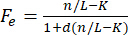
Where:
Fe = estimated refractive power of the aphakic eye (spherical equivalent, in diopters)
L = axial length (in meters)
K = mean corneal curvature (in diopters)
n = refractive index of aqueous humor (1.336)
d = vertex distance (0.012 m for adults; 0.009 m for children in this protocol)
- Positioning and eyelid retraction
- Have the parent hold the infant in a cradling position. Position awake children in a seated posture and sleeping or sedated children in a supine or semi-recumbent position.
- Position an assistant beside the parent to gently retract the infant's upper eyelid with a medical cotton swab. Ensure full exposure of the pupil.
- If no dedicated assistant is available, instruct the parent to gently retract the infant's upper eyelid with a clean finger (see Figure 1).
- Stabilize the infant's head throughout the procedure. Prevent head movement from altering the retinoscopy light path and compromising measurement accuracy.
NOTE: Children in a sleep state frequently exhibit upward rotation of the eyeball (Bell's phenomenon). When this occurs, release the eyelid, adjust the infant's posture, and wait until the eye returns to a neutral position before resuming retinoscopy.
4. Retinoscopy procedure (spectacle-over-refraction method)
- Base frame lens placement
- Select the base frame lens whose power and interpupillary distance most closely match the calculated Fe value and the infant's interpupillary distance. For example, if the calculated Fe is +20.50 D and the interpupillary distance is 41 mm, select a +20.00 D base frame lens with a 42 mm interpupillary distance. Thoroughly disinfect the lens with disinfectant wipes before use.
- Place the base frame lens on the infant. Maintain the vertex distance and ensure the lens is parallel to the cornea and free of displacement.
NOTE: Aphakic eyes are highly sensitive to vertex distance. Even minor changes in distance produce measurable variations in spherical equivalent. The vertex distance must be strictly fixed throughout the procedure.
- Initial retinoscopy setup
- Turn on the retinoscope light source. Direct the light beam toward the center of the pupil of the eye being examined.
- Begin placing retinoscopy lenses in front of the base frame lens. Adjust the retinoscope angle so that the light beam enters precisely along the infant's visual axis (see Figure 2).
NOTE: Maintain a fixed working distance between the retinoscope and the examined eye; we routinely use 50 cm (67 cm may also be used if held constant and applied consistently in the calculation). Do not alter the working distance throughout the procedure. Keep both eyes open and observe the retinal reflex and shadow movement patterns in the pupil area through the retinoscope (see Figure 3).
- Reflex observation and assessment
- Observe the direction of the reflex movement. Distinguish between "with" movement, "against" movement, and the neutralization point.
- Observe the speed of the reflex movement. Estimate the distance from the current state to the neutralization point.
- Observe the brightness of the reflex. Confirm that the reflected light is clear and exclude interference from corneal dryness or refractive media opacities.
- Rotate the streak retinoscope slowly, scanning from 0° to 180°. Identify the two principal meridian directions.
NOTE: A very dim retinoscopy reflex may indicate that the eye is far from the neutralization point, requiring a high-power plus or minus lens. If this is suspected, reduce the working distance or experimentally add a high-power lens (e.g., ±5.00 D) in front of the frame lens and re-observe. Then replace the base frame lens with one of higher or lower power and resume retinoscopy.
- Spherical lens neutralization of principal meridians
- Select the first principal meridian and observe the direction of reflex movement.
- If the reflex shows "with" movement, add plus spherical lenses in front of the base frame lens in increments of +0.25 D to +0.50 D. Continue adding lenses until complete neutralization is achieved.
- If the reflex shows "against" movement, add minus spherical lenses in front of the base frame lens in increments of −0.25 D to −0.50 D. Continue adding lenses until complete neutralization is achieved.
- Record the neutralizing spherical lens power for the first principal meridian and the axis.
NOTE: After neutralizing the refractive error along the principal meridian through the base frame with a streak retinoscope, the examiner holds the streak steady in its final orientation to maintain meridian alignment. A trial frame is then placed parallel to the base frame, and the streak orientation is carefully transferred to the axis scale of the trial frame. The axis is read directly from the calibrated scale on the trial frame. This approach allows reliable axis measurement without requiring a trial frame during the initial neutralization phase. - Rotate the streak light to align with the second principal meridian. Repeat steps 4.4.1 through 4.4.4 and record the neutralizing spherical lens power and the axis.
NOTE: Since retinoscopy is performed in front of the base frame lens, using a cylindrical lens to neutralize the second meridian is impractical: without a trial frame, accurate positioning of the cylinder axis is difficult. Additionally, stacking three lenses (base frame, spherical, and cylindrical) reduces the brightness of the retinoscopy streak. Therefore, use only spherical lenses to neutralize each of the two principal meridians separately. If the reflex is distorted by irregular corneal astigmatism and no axis can be identified, use the keratometry readings to estimate the principal meridians.
- Intermittent eye rest
- If a single retinoscopy session exceeds 30 s, instruct the assistant to release the eyelid. Allow the infant to close the eye and rest briefly.
- After the rest period, retract the eyelid to re-expose the pupil. Confirm that the base frame lens position has not shifted, then resume retinoscopy.
NOTE: Ensure that the distance between the base frame lens and the eyeball remains fixed. Avoid frequent repositioning of the base frame lens. Each time retinoscopy lenses are changed, confirm that the base frame lens remains in position and press the retinoscopy lens firmly against the base frame lens. Prolonged eye opening causes corneal dryness, which impairs the retinoscopy reflex.
- Examination of the contralateral eye
- After completing retinoscopy of the first eye, maintain the infant's sedated or sleep state unchanged.
- Perform retinoscopy of the contralateral eye following steps 4.1 through 4.5.
5. Calculation and documentation of results
- For purely spherical refractive errors, calculate the final refractive power using the following formula:

Where:
Fa — final spherical equivalent refractive power (in diopters)
SEb — base frame lens power (in diopters)
SE — neutralizing power (in diopters)
d — retinoscopy working distance (in meters), - For astigmatic refractive errors, calculate the final results using the following formulas:


Where:
Fa — final spherical equivalent refractive power (in diopters)
FaS — final spherical power (in diopters)
FaC — final cylindrical power (in diopters)
SEb — base frame lens power (in diopters)
SE1, SE2 — neutralizing powers of the two principal meridians. SE1, corresponds to the more‑positive (or less‑negative) neutralizing power, while SE2 corresponds to the relatively more‑negative (or less‑positive) neutralizing power (in diopters)
d — retinoscopy working distance (in meters)
axis — astigmatic axis (in degrees), - Document the retinoscopy working distance (d), base frame lens power (SEb), and the neutralizing powers of the two principal meridians (SE1, SE2) .
- Enter the final spherical power, cylindrical power, and axis into the medical record system in a standardized format. The final refractive result is expressed in standard sphero-cylindrical notation: FeS / FeC × axis. For example, +20.00 / −1.50 × 180. Using the minus-cylinder convention adopted here, the more positive or less negative meridian is taken as the sphere, and the cylinder is negative, with its axis aligned to the SE2 meridian.
6. Quality control and data verification
- After completing retinoscopy and calculating the final spherical and cylindrical refractive power, compare the derived retinoscopy spherical‑equivalent value with the spherical‑equivalent estimate output from the Vergence formula. If the absolute difference between these two values exceeds 3.00 D, trigger the mandatory verification procedure described below.
- Assign a second qualified, experienced optometrist who is blinded to the initial retinoscopy result to repeat the full static retinoscopy procedure from step 4.1 through step 5.4, including base‑lens placement, reflex observation, principal‑meridian neutralization, and result calculation.
- Compute the final refractive outcome by taking the arithmetic average of the two independent retinoscopy measurements (initial examination and repeated examination by the second optometrist).
NOTE: The Vergence‑formula estimate is not used for computing the final clinical prescription; it only serves as an alarm trigger for quality‑control purposes. - Document both retinoscopy results and the final averaged refractive power in the medical record. Use this averaged result as the basis for clinical spectacle correction.
Resultaten
Using this standardized protocol, we retrospectively reviewed all consecutive pediatric patients who underwent congenital cataract extraction in our center during 2024. All medical records were screened according to pre‑defined inclusion and exclusion criteria. Inclusion criteria: (1) age ranging from 2 months to 4 years, regardless of sex; (2) confirmed diagnosis of congenital cataract after cataract extraction; (3) postoperative retinoscopy refraction results available; (4) complete preoperative axial‑length and keratometry data for Vergence‑formula calculation. Exclusion criteria: (1) co‑existing other ocular disorders (congenital glaucoma, retinopathy, optic‑nerve dysplasia, corneal opacification, etc.) that could interfere with refractive measurement; (2) systemic diseases (neurological, metabolic, or cardiopulmonary disorders) precluding sedated retinoscopy; (3) known allergy or contraindication to intranasal dexmedetomidine; (4) incomplete follow‑up records lacking key biometric or retinoscopy results. After screening, 64 children (102 eyes) fulfilled the criteria. For the primary statistical analysis of this study, the right eye was selected for bilateral cases; for children with unilateral cataract, the only surgically‑operated eye was retained. This selection strategy yielded a dataset of 64 independent eyes for all primary analyses. The cohort included 30 males and 34 females, with ages ranging from 6 months to 4 years (mean 1.45 ± 0.90 years). All children underwent retinoscopy within 3 days after cataract surgery under dexmedetomidine sedation due to their inability to cooperate independently, and all procedures were performed by the same experienced optometrist. Preoperative biometric data (axial length and corneal curvature) were retrieved from the most recent preoperative measurements for Vergence formula estimation.
Descriptive comparison
The mean refractive power obtained by static retinoscopy was 22.61 ± 3.24 D, and the mean estimated by the Vergence formula was 22.45 ± 3.88 D (mean difference: +0.16 D; 95% CI: −0.28 to +0.59 D). The two methods showed a strong positive correlation (r = 0.8963, R2 = 0.803, P < 0.001; see Figure 4).
Agreement analysis
The ICC(2,1) for absolute agreement was 0.883 (95% CI: 0.824–0.921), indicating good agreement. Bland-Altman analysis (see Figure 5) showed 95% limits of agreement from −3.24 D to +3.56 D, with 61 of 64 eyes (95.3%) within these limits. Regression of the difference against the mean revealed a significant proportional bias (slope = −0.189, P = 0.0021): at higher refractive powers, the two methods converged, whereas at lower powers, retinoscopy yielded slightly higher values. To formally assess equivalence, TOST was performed with bounds of ±1.50 D and ±2.00 D; both were significant (p < 0.001), confirming that the difference between methods fell within clinically acceptable limits.
Error distribution
The mean absolute error was 1.39 D. Cumulative analysis showed that 28.1% of eyes had an error ≤0.50 D, 42.2% ≤1.00 D, 57.8% ≤1.50 D, 70.3% ≤2.00 D, 81.2% ≤2.50 D, and 92.2% ≤3.00 D (see Figure 6). Given that aphakic refractive power in this cohort typically exceeded +20 D and base frame lens power intervals were 2.00 D, this supports the clinical utility of the Vergence formula as a quality control reference for base frame lens selection and preliminary result assessment.
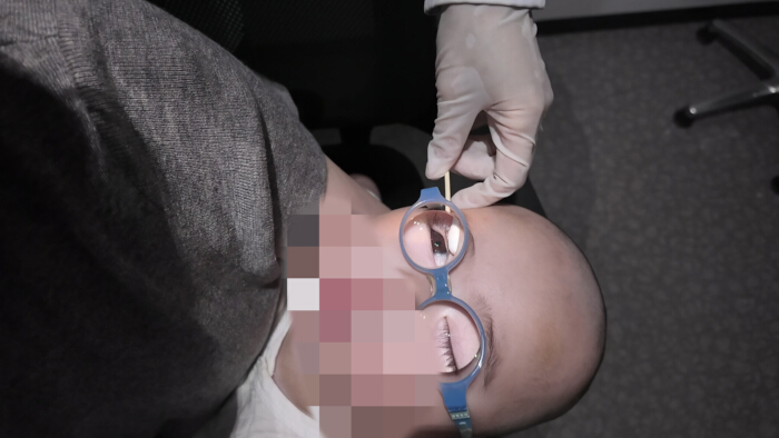
Figure 1: Demonstration of eyelid retraction and positioning for infant retinoscopy. A sleeping child volunteer, wearing the base frame spectacles, is positioned supine; no sedation was used. An assistant gently retracts the infant's upper eyelid with a medical cotton swab to ensure full exposure of the pupil. The infant is in a supine position, and the parent holds the infant's head steady. Proper eyelid retraction is critical for unobstructed visualization of the retinal reflex during static retinoscopy. Please click here to view a larger version of this figure.
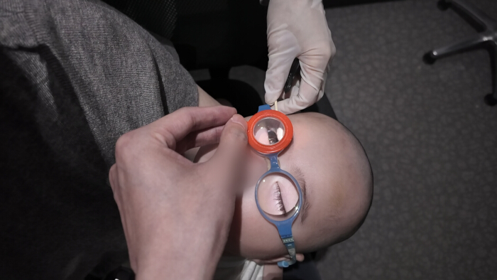
Figure 2: Demonstration of spectacle-over-refraction technique with base frame lens. Retinoscopy lenses are placed in front of the base frame spectacle lens during retinoscopy. The retinoscope beam is aligned along the infant's visual axis, entering through the center of the pupil. This spectacle-over-refraction method allows neutralization of high refractive errors in aphakic eyes by combining the base frame power with additional trial lenses. Please click here to view a larger version of this figure.
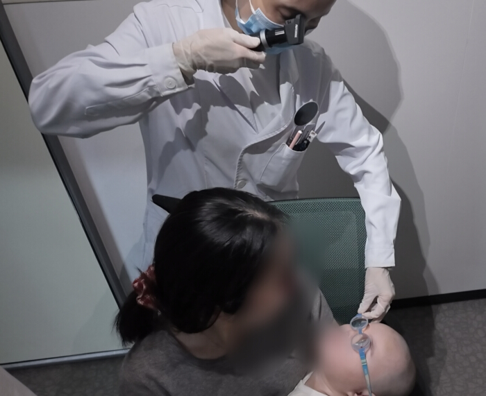
Figure 3: Demonstration of static retinoscopy at a standardized working distance. An optometrist performs static retinoscopy at a standardized working distance of 50 cm from the infant's eye. The examiner observes the retinal reflex and shadow movement patterns in the pupil area through the retinoscope, keeping both eyes open. The same fixed distance is entered into the Vergence-formula calculation. Please click here to view a larger version of this figure.
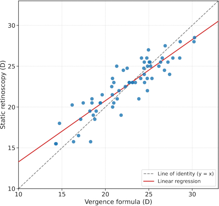
Figure 4: Scatter plot of refractive powers measured by static retinoscopy versus values calculated by the Vergence formula (n = 64 eyes). The dashed gray line represents the line of identity (y = x), and the solid red line represents the linear regression fit. A strong positive linear relationship was observed (r = 0.896, R2 = 0.803, p < 0.001), indicating close agreement between the two methods across the measured range of aphakic refractive powers. Please click here to view a larger version of this figure.
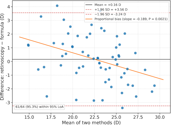
Figure 5: Bland-Altman plot of the agreement between static retinoscopy and the Vergence formula. The middle solid line represents the mean difference (+0.16 D; retinoscopy minus formula), and the upper and lower dashed lines represent the 95% limits of agreement (−3.24 D to +3.56 D). Sixty-one of 64 eyes (95.3%) fell within these limits. The orange line depicts regression of the difference on the mean, revealing a significant proportional bias (slope = −0.189, p = 0.002). Please click here to view a larger version of this figure.
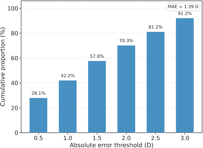
Figure 6: Cumulative distribution of absolute errors between static retinoscopy and the Vergence formula. Bars show the proportion of eyes with absolute error within each threshold: 28.1% (≤0.50 D), 42.2% (≤1.00 D), 57.8% (≤1.50 D), 70.3% (≤2.00 D), 81.2% (≤2.50 D), and 92.2% (≤3.00 D). The mean absolute error was 1.39 D. Please click here to view a larger version of this figure.
Discussie
Although portable autorefractors have gained increasing popularity in clinical practice, age remains a critical consideration when selecting a measurement technique. For older children or those with adequate cooperation, portable autorefractors such as the Retinomax can serve as an effective alternative. Studies have demonstrated that cooperation improves significantly in children over 36 months of age, and measurements obtained with devices such as the Retinomax show good agreement with cycloplegic retinoscopy16. However, for children under 3 years of age, particularly those with aphakia following congenital cataract surgery, the high refractive power often exceeds the measurement range of portable autorefractors, and poor fixation ability renders stable and reliable results difficult to obtain17. Furthermore, although photorefraction can be used for large-scale population screening, its accuracy in populations with high refractive errors and aphakia remains controversial, making it insufficient to meet the demands of precise clinical refractive assessment12. For children who cannot cooperate with examination, intranasal dexmedetomidine sedation has been proven to be a safe and effective option for ophthalmic outpatient examinations. At a dose of 3 µg/kg, the success rate for keratometry and axial length measurements reaches 92%–96%, with stable vital signs throughout the procedure and no significant adverse events13.
Static retinoscopy is the clinical gold standard for refractive assessment in children and young children14. However, the reliability of its results is highly dependent on operator experience. The current standardized workflow reduces operator dependence by unifying operational protocols, thereby promoting consistent execution of the entire retinoscopy procedure. The incorporation of the Vergence formula to estimate refractive power as a quality control reference further enhances the stability and credibility of the examination results. Although the 95% limits of agreement (−3.24 to +3.56 D) are wide and do not indicate interchangeability, this degree of disagreement is consistent with the intended use of the formula for base‑frame selection only. The base‑frame provides merely a starting platform and is never prescribed; any error in the initial base‑frame setting is corrected by subsequent retinoscopy performed to reach the true reflex endpoint. The wider limits relative to Tian et al. (−1.37 to +2.00 D;15) reflect the younger, non-cooperative children (mean 1.45 years) spanning a higher dioptric range (approximately 14–31 D), where biometry is intrinsically less precise, and a significant proportional bias (slope = −0.189) is present. The mean absolute error was 1.39 D, and 92.2% of eyes differed by ≤3.00 D; larger discrepancies trigger an independent second retinoscopy rather than acceptance of the formula value.
According to previous studies, the Vergence formula was specifically designed for aphakic eyes in infants and young children, with its theoretical basis grounded in treating the cornea and vitreous cavity as a thin lens combination15. The advantage of this formula is that it requires only two basic parameters, axial length and corneal curvature, rendering the calculation relatively straightforward, and it has been shown to possess good predictive accuracy in clinical practice. This workflow innovatively combines the Vergence formula with standardized retinoscopy procedures: the formula-based method rapidly narrows the approximate refractive range and improves retinoscopy efficiency, while standardized retinoscopy ensures the clinical accuracy of the final results. The two approaches form a complementary rather than a substitutive relationship. This is consistent with the conclusion of related studies that objective biometric parameter measurement can assist in improving the reliability of traditional refractive measurement methods18.
Another distinctive feature of this protocol is the base‑frame‑supported spectacle‑over‑refraction design. Retinoscopy is performed through the base frame worn by the child. When spectacles are dispensed clinically, selecting the identical frame model used during retinoscopy narrows vertex‑distance discrepancy between the examination setting and real‑life wearing condition, thereby reducing systematic dioptric errors originating from vertex‑distance mismatch.
The standardized retinoscopy workflow has several limitations. First, for uncooperative young children, the refraction procedure requires sedation or natural sleep, which necessitates appropriate anesthesia conditions and monitoring capabilities, thereby limiting its widespread application in routine outpatient settings where children are fully awake. However, the workflow requires only widely available equipment and no high-end instruments, making it feasible for implementation in institutions with appropriate capabilities. Second, the vertex distance of 8–10 mm used in this workflow is an anatomical inference based on the flat nasal bridge and distinct facial anatomy of children compared with adults. Currently, direct measurement data on vertex distance in large infant cohorts are lacking, and targeted studies are warranted to further validate the applicability of this parameter. Third, the present study was performed by a single experienced optometrist who was not masked to the Vergence-formula estimate during the initial examination, and neither inter- nor intra-examiner reproducibility was formally assessed. Inter‑examiner agreement across operators with varying levels of clinical experience was not evaluated. Whether this written SOP yields consistent results when used by different examiners remains an open question and requires independent external validation by other clinical groups in future work. Although the detailed step-by-step instructions are intended to minimize operator-dependent variability, performance across examiners of different experience levels remains unknown. Adding measurements by multiple examiners prospectively would prolong sedation purely for research purposes, which we considered unjustified in this vulnerable cohort, and repeated intra-examiner measurements were not part of the retrospective clinical workflow. External real-world validation from other clinical sites and future masked, multi-examiner studies would help strengthen the evidence base. Additionally, this study represents a single-center experience, and future multicenter, large-sample studies are needed to further verify the generalizability and reliability of this workflow.
Overall, standardized retinoscopy is not limited by device range and can be used even with complex ocular conditions after congenital cataract surgery. Supplemented by objective formula-derived data for comprehensive quality control, this approach preserves the clinical utility of the traditional examination while compensating for the experience-dependent limitations inherent to a purely subjective assessment. This workflow offers a structured approach to refractive assessment in children with aphakia; its reproducibility across examiners and centers remains to be tested in future work.
Openbaarmakingen
The authors have no conflicts of interest to declare.
Materialen
| Naam | Bedrijf | Catalogusnummer | Opmerkingen |
|---|---|---|---|
| Aphakic ophthalmic single vision lenses | Meijing Optical | CCL-A congenital cataract lenses | Custom high-power plus lenses for aphakia; 1.60 index; AR coating; power range +4.00 D to +30.00 D |
| Contact A-scan biometer | Quantel Medical | AXIS NANO | Portable A-scan ultrasound biometer for axial length measurement; 11 MHz probe; 0.01 mm accuracy |
| Dexmedetomidine hydrochloride injection | Chenxin Pharmaceutical Co., Ltd. | Chenjiao (2 mL:0.2 mg) | Prescription sedative; intranasal administration per institutional protocol; 3 μg/kg dosage |
| Handheld autorefractor/keratometer | NIDEK | HandyRef-K | Portable autorefractor and keratometer for baseline refractive assessment |
| Hand-held streak retinoscope | Keeler | 1302-P-1011 | Professional Streak Retinoscope, 3.6V rechargeable lithium battery; adjustable working distance |
| Infant spectacle frame | TOMATO Eyewear (Jiangsu Tomato Eyewear) | KB40 series | Infant size frames for high plus lenses; PD ~40 mm range; silicone nose pad and temple tips; adjustable length |
| Standard trial lens set | Shanghai Riyue Optical Instrument Co., Ltd. | SL-232 | Complete set of spherical and cylindrical lenses for retinoscopy; ISO 9001 certified |
Referenties
- Tătaru CI, et al. Congenital cataract - clinical and morphological aspects. Rom J Morphol Embryol. 2020;61(1):105-112.
- Bremond-Gignac D, Daruich A, Robert MP, Valleix S. Recent developments in the management of congenital cataract. Ann Transl Med. 2020;8(22):1545.
- Lenhart PD, Lambert SR. Current management of infantile cataracts. Surv Ophthalmol. 2022;67(5):1476-1505.
- Talebnejad MR, et al. The prevalence and causes of low vision and visual impairment in school-aged children: the Shiraz Pediatric Eye Study. J Curr Ophthalmol. 2022;34(3):333-340.
- Groot ALW, et al. Meta-analysis of ocular axial length in newborns and infants up to 3 years of age. Surv Ophthalmol. 2022 Mar-Apr;67(2):342-352.
- Li L, et al. Changes in eye axis length and corneal curvature in central China from the perspective of birth year. Eye (Lond). 2025;39(7):1373-1379.
- VanderVeen DK, et al. Refractive change at 5 years in the Toddler Aphakia and Pseudophakia Study. Ophthalmology. 2025;132(5):610-616.
- Mirzajani A, Amini Vishteh R, Khalilian M. Introducing a new method of retinoscopy for refraction of infants and young children: the "Mirza" tele lens retinoscopy. J Optom. 2021;14(3):254-262.
- Lin D, et al. Postoperative myopic shift and visual acuity rehabilitation in patients with bilateral congenital cataracts. Front Med (Lausanne). 2024;11:1406287.
- Arslantürk Eren M, Nalcı Baytaroğlu H, Atilla H. Comparison of Spot Vision Screener and tabletop autorefractometer with retinoscopy in the pediatric population. Turk J Ophthalmol. 2024;54(2):56-62.
- Rydberg A, et al. Accuracy of noncycloplegic retinoscopy, Retinomax autorefractor, and SureSight vision screener for detecting significant refractive errors. Optom Vis Sci. 2015;92(2):215-225.
- Schaafsma JD, Arnold RW. Pre-cycloplegic exam benefit of photoscreening and accommodation-relaxing skiascopy. Clin Ophthalmol. 2024;18:833-846.
- Chen C, et al. Study of feasibility and safety of higher-dose dexmedetomidine in special outpatient examination of pediatric ophthalmology. J Ophthalmol. 2019;2019:2560453.
- Lin L et al. Accuracy of newer generation intraocular lens power calculation formulas in pediatric cataract patients. Graefes Arch Clin Exp Ophthalmol. 2023;261(4):1019-1027.
- Tian L, et al. Vergence formula for estimating the refractive status of aphakic eyes in pediatric patients. Front Med (Lausanne). 2022;9:861745.
- Gole GA, Schluter PJ, Hall J, Colville D. Comparison of the Retinomax autorefractor with hand-held retinoscopy in 1-year-old infants. Clin Exp Ophthalmol. 2003;31(4):341-347.
- Mittelviefhaus H, Gentner C. Messungenauigkeiten der Keratometrie bei der Intraokularlinsenberechnung für Säuglinge [Errors in keratometry for intraocular lens implantation in infants]. Ophthalmologe. 2000;97(3):186-188.
- Han C, et al. A 2-year cohort study of ocular bioparameters in non-myopic primary schoolchildren with different refractive states. Medicine (Baltimore). 2026;105(5):e47486.
Herprints en machtigingen
Dit artikel is gepubliceerd
Video binnenkort beschikbaar