Przedstawiamy tutaj kompleksową baterię zadań do oceny widzenia binokularnego. Zadania obejmują stereookość obwodową i ruch w głębi, które nie są oceniane przez obecne testy kliniczne. Włączenie tych zadań zapewni dogłębną ocenę binokularności osoby.
Subskrypcja JoVE jest wymagana do oglądania tego materiału. Zaloguj się lub rozpocznij bezpłatny okres próbny.
Artykuł metodologiczny
Zestaw ilościowych testów widzenia binokularnego dla dorosłych: Protokoły testowania
W tym artykule
Podsumowanie
Streszczenie
Fuzja sensoryczna i stereozja to dwa kluczowe elementy widzenia binauralnego głębi. Obecne testy kliniczne nie zapewniają ilościowego pomiaru fuzji sensorycznej i oceniają ograniczone aspekty stereozji. Co ważniejsze, pacjenci, którzy nie wykazują żadnej fuzji binauralnnej w standardowych testach klinicznych, mogą wykazywać integrację binauralnną i postrzegać głębię na podstawie różnicy binauralnnej i wskazówek ruchowych, co sugeruje, że obecne testy są niewystarczające do zbadania wszystkich aspektów binauralnności. Prezentowana jest bateria ośmiu testów, aby zapewnić kompleksową ocenę widzenia binauralnnego. Testy te są zaprojektowane tak, aby udoskonalić i rozszerzyć istniejące oceny poprzez ukierunkowanie na różne komponenty przetwarzania binauralnnego. Szczególnie dominacja okulorna liter i integracja ruchu w kratkę zapewniają ilościowe miary fuzji sensorycznej. Integracja binauraln w widzeniu obwodowym jest oceniana za pomocą testów stereozji obwodowej drobnej i gruboziarnistej, dynamicznej lokalnej stereozji i paralaksy ruchowej. Ruch w głębi jest oceniany za pomocą dynamicznej lokalnej stereozji, zadania Pulfrich i paralaksy ruchowej. Dodatkowy test stereozji da Vinci zapewnia jakościową opinię o integracji binauralnnej poprzez pomiar różnicy okluzji między dwoma oczami. Wyniki przykładowe są pokazane od uczestnika o normalnym widzeniu. Modyfikacje obecnego protokołu mogą być dokonane, aby dostosować się do konkretnych potrzeb i wspierać zastosowania w szerszych populacjach.
Wprowadzenie
Binarne widzenie umożliwia dokładne postrzeganie głębi poprzez trzy hierarchiczne procesy. Pierwszym krokiem jest jednoczesne postrzeganie, w którym obserwator postrzega informacje z obu oczu. Następnie zachodzi fuzja ruchowa i sensoryczna, gdy oba oczy są wyprostowane i odbierają przestrzennie skorelowane obrazy. Stereoezja jest najwyższym poziomem fuzji sensorycznej i opiera się na przetwarzaniu różnicy binokularnej1,2. Dostępny jest szereg testów klinicznych do oceny jednoczesnego postrzegania i fuzji ruchowej, na przykład synoptofory, fuzja zbieżności za pomocą pryzm i testy Worth 4-Dot (W4D)3,4,5. Jednak ilościowa ocena fuzji sensorycznej i stereoezji poza centralnym widzeniem pozostaje ograniczona. Celem tej baterii testów jest ocena aspektów przetwarzania binokularnego, które obecnie nie są oceniane.
Fuzję sensoryczną ocenia się klinicznie za pomocą narzędzi takich jak test W4D i soczewki prążkowane Bagolini3,4,5. Metody te skutecznie określają, czy pacjent osiąga fuzję binokularną lub tłumi dane z jednego oka. Jednak są one jakościowe i nie są w stanie rozróżnić pacjentów o różnych zdolnościach do fuzji sensorycznej. Proponowana bateria testów zapewnia ilościową miarę fuzji sensorycznej i tłumienia interokularnego6 na podstawie testu dominacji wzroku na litery i integracji ruchu w układzie kratkowym7,8,9.
Stereoezję można ocenić za pomocą różnych testów klinicznych, w tym testów stereo TNO, Randot i Frisby3,4,5. Chociaż te instrumenty zapewniają ilościowe miary stereoostry, są one nadal ograniczone pod wieloma względami10,11. Po pierwsze, te drukowane testy oferują tylko stały zakres poziomów różnicy, rozdzielone dyskretnymi krokami. Po drugie, stereoezja jest oceniana tylko w obrębie centralnego widzenia, gdzie rozdzielczość wzroku jest najwyższa12,13,14. Po trzecie, formaty drukowane mierzą tylko statyczną stereoezję. Wiadomo, że przetwarzanie różnicy binokularnej i informacji o ruchu jest powiązane na poziomie neuronowym15; dlatego ważne jest opracowanie testów, które uwzględniają wskazówki dotyczące ruchu.
Wszystkie zadania w proponowanej baterii mają na celu zajęcie się tymi ograniczeniami poprzez zastosowanie rygorystycznych metod psychofizycznych, w tym procedur adaptacyjnych i metody stałych bodźców. Bateria składa się z ośmiu testów (Tabela 1), których celem są różne komponenty postrzegania głębi. Zadania są zaprojektowane tak, aby zapewnić precyzyjne oszacowanie indywidualnych progów fuzji sensorycznej, stereoezji w widzeniu obwodowym16,17,18 oraz postrzegania ruchu w głębi19,20,21,22.
Ten protokół wymaga minimalnego wyposażenia, a większość komponentów można łatwo zastąpić standardowymi komercyjnymi materiałami lub kupić od specjalistycznego dostawcy w przypadku stereoskopu lustrzanego. Box odpowiedzi jest opcjonalny i można go wydrukować w technologii 3D. Pełna bateria testów jest najlepiej przystosowana do środowisk badawczych, gdzie dłuższy czas testowania jest wykonalny (około 85 minut, plus instrukcje, próby praktyczne i przerwy). Niemniej jednak, podzbiory tych testów mogą dać szybkie i cenne wglądy w określone aspekty widzenia binokularnego (Tabela 1) dla badaczy i lekarzy, szczególnie stereoezja obwodowa i ruch w głębi, domeny, które obecnie nie są testowane w warunkach klinicznych. Dzięki podstawowemu zrozumieniu zasad psychofizyki i minimalnym umiejętnościom programowania w Pythonie, te zadania można modyfikować, aby dostosować je do różnych potrzeb badawczych lub klinicznych.
| Zadanie | Cechy | Zekscentryczność stymulusowa | Metodologia | Oficjalny czas trwania testu (min) |
| Dominancja wzroku na litery | Fuzja sensoryczna | 3,75° | Dwie drabinki 1-w-górę-1-w-dół & metoda stałych stymulusów | 7 |
| Integracja ruchu w układzie kratkowym | Fuzja sensoryczna | 0° | 3 próby trwające jedną minutę | 3 |
| Drobna stereoezja obwodowa | Stereoezja centralna & obwodowa | 0° & 10° |
Dostęp ograniczony. Zaloguj się lub rozpocznij wersję próbną, aby wyświetlić tę treść.
Protokół
Protokół tego badania został zatwierdzony przez Biuro Etyki Badań Uniwersytetu w Waterloo (44843) oraz Radę Etyki Badań McMaster (1733). Wszystkie procedury zostały przeprowadzone zgodnie z zasadami określonymi w Deklaracji Helsińskiej. Przed przystąpieniem do udziału w badaniu uzyskano pisemną świadomą zgodę. Specyfikacje dotyczące bodźca testowego znajdują się w Pliku uzupełniającym 1.
1. Przygotowanie konfiguracji sprzętowej
- Przygotować lustrzany stereoskop zgodnie z Rysunkiem 1.
- Ustawić monitor w odległości widzenia wynoszącej 99,5 cm.
UWAGA: Odległość widzenia jest sumą trzech składowych: odległości od monitora do jednego z dużych luster, odległości między lustrem dużym a małym oraz odległości od jednego z małych luster do podstawki pod brodę. Przy tej odległości każdy piksel na monitorze pod kątem

- Zmieniać odległość widzenia w zależności od konkretnego sprzętu, interesujących ekscentryczności oraz pożądanego rozmiaru piksela.
- Przeprowadzić kalibrację gamma monitora oddzielnie dla luminancji oraz kolorów czerwonego, zielonego i niebieskiego.
- Zmierzyć szerokość ekranu w centymetrach.
- Otworzyć PsychoPy Coder. Kliknąć ikonę Monitor na górze w celu przeprowadzenia kalibracji i konfiguracji monitora.
- W wyskakującym oknie PsychoPy Monitor Center kliknąć New, aby utworzyć nowy monitor. Wprowadzić prawidłową odległość widzenia, rozmiar ekranu w pikselach, szerokość ekranu, minimalną i maksymalną luminancję oraz wartości gamma dla skali szarości oraz kolorów czerwonego, zielonego i niebieskiego. Kliknąć Save.
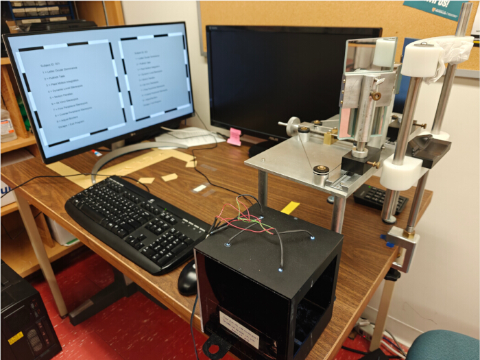
Rycina 1: Układ eksperymentalny. Układ eksperymentalny zawierający monitor, stereoskop lustrzany, podpierkę do brody, panel odpowiedzi, klawiaturę, mysz oraz klawiaturę numeryczną dla uczestników. Kliknij tutaj, aby wyświetlić powiększoną wersję tej ryciny.
2. Rozpoczęcie eksperymentu
UWAGA: Ten krok zawiera ogólne instrukcje przeprowadzania testów próbnych i testów formalnych. Szczegółowe instrukcje dla każdego zadania znajdują się w krokach 3–10.
- Otwórz plik BinocularBattery.py w programie PsychoPy Coder.
- Zmodyfikuj w skrypcie informacje o monitorze, w tym nazwę monitora, częstotliwość odświeżania oraz rozdzielczość, zgodnie z parametrami używanego monitora.
- Kliknij przycisk Run experiment u góry ekranu.
- Poleć uczestnikowi spojrzenie przez stereoskop lustrzany.
- Zapytaj uczestnika, czy widzi na środku krzyż składający się z linii pionowej i poziomej. Jeśli nie, zapytaj uczestnika, czy linia pionowa powinna zostać przesunięta w lewo, czy w prawo.
- Użyj klawiszy strzałek lewo i prawo, aby przesunąć linię pionową w danym kierunku, aż uczestnik zobaczy linię pionową w środku linii poziomej. Naciśnij Enter, aby kontynuować.
- Przypisz i wprowadź w programie identyfikator uczestnika. Na ekranie menu głównego (Main Menu) użyj klawiatury numerycznej, aby wybrać poszczególne zadania.
- Zacznij od przeprowadzenia testu praktycznego.
- Przekaż instrukcje do każdego zadania, zgodnie z opisem w krokach 3–10 poniżej.
- Ukończ test praktyczny i oceń wyniki.
UWAGA: Niektóre zadania nie zapewniają oceny dokładności (tj. dominacja oczna dla liter, integracja ruchu w kratkę oraz efekt Pulfricha). W celu uzyskania wskazówek dotyczących interpretacji wyników dla tych zadań zapoznaj się z sekcją Wyniki reprezentatywne (Representative Results). - Jeśli wyniki praktyki są niższe niż 80% poprawnych odpowiedzi (dla praktyk opartych na dokładności) lub są nieoczekiwane (dla zadań bez oceny dokładności), naciśnij Enter i wybierz test, aby ponownie rozpocząć praktykę.
- Przejdź do przeprowadzenia formalnego testu, gdy wyniki uczestnika przekraczają 80% poprawnych odpowiedzi (dla praktyk opartych na dokładności) lub są spójne i interpretowalne (dla zadań bez oceny dokładności) w dwóch ostatnich testach praktycznych.
UWAGA: Jeśli wyniki uczestnika pozostają poniżej 80% poprawnych odpowiedzi lub są niespójne w kilku testach praktycznych mimo prawidłowych instrukcji, eksperymentator powinien przerwać zadanie, ponieważ wyniki mogą być niewiarygodne.
- Przeprowadź formalny test.
- Poleć uczestnikowi wykonanie zadania.
UWAGA: Podczas testowania kluczowe jest przypominanie uczestnikowi o utrzymywaniu fiksacji wzroku na krzyżyku fiksacyjnym, jeśli jest on obecny. - Po zakończeniu oceń wyniki formalnego testu.
UWAGA: Jeśli wyniki wydają się nietypowe, można przeprowadzić drugi formalny test w celu potwierdzenia odkryć. - Naciśnij Enter, aby powrócić do menu głównego i ponownie wybrać test.
- Poleć uczestnikowi wykonanie zadania.
- Zacznij od przeprowadzenia testu praktycznego.
3. Wykonanie testu dominacji ocznej z wykorzystaniem liter
UWAGA: Jeden formalny test składa się z dwóch schodkowych procedur 1-up-1-down oraz metody stałych bodźców.
- Zaprezentuj bodziec literowy do badania dominacji ocznej, jak pokazano na Rysunku 2A. Poinstruuj uczestnika, aby utrzymał fiksację na centralnym krzyżyku, zwracając uwagę na luminancję dwóch liter przedstawionych powyżej i poniżej krzyżyka fiksacyjnego.
- Poinstruuj uczestnika, aby używał klawiszy 8 lub 5 na klawiaturze numerycznej w celu wskazania, która litera (górna czy dolna) wydawała się jaśniejsza podczas fiksacji centralnego krzyżyka.
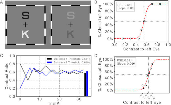
Rycina 2: Zadanie oceny dominacji ocznej dla liter oraz wyniki. (A) Bodziec testowy. (B) Wyniki testu próbnego z zastosowaniem metody stałych bodźców. (C) Wyniki z dwóch procedur schodkowych. Linie czarna i niebieska reprezentują kontrasty na każdym stopniu dwóch procedur schodkowych. Kolorowe słupki reprezentują progi uzyskane z odpowiednich procedur. (D) Funkcja psychometryczna z metody stałych bodźców. Kropki wskazują procent prób, w których uczestnik ocenił literę prezentowaną lewemu oku jako jaśniejszą dla każdego testowanego kontrastu. Czerwona krzywa reprezentuje dopasowaną krzywą psychometryczną. Przecięcie linii przerywanych przedstawia szacowany punkt subiektywnej równości (PSE). Kliknij tutaj, aby wyświetlić powiększoną wersję tej ryciny.
4. Uruchomienie integracji ruchu plaid
UWAGA: Należy wykorzystać wstępną demonstrację, aby zapoznać uczestnika z przebiegiem badania, a następnie nacisnąć Enter, aby rozpocząć próbę praktyczną.
- Zaprezentuj bodziec w postaci ruchomej kratki, zgodnie z Rysunkiem 3A. Poleć uczestnikowi obserwowanie ruchomych kratek, które mogą poruszać się w jednym z pięciu kierunków, zgodnie ze wskazaniami na ekranie, lub jednocześnie w kierunku lewym i prawym (ruch rozszczepiony).
- Poproś uczestnika o ustne zgłoszenie postrzeganego kierunku ruchu oraz wszelkich zmian w postrzeganym ruchu podczas oglądania bodźca.
- Naciśnij i przytrzymaj odpowiednią liczbę (1–5 dla każdego kierunku zgodnie z oznaczeniami lub 0 dla ruchu rozszczepionego), aż uczestnik zgłosi inną liczbę.
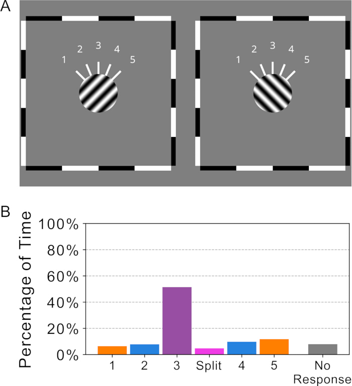
Rycina 3: Zadanie integracji ruchu kratowego i wyniki. (A) Bodziec testowy. (B) Reprezentatywne wyniki. Kolorowe słupki przedstawiają procent czasu, w którym uczestnik zgłaszał każdy z oznaczonych kierunków postrzeganego ruchu, w tym split, gdy oba kierunki (lewy i prawy) były widoczne jednocześnie. Kliknij tutaj, aby wyświetlić powiększoną wersję tej ryciny.
5. Przeprowadzenie badania precyzyjnej stereopsji obwodowej
- Prezentuj bodźce stereopsji obwodowej i centralnej, zgodnie z przedstawionymi na Rysunku 4A,B. Wykorzystaj zadanie Stationary, aby zapoznać uczestnika z procedurą.
- Poleć uczestnikowi fiksację wzroku na krzyżyku, niezależnie od miejsca jego wyświetlenia na ekranie.
- UWAGA: Obserwuj ruchy gałek ocznych uczestnika, aby upewnić się, że jego wzrok podąża za krzyżykiem fiksacyjnym.
- Zapytaj uczestnika, czy czarny kwadrat znajduje się za białą ramką, przed nią, czy w tej samej płaszczyźnie.
- Naciśnij 1, jeśli czarny kwadrat posiada głębię, lub 2, jeśli nie.
- Wykorzystaj zadanie Regular, aby przećwiczyć zadanie wymuszonego wyboru z dwóch przedziałów (2-interval forced-choice task).
- Poinformuj uczestnika, że bodziec pojawi się dwukrotnie, a głębia będzie widoczna w jednym z tych dwóch błysków.
- Poleć uczestnikowi wskazanie, w którym z dwóch błysków dostrzegł głębię.
- Naciśnij 1 lub 2 w zależności od wyboru uczestnika.
- Przeprowadź formalny test, stosując te same instrukcje, co w ćwiczeniu Regular.
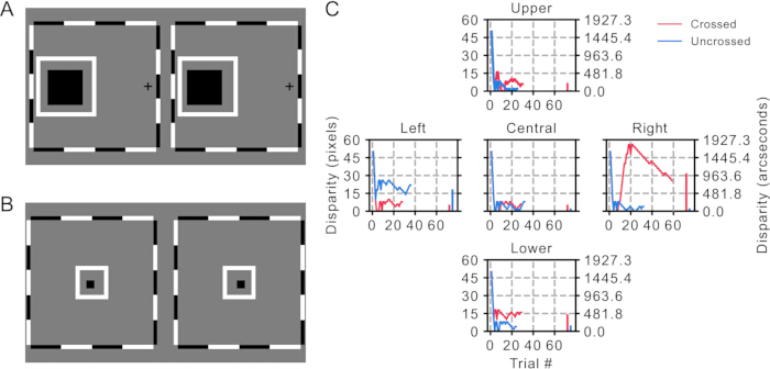
Rycina 4: Zadanie i wyniki badania precyzyjnej stereopsji peryferyjnej. (A) Bodziec testowy dla zadania peryferyjnego. (B) Bodziec testowy dla zadania centralnego. (C) Reprezentatywne wyniki. Krzywe czerwona i niebieska przedstawiają dane z procedury schodkowej dla dysparytetów skrzyżowanych i nieskrzyżowanych odpowiednio, w każdym punkcie (ekscentryczność 0° i 10°). Kolorowe słupki po prawej stronie każdego wykresu wskazują progi uzyskane z poszczególnych procedur schodkowych. Kliknij tutaj, aby wyświetlić większą wersję tej ryciny.
6. Przeprowadzenie wstępnej oceny stereopsji obwodowej
UWAGA: Naciśnij pasek spacji, aby rozpocząć próbę.
- Wyświetl bodziec do badania grubej stereopsji, zgodnie z Rysunkiem 5A. Poinstruuj uczestnika, aby utrzymywał fiksację wzroku na krzyżyku.
- Po zniknięciu gwiazd zadaj uczestnikowi następujące pytania: czy widział pan/pani głębię?; w przypadku odpowiedzi tak: czy gwiazda/gwiazdy znajdowały się za czy przed księżycami? oraz ile gwiazd zostało wyświetlonych?
- Używaj strzałek w górę i w dół do wyboru odpowiedzi oraz strzałek w lewo i w prawo do nawigacji między pytaniami.
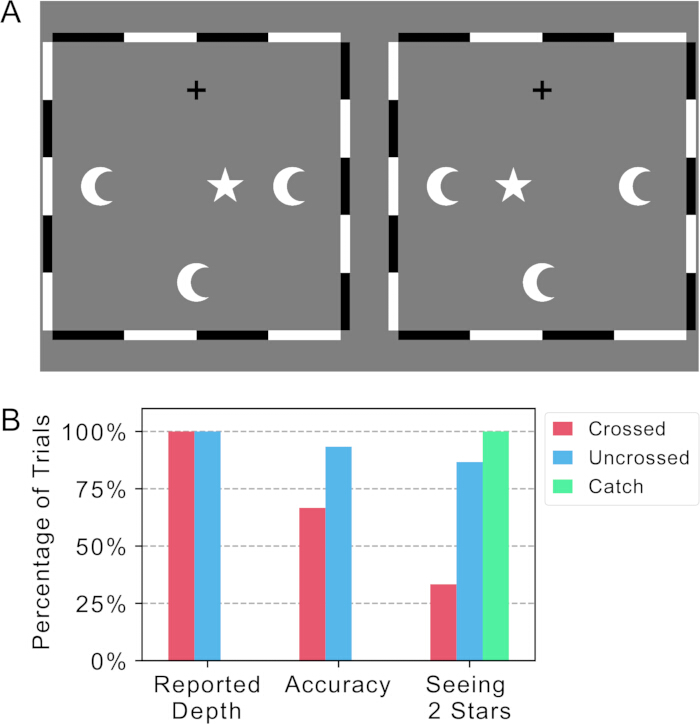
Rysunek 5: Zadanie dotyczące grubej stereopsji peryferyjnej oraz wyniki. (A) Bodziec testowy. (B) Reprezentatywne wyniki. Kolory czerwony, niebieski i zielony reprezentują odpowiednio dane z prób skrzyżowanych, nieskrzyżowanych i kontrolnych (catch trials). Słupki po lewej stronie przedstawiają procent prób, w których zgłoszono głębię. Środkowe słupki reprezentują proporcję poprawnych odpowiedzi dotyczących kierunku głębi w próbach, w których zgłoszono głębię. Słupki po prawej stronie pokazują procent prób, w których zgłoszono dwie gwiazdki. Kliknij tutaj, aby wyświetlić większą wersję tego rysunku.
7. Przeprowadzenie dynamicznej lokalnej stereopsji
UWAGA: Test ten zawiera cztery próby treningowe, które mają na celu zapewnienie instruktażu krok po kroku, aby pomóc czytelnikom zapoznać się z zadaniem.
- Wyświetl bodziec stereopsji dynamicznej oraz interfejs zadania, zgodnie z przedstawionymi na Rysunku 6A,B. Rozpocznij od uruchomienia zadania treningowego 1 (pojedyncza lokalizacja, pojedynczy interwał).
- Poleć uczestnikom patrzenie w centrum, zwracając jednocześnie uwagę na wskazaną lokalizację celu.
UWAGA: Aby pomóc uczestnikowi zrozumieć charakter bodźca, pozwól mu patrzeć bezpośrednio na lokalizację celu wyłącznie podczas treningu. - Zapytaj uczestnika, czy kwadrat przybliżył się do niego, czy oddalił.
- Naciśnij 1, jeśli uczestnik dostrzegł ruch w głąb, lub 2, jeśli nie.
UWAGA: Ruch poziomy nie jest traktowany jako ruch w głąb. - W przypadku pacjentów z lepszym widzeniem obwodowym niż centralnym, zalec niesienie pacjentowi, aby korzystał z widzenia obwodowego podczas obserwacji lokalizacji celu.
- Jeśli uczestnik jest w stanie dostrzec ruch w głąb, przejdź do następnego zadania treningowego.
- Poleć uczestnikom patrzenie w centrum, zwracając jednocześnie uwagę na wskazaną lokalizację celu.
- Uruchom zadanie treningowe 2 (pojedyncza lokalizacja, wymuszony wybór z dwóch interwałów).
- Poinformuj uczestnika, że bodziec rozbłyśnie teraz dwukrotnie.
- Zapytaj uczestnika, który z dwóch rozbłysków zawiera ruch w głąb we wskazanej lokalizacji celu.
- Naciśnij 1 lub 2 w zależności od wyboru uczestnika.
- Jeśli uczestnik nie jest w stanie niezawodnie dostrzec ruchu w głąb, ponownie przekaż instrukcje i pozwól mu patrzeć bezpośrednio na lokalizację celu.
- Jeśli uczestnik jest w stanie dostrzec ruch w głąb, przejdź do następnego zadania treningowego.
- Uruchom zadanie treningowe 3 (dwie lokalizacje, wymuszony wybór z dwóch interwałów).
- Przekaż te same instrukcje co w zadaniu 2, z wyjątkiem tego, że cel może znajdować się teraz w jednej z dwóch lokalizacji.
- Uruchom zadanie treningowe 4 (pełny trening).
- Przekaż te same instrukcje co w zadaniu 3, z wyjątkiem tego, że cel może znajdować się teraz w dowolnej z czterech lokalizacji obwodowych.
- Po przygotowaniu uruchom testy Dynamic Periphery oraz Dynamic Center w ramach formalnego badania.
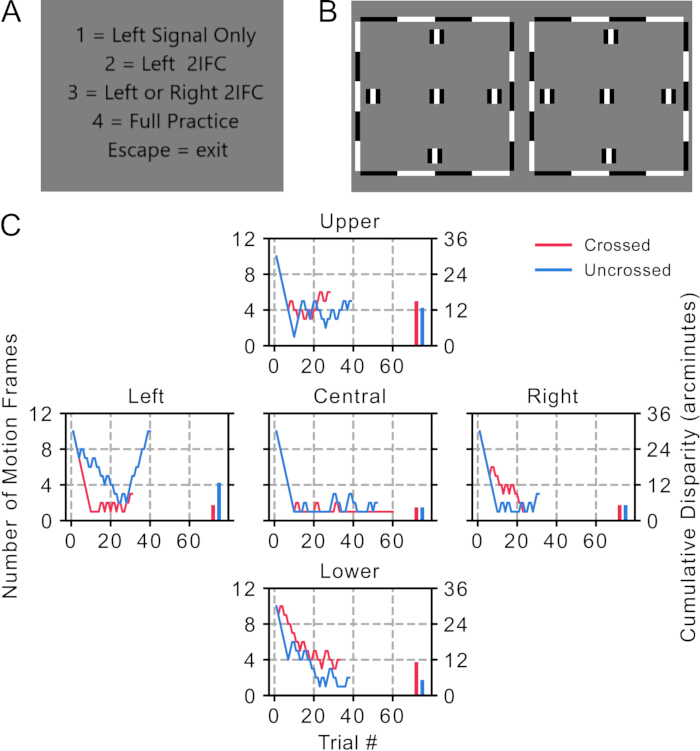
Rysunek 6: Zadanie dynamicznej lokalnej stereopsji i wyniki. (A) Menu do ćwiczeń krok po kroku. (B) Bodziec testowy. (C) Reprezentatywne wyniki. Krzywe czerwona i niebieska przedstawiają dane z procedury schodkowej odpowiednio dla dysparytetów skrzyżowanych i nieskrzyżowanych w każdym położeniu siatkówkowym (ekscentryczność 0° i 5°). Kolorowe słupki po prawej stronie każdego wykresu wskazują progi uzyskane z odpowiednich procedur schodkowych. Kliknij tutaj, aby zobaczyć większą wersję tego rysunku.
8. Wykonanie zadania Pulfricha
UWAGA: Jasność jednej połowy ekranu została zmniejszona (Rysunek 7A), aby zasymulować działanie filtrów neutralnych (ND).
- Wyświetl bodziec zadania Pulfricha, jak pokazano na Rysunku 7A. Potwierdź, czy uczestnik dostrzega głębię oraz czy księżyce poruszają się przed gwiazdami czy za nimi.
- Poproś uczestnika o użycie klawisza / lub *, aby przesunąć stacjonarny księżyc na dole ekranu do przodu lub do tyłu, aż będzie on wydawał się znajdować na tej samej głębokości co poruszające się księżyce.
- Opcjonalnie: poproś uczestnika o wskazanie postrzeganej głębokości za pomocą pola odpowiedzi (Rysunek 7B).
- Przedstaw pole odpowiedzi uczestnikowi w taki sposób, aby środek pola znajdował się w odległości około 40 cm od jego oczu.
- Poproś uczestnika o przesuwanie suwaka po obu stronach pola, aż księżyce wewnątrz pola będą wydawać się znajdować na tej samej głębokości co poruszające się księżyce na ekranie komputera.
- Wpisz odczyt z suwaka na ekranie komputera. Naciśnij +, aby przejść dalej.
UWAGA: Pudełko odpowiedzi fizycznej zostało opisane w Pliku uzupełniającym 2.
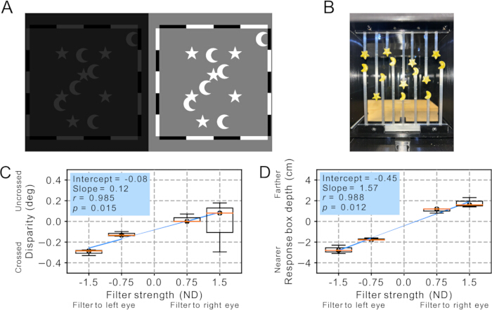
Rycina 7: Zadanie Pulfricha i wyniki. (A) Bodziec testowy. Wszystkie księżyce są w ruchu z wyjątkiem tego na dole. (B) Widok z wnętrza skrzynki odpowiedzi, pokazujący gwiazdy przymocowane do sufitu i księżyce na szynie. (C) Wyniki zadania dopasowania dysparytetu. Mediany są oznaczone czarnymi kropkami i pomarańczowymi liniami. Wykresy pudełkowe przedstawiają zakres (minimum i maksimum) oraz rozstęp międzykwartylny. Należy zauważyć, że ponieważ dla każdej siły filtra przeprowadzono tylko trzy próby, mediany i wartości ekstremalne odzwierciedlają wszystkie dane dla każdego warunku. Niebieskie linie przedstawiają regresje liniowe wykonane przy użyciu danych mediany dla wszystkich sił filtra. (D) Wyniki zadania dopasowania głębi fizycznej. Mediany są oznaczone czarnymi kropkami i pomarańczowymi liniami. Wykresy pudełkowe przedstawiają zakres (minimum i maksimum) oraz rozstęp międzykwartylny. Należy zauważyć, że ponieważ dla każdej siły filtra przeprowadzono tylko trzy próby, mediany i wartości ekstremalne odzwierciedlają wszystkie dane dla każdego warunku. Niebieskie linie przedstawiają regresje liniowe wykonane przy użyciu danych mediany dla wszystkich sił filtra. Kliknij tutaj, aby wyświetlić większą wersję tej ryciny.
9. Wykonaj paralaksę ruchu
- Wyświetl bodźce paralaksy ruchowej, zgodnie z przedstawionym na rysunku 8A,B. Poleć uczestnikowi fiksację wzroku na centralnym krzyżyku.
- Naciśnij Enter, aby rozpocząć próbę.
- Poleć uczestnikowi wskazanie kwadratu, który podczas obrotu wydawał się znajdować na wyższym poziomie głębi, przy jednoczesnym fiksowaniu wzroku na centralnym krzyżyku.
- Naciśnij liczbę odpowiadającą temu kwadratowi.
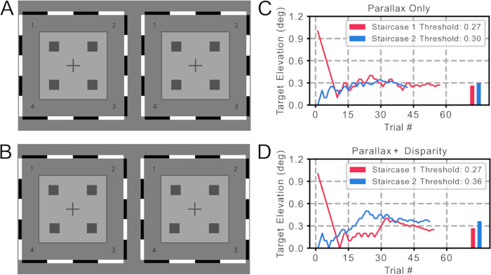
Rysunek 8: Zadanie z paralaksą ruchu i wyniki. (A) Bodziec testowy dla zadania „paralaksa”. (B) Bodziec testowy dla zadania „paralaksa + dysparytet”. Należy zauważyć, że dolny lewy kwadrat w tym przykładzie (numer „4”) zawiera wskazówkę dysparytetu binokularnego. (C) Reprezentatywne wyniki z zadania „paralaksa”. Linie czerwona i niebieska przedstawiają surowe dane z dwóch procedur schodkowych. Kolorowe słupki reprezentują progi uzyskane z odpowiednich procedur schodkowych. (D) Reprezentatywne wyniki z zadania „paralaksa + dysparytet”. Linie czerwona i niebieska przedstawiają surowe dane z dwóch procedur schodkowych. Kolorowe słupki reprezentują progi uzyskane z odpowiednich procedur schodkowych. Kliknij tutaj, aby wyświetlić powiększoną wersję tego rysunku.
10. Przeprowadzenie testu stereopsji da Vinci
- Przedstaw konfigurację bodźca dichoptycznego zgodnie z Rysunkiem 9A. Poleć uczestnikowi porównanie głębi dwóch pasków względem dużego pudełka i wskazanie, który pasek wydaje się znajdować przed pudełkiem.
- Naciśnij 8 lub 5 dla górnego lub dolnego paska, lub naciśnij Enter, jeśli wszystkie wydają się znajdować w tej samej płaszczyźnie.
- Podczas ćwiczeń, jeśli uczestnik dostrzeże głębię w próbie kontrolnej, ponownie udziel instrukcji, aby upewnić się, że rozumie zadanie.
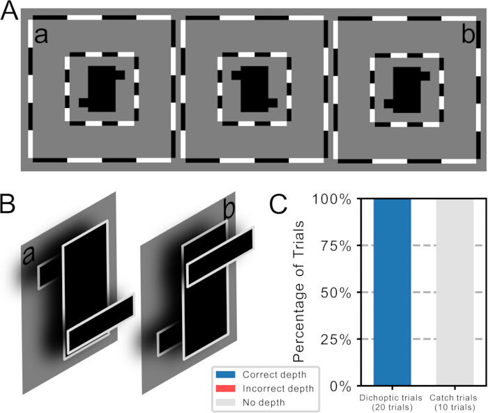
Rysunek 9: Zadanie stereopsji Da Vinci i wyniki (A) Ilustracja bodźca testowego, przedstawiająca dwie pary bodźców dichoptycznych. (B) Efekty percepcyjne po fuzji. Poprzez zeskrywanie oczu, fuzja bodźców lewego i środkowego wytwarza iluzję pokazaną w (a), a fuzja bodźców środkowego i prawego wytwarza iluzję pokazaną w (b). Należy zauważyć, że podczas każdej próby wyświetlana jest tylko jedna para bodźców. (C) Reprezentatywne wyniki. Niebieskie, czerwone i szare słupki wskazują procent prób, w których uczestnicy zgłosili poprawną głębię, błędną głębię lub brak głębi. Zauważmy, że w tym przykładzie wszystkie odpowiedzi były poprawne, dlatego nie przedstawiono czerwonego słupka. Kliknij tutaj, aby wyświetlić powiększoną wersję tego rysunku.
Dostęp ograniczony. Zaloguj się lub rozpocznij wersję próbną, aby wyświetlić tę treść.
Wyniki
Dane reprezentatywne zostały zebrane od 20-letniego uczestnika o normalnym widzeniu (ostrość wzroku 0,0 logMAR dla każdego oka; stereoskuność 40 arcsec).
Dominacja oka na litery
Wyniki z dwóch schodków i metody stałych stymulacji (Rysunek 2C,D) wskazują na nieznacznie silniejszą dominację prawego oka dla tego uczestnika.
Oczekiwane wyniki: Każdy schodek powinien stopniowo zbliżać się do progu, a o...
Dostęp ograniczony. Zaloguj się lub rozpocznij wersję próbną, aby wyświetlić tę treść.
Dyskusja
Ta nowa bateria testów została opracowana w celu oceny wielu aspektów widzenia binokularnego, począwszy od zarazem percepcji i fuzji, aż po odrębne mechanizmy leżące u podstaw stereopsji. Cały protokół trwa co najmniej 85 minut, zapewniając kompleksową ocenę postrzegania głębi. Testy mogą być wykonywane indywidualnie lub w połączeniu, w zależności od potrzeb badawczych lub klinicznych. Pełna bateria testów jest najlepiej przystosowana do środowisk badawczych, gdzie możliwe są dłuższe okresy testowania. Niemniej jednak, p...
Dostęp ograniczony. Zaloguj się lub rozpocznij wersję próbną, aby wyświetlić tę treść.
Oświadczenia
Wszyscy autorzy deklarują brak konfliktu interesów.
Podziękowania
To dzieło było wspierane przez grant z Canadian Institutes of Health Research (CIHR) (PJT 183761) dla EN, XG, DM, DIS i BT. Dziękujemy Grupie Badawczej REWIRE-D (rewire-d.ca) za ich wkład w rozwój tej pracy.
Dostęp ograniczony. Zaloguj się lub rozpocznij wersję próbną, aby wyświetlić tę treść.
Materiały
| Nazwa | Firma | Numer katalogowy | Komentarze |
|---|---|---|---|
| Codes | N/A | https://git.uwaterloo.ca/hvnl/binocularbatterycodes_manuscript/-/tree/9f7ff9eb1705d50060ca46a8ac2d2d1848349368/ | Kody użyte do generowania stymuli i uruchomienia protokołu są dostępne w repozytorium podanym w kolumnie "Numer katalogowy". |
| Komputer | Alienware, Dell Inc., Texas, USA | XPS 8930 | Intel Core i7-9700 CPU, 64 GB RAM |
| Podpora na czoło i podbródek | N/A | N/A | Wykonane na zamówienie, używane do utrzymania pozycji głowy i odległości widzenia |
| Stereoskop z lustrem | N/A | N/A | Wykonane na zamówienie |
| Monitor | LG Electronics, Seoul, Korea | LG 27UP600-W | Rozdzielczośc: 3840 × 2160 pikseli, szerokość ekranu: 59,5 cm, częstotliwość odświeżania: 60 Hz, czas reakcji: 5 ms; szarość tła ustawiona na 340 cd/m2 |
| Mysz i klawiatura | N/A | N/A | Do użytku eksperymentatora |
| Klawiatura numeryczna | N/A | N/A | Do użytku uczestnika |
| Środowisko programistyczne | N/A | https://www.psychopy.org/ | PsychoPy 2022.2.4 (na Python 3.6.6), działający na Windows 10 (Microsoft Corporation, Redmond, WA, USA) |
| Skanowaczka | N/A | N/A | Wykonane na zamówienie, patrz Schematy w Dokumentach uzupełniających dla wymaganych części |
Bibliografia
- Ali, R. S. . Relationship between ocular sensory dominance and stereopsis. , (2016).
- Cibis, G. W., Ing, M. R. Binocular vision. Duane’s Ophthalmology. , (2013).
- Barrett, B. T. Assessment of binocular vision and accommodation. Clinical procedures in primary eye care. , (2014).
- Lee, J., McIntyre, A. Clinical tests for binocular vision. Eye. 10 (2), 282-285 (1996).
- von Noorden, G. K., Campos, E. C. . Binocular vision and ocular motility: theory and management of strabismus. , (2002).
- de Belsunce, S., Sireteanu, R. The time course of interocular suppression in normal and amblyopic subjects. Invest Ophthalmol Vis Sci. 32 (9), 2645-2652 (1991).
- Hupé, J. M., Signorelli, C. M., Alais, D. Two paradigms of bistable plaid motion reveal independent mutual inhibition processes. J Vis. 19 (4), 5 (2019).
- Riesen, G., Norcia, A. M., Gardner, J. L. Humans perceive binocular rivalry and fusion in a tristable dynamic state. J Neurosci. 39 (43), 8527-8537 (2019).
- Dieter, K. C., Sy, J. L., Blake, R. Individual differences in sensory eye dominance reflected in the dynamics of binocular rivalry. Vision Res. 141, 40-50 (2017).
- O’Connor, A. R., Tidbury, L. P. Stereopsis: are we assessing it in enough depth?. Clin Exp Optom. 101 (4), 485-494 (2018).
- Tittes, J., et al. Assessment of stereovision with digital testing in adults and children with normal and impaired binocularity. Vision Res. 164, 69-82 (2019).
- Hubel, D. H., Wiesel, T. N. Receptive fields, binocular interaction and functional architecture in the cat’s visual cortex. J Physiol. 160, 106-154 (1962).
- Rovamo, J., Virsu, V. An estimation and application of the human cortical magnification factor. Exp Brain Res. 37 (3), (1979).
- Spillmann, L., Ransom-Hogg, A., Oehler, R. A comparison of perceptive and receptive fields in man and monkey. Hum Neurobiol. 6 (1), 51-62 (1987).
- Nadler, J. W., Barbash, D., Kim, H. R., Shimpi, S., Angelaki, D. E., et al. Joint representation of depth from motion parallax and binocular disparity cues in macaque area MT. J Neurosci. 33 (35), 14061-14074 (2013).
- Verghese, P. The utility of peripheral stereopsis. Front Neurosci. 17, 1217993 (2023).
- Witz, N., Hess, R. F. Mechanisms underlying global stereopsis in fovea and periphery. Vision Res. 87, 10-21 (2013).
- Yildirim, C., Mehmet Mutlu, F., Chen, Y., Altinsoy, H. I. Assessment of central and peripheral fusion and near and distance stereoacuity in intermittent exotropic patients before and after strabismus surgery. Am J Ophthalmol. 128 (2), 222-230 (1999).
- Hess, R. F., Mansouri, B., Thompson, B., Gheorghiu, E. Latent stereopsis for motion in depth in strabismic amblyopia. Invest Ophthalmol Vis Sci. 50 (10), 5006 (2009).
- Mollenhauer, K. A., Haase, W. Dynamic stereopsis in patients with impaired binocular function. Strabismus. 8 (4), 275-281 (2000).
- Watanabe, Y., Kezuka, T., Harasawa, K., Usui, M., Yaguchi, H., et al. A new method for assessing motion-in-depth perception in strabismic patients. Br J Ophthalmol. 92 (1), 47-50 (2008).
- Tredici, T. D., von Noorden, G. K. The Pulfrich effect in anisometropic amblyopia and strabismus. Am J Ophthalmol. 98 (4), 499-503 (1984).
- Bossi, M., Hamm, L. M., Dahlmann-Noor, A., Dakin, S. C. A comparison of tests for quantifying sensory eye dominance. Vision Res. 153, 60-69 (2018).
- Bossi, M., et al. Binocular therapy for childhood amblyopia improves vision without breaking interocular suppression. Invest Ophthalmol Vis Sci. 58 (7), 3031-3041 (2017).
- Elliott, D. B. . Clinical procedures in primary eye care. , (2014).
- Lanska, D. J., Lanska, J. M., Remler, B. F. Description and clinical application of the Pulfrich effect. Neurology. 84 (22), 2274-2278 (2015).
- Read, J. C. A., Cumming, B. G. Effect of interocular delay on disparity-selective V1 neurons: relationship to stereoacuity and the Pulfrich effect. J Neurophysiol. 94 (2), 1541-1553 (2005).
- Carney, T., Paradiso, M. A., Freeman, R. D. A physiological correlate of the Pulfrich effect in cortical neurons of the cat. Vision Res. 29 (2), 155-165 (1989).
- Wu, Y., et al. Two patterns of interocular delay revealed by spontaneous motion-in-depth Pulfrich phenomenon in amblyopes with stereopsis. Invest Ophthalmol Vis Sci. 61 (3), 22 (2020).
- Reynaud, A., Hess, R. F. An unexpected spontaneous motion-in-depth Pulfrich phenomenon in amblyopia. Vision. 3 (4), 54 (2019).
- Kim, H. R., Angelaki, D. E., DeAngelis, G. C. The neural basis of depth perception from motion parallax. Philos Trans R Soc B Biol Sci. 371 (1697), 20150256 (2016).
- Rogers, B. Revisiting motion parallax as a source of 3-D information. Perception. 45 (11), 1267-1278 (2016).
- Tsirlin, I., Wilcox, L. M., Allison, R. S. da Vinci decoded: does da Vinci stereopsis rely on disparity?. J Vis. 12 (12), 2 (2012).
- Blake, R., Wilson, H. Binocular vision. Vision Res. 51 (7), 754-770 (2011).
- Tsirlin, I., Wilcox, L. M., Allison, R. S. A computational theory of da Vinci stereopsis. J Vis. 14 (7), 5 (2014).
- Chen, X., Hall, K., Bobier, W. R., Thompson, B., Chakraborty, A. Transcranial random noise stimulation and exercise do not modulate ocular dominance plasticity in adults with normal vision. J Vis. 22 (10), 14 (2022).
- Chen, X., Bobier, W., Thompson, B. Short-term ocular dominance plasticity is not modulated by visual cortex tDCS but increases with length of monocular deprivation. Sci Rep. 13 (1), 6666 (2023).
- Li, J., et al. The role of suppression in amblyopia. Invest Ophthalmol Vis Sci. 52 (7), 4169 (2011).
- Hess, R. F., Thompson, B., Baker, D. H. Binocular vision in amblyopia: structure, suppression and plasticity. Ophthalmic Physiol Opt. 34 (2), 146-162 (2014).
- Christian, L. W., Nandakumar, K., Hrynchak, P. K., Irving, E. L. Visual and binocular status in elementary school children with a reading problem. J Optom. 11 (3), 160-166 (2018).
- Grisham, D., Powers, M., Riles, P. Visual skills of poor readers in high school. Optometry. 78 (10), 542-549 (2007).
- Mesfin, Y., Kong, A., Backus, B. T., Deiner, M., Ou, Y., et al. Pilot study comparing a virtual reality–based visual field test to standard perimetry in children. J AAPOS. 28 (3), 103933 (2024).
- Denkinger, S., et al. The eRDS v6 stereotest and the vivid vision stereo test: two new tests of stereoscopic vision. Transl Vis Sci Technol. 12 (3), 1 (2023).
- Kaernbach, C. Simple adaptive testing with the weighted up-down method. Percept Psychophys. 49 (3), 227-229 (1991).
- García-Pérez, M. A. A cautionary note on the use of the adaptive up-down method. J Acoust Soc Am. 130 (4), 2098-2107 (2011).
- I-BiT study group. Interactive binocular treatment (I-BiT) for amblyopia: pilot study of a 3D shutter glasses system. Eye. 27 (9), 1077-1083 (2013).
- Saraiva, A. A., Nogueira, A. T., Ferreira, N. M. F., Valente, A. Application of virtual reality for treatment of strabismus and amblyopia. , (2018).
- Chan, H. S., Tang, Y. M., Do, C. W., Wong, H. H. Y., Chan, L. Y., et al. Design and assessment of vision training using virtual reality. Digit Health. 9, 20552076231176638 (2023).
- Sun, X., Varshney, A. Investigating perception time in far peripheral vision. , (2018).
- Vera, J., Glazier, A. N., Dunbar, M. T., Ripkin, D., Nafey, M. Evaluating validity of virtual reality headsets for visual field testing. Vision. 9 (4), 80 (2025).
Dostęp ograniczony. Zaloguj się lub rozpocznij wersję próbną, aby wyświetlić tę treść.
Przedruki i uprawnienia
