This study examines the association between admission serum homocysteine and clinically adjudicated discharge neurocognitive dysfunction after acute cerebrovascular disease in a high-altitude setting.
Artykuł badawczy
Association of Admission Serum Homocysteine with Discharge Neurocognitive Dysfunction after Acute Cerebrovascular Disease in a High-Altitude Setting
* These authors contributed equally
W tym artykule
Podsumowanie
Streszczenie
Discharge-phase neurocognitive abnormalities may affect rehabilitation participation, self-management, and discharge safety after acute cerebrovascular disease, but early clinically accessible correlates remain incompletely defined. We examined the association between admission serum homocysteine and clinically adjudicated discharge neurocognitive dysfunction in a high-altitude clinical setting. This single-center retrospective cohort included 227 adults admitted for acute ischemic stroke, intracerebral hemorrhage, or transient ischemic attack. The first available serum homocysteine measurement within 24 h of hospital arrival was analyzed continuously and by cohort-derived tertiles. The primary outcome was neurocognitive dysfunction documented within 24–48 h before discharge and adjudicated independently by two reviewers blinded to homocysteine levels; standardized cognitive instruments were not systematically available. Logistic regression was sequentially adjusted for demographic, renal, neurological, neuroimaging, and cerebrovascular phenotype-related factors. Among 227 patients, 98 (43.2%) had discharge neurocognitive dysfunction. Admission homocysteine was higher in patients with dysfunction than in those without dysfunction (18.3 ± 6.2 vs. 12.4 ± 4.7 µmol/L, P < 0.001), and dysfunction prevalence increased from 22.4% in T1 to 38.5% in T2 and 69.9% in T3 (P for trend < 0.001). In the primary fully adjusted model, each 1 µmol/L increase in homocysteine was associated with higher odds of dysfunction (OR 1.14, 95% CI 1.07–1.21, P < 0.001), and T3 versus T1 remained associated with the outcome (OR 4.36, 95% CI 2.00–9.51, P < 0.001). Estimates were similar in prespecified sensitivity analyses, including exclusion of delirium or positive confusion assessment method for the intensive care unit (CAM-ICU) screening and restriction to acute ischemic stroke. Higher admission homocysteine was associated with discharge-phase neurocognitive dysfunction, supporting prospective evaluation of homocysteine as a potential marker of short-term neurocognitive vulnerability rather than a validated predictor of persistent post-stroke cognitive impairment.
Wprowadzenie
Acute cerebrovascular disease, including acute ischemic stroke, intracerebral hemorrhage, and transient ischemic attack, is a major source of early neurological disability and prolonged post-discharge care needs1. Clinical assessment during hospitalization has traditionally emphasized survival, lesion stabilization, motor recovery, and global neurological severity, commonly reflected by measures such as the National Institutes of Health Stroke Scale (NIHSS)2. However, the transition from inpatient care to discharge also depends on a patient’s ability to understand instructions, participate in rehabilitation, retain treatment information, and maintain basic cognitive-functional safety3,4. Neurocognitive abnormalities present near discharge may therefore represent a clinically meaningful dimension of short-term neurological vulnerability that is not fully captured by conventional severity assessment alone5. Prior work has shown that cognitive deficits can persist even after apparently mild stroke and that neurological severity measures are related to cognitive outcomes, supporting the need to characterize discharge-phase neurocognitive status more explicitly in acute cerebrovascular populations5.
The identification of discharge neurocognitive dysfunction in routine care is challenging6. Formal cognitive screening during acute hospitalization may be constrained by aphasia, fatigue, impaired attention, fluctuating arousal, motor limitations, or the rapidly evolving neurological state of patients admitted through emergency pathways. In practice, clinically relevant neurocognitive dysfunction is often recognized through convergent documentation from physicians, nurses, and rehabilitation teams, including persistent disorientation, impaired comprehension, difficulty following multi-step instructions, or concerns that cognitive-functional limitations may affect discharge education and self-management. These observations are particularly important when patients also carry markers of neurological or structural brain vulnerability, such as higher admission NIHSS scores, reduced renal function reflected by lower estimated glomerular filtration rate, greater white matter hyperintensity burden, increased numbers of lacunes, or cortical atrophy7. Such features may indicate that discharge neurocognitive status emerges from the interaction between acute event severity and pre-existing cerebral reserve, rather than from lesion type alone8. Evidence from stroke cohorts has linked white matter hyperintensity burden with post-stroke cognitive performance, reinforcing the relevance of structural brain vulnerability when evaluating short-term neurocognitive outcomes9.
Homocysteine is a plausible biomarker for this clinical problem because it lies at the intersection of vascular injury, metabolic stress, and neurological vulnerability10. Elevated circulating homocysteine has been associated with adverse cerebrovascular prognosis and with cognitive impairment after ischemic stroke11. In post-stroke cohorts, higher acute-phase homocysteine concentrations have been linked to subsequent cognitive impairment12, while other studies have reported associations between elevated homocysteine and poorer functional outcomes after acute ischemic stroke. These findings suggest that admission homocysteine may capture a biologically relevant component of early neurovascular stress13. Nevertheless, most available evidence has focused on delayed cognitive outcomes or broader functional prognosis rather than on neurocognitive dysfunction documented immediately before hospital discharge14. Whether admission homocysteine is associated with this clinically proximal discharge-phase outcome, after accounting for age, education, renal function, admission NIHSS, white matter hyperintensity burden, and acute cerebrovascular phenotype, remains insufficiently defined.
The high-altitude setting provides a distinct clinical context in which to examine this association. This study was conducted in Xining, Qinghai Province, at approximately 2,275 m above sea level, where patients with acute cerebrovascular disease are managed within a distinct environmental context15. Multiregional Chinese data have shown that acute ischemic stroke profiles vary across altitude strata, indicating that stroke populations in plateau regions may not be fully interchangeable with low-altitude cohorts16. In parallel, residence altitude has been associated with differences in serum folate levels among Tibetan adults17, a finding that is pertinent because folate-dependent one-carbon metabolism is closely related to homocysteine homeostasis18. These observations do not establish a causal altitude-homocysteine pathway but provide a rationale for evaluating the association in a high-altitude clinical population while interpreting altitude as contextual rather than causal13.
To address these gaps, we conducted a retrospective cohort study of patients admitted through the emergency department for acute ischemic stroke, intracerebral hemorrhage, or transient ischemic attack at a tertiary hospital in a high-altitude region. We examined whether serum homocysteine measured within 24 h of admission was associated with discharge neurocognitive dysfunction adjudicated from prespecified discharge-phase clinical documentation. We further assessed whether the association showed a graded pattern across cohort-derived homocysteine tertiles, persisted after adjustment for prespecified demographic, renal, neurological, neuroimaging, and cerebrovascular phenotype-related factors, and remained directionally consistent across sensitivity and exploratory subgroup analyses. By focusing on an early routinely available biomarker and a clinically proximal neurocognitive outcome, this study sought to clarify whether admission homocysteine may help characterize short-term neurocognitive vulnerability during hospitalization for acute cerebrovascular disease.
Protokół
Ethics approval and data confidentiality
This retrospective study was approved by the Research Ethics Committee of Qinghai Provincial People's Hospital (Approval Number: (2025)-259-02). The requirement for individual informed consent was waived because the study used routinely collected retrospective clinical data, involved no direct patient contact or additional intervention, and all records were de-identified before analysis. All study procedures were conducted in accordance with applicable institutional and national requirements for retrospective clinical research and the ethical principles of the Declaration of Helsinki. To protect patient privacy, all extracted data were de-identified before analysis and were accessible only to authorized study personnel. Direct personal identifiers were not included in the analytic dataset, figure generation, or statistical modeling. Clinical records used for discharge neurocognitive adjudication were reviewed solely for research purposes within the approved protocol, and all analyses were conducted on aggregated data to minimize the risk of re-identification.
Study design, setting, and reporting framework
This single-center retrospective cohort study was conducted at Qinghai Provincial People's Hospital, Xining, Qinghai Province, China, a high-altitude region located at approximately 2,275 m above sea level. The study focused on patients admitted through the emergency department for acute ischemic stroke, intracerebral hemorrhage, or transient ischemic attack and was designed to evaluate the association between admission serum homocysteine levels and clinically adjudicated discharge neurocognitive dysfunction. The high-altitude location was treated as an important clinical context for the study rather than as an exposure or effect modifier, because the present cohort did not include a lower-altitude comparison group or individual measures of long-term altitude exposure.
The study was developed and reported in accordance with the Strengthening the Reporting of Observational Studies in Epidemiology (STROBE) recommendations for cohort studies. Given the retrospective use of routinely collected inpatient clinical, laboratory, imaging, nursing, and rehabilitation documentation, emphasis was placed on transparent cohort derivation, prespecified outcome adjudication, explicit exposure classification, and multivariable analytical strategies to reduce bias and improve reproducibility.
Study population, eligibility criteria, and data sources
Adult patients admitted through the emergency department for an acute cerebrovascular event between January 2021 and December 2025 were retrospectively screened. Eligible events included acute ischemic stroke and intracerebral hemorrhage diagnosed by attending neurologists on the basis of the acute clinical presentation and brain computed tomography or magnetic resonance imaging, as well as transient ischemic attack diagnosed clinically after appropriate neuroimaging evaluation during the index admission. Patients were considered eligible if they had an admission serum homocysteine measurement obtained within 24 h of hospital arrival and sufficient discharge-phase clinical documentation to permit prespecified neurocognitive outcome adjudication.
Patients were excluded if admission homocysteine data were unavailable within the required time window, discharge-phase records were insufficient for neurocognitive adjudication, severe neurological or systemic conditions precluded meaningful discharge evaluation, including persistent coma, early transfer to intensive care, or severe encephalopathy, or if a pre-existing major neurocognitive disorder or severe dementia had been documented before the index event. Patients with missing key covariates required for the planned multivariable analyses were also excluded. The primary analyses therefore used complete cases for the prespecified model covariates; 11 patients excluded during cohort construction had missing key covariate data, and no imputation was applied to these excluded observations. Clinical data were extracted from electronic medical records, laboratory information systems, radiology and picture archiving systems, nursing documentation, rehabilitation assessments, and discharge summaries. These data sources were used to define the analytic cohort, characterize acute cerebrovascular phenotypes, capture covariates, and support discharge neurocognitive adjudication.
Exposure assessment: admission serum homocysteine
The primary exposure of interest was the serum homocysteine concentration measured during the acute admission period. For all included patients, the first available serum homocysteine measurement obtained within 24 h of hospital arrival was used as the admission exposure and was abstracted from the hospital laboratory information system. Fasting status before homocysteine measurement was not uniformly documented or standardized in the available retrospective records; accordingly, admission homocysteine was interpreted as an early acute-phase measurement rather than a standardized fasting baseline concentration. Homocysteine was analyzed both as a continuous variable, expressed per 1 µmol/L increase, and categorically using cohort-derived tertiles to characterize graded exposure-response patterns.
For categorical analyses, patients were stratified according to cohort-derived tertiles of admission homocysteine: T1, ≤11.5 µmol/L; T2, 11.6–15.8 µmol/L; and T3, ≥15.9 µmol/L. These tertiles were used to describe the gradient of discharge neurocognitive dysfunction across increasing homocysteine burden and to support complementary logistic regression analyses comparing intermediate and high exposure categories with the lowest tertile as the reference group. The tertile thresholds were derived from the distribution of homocysteine values in the analytic cohort and were not intended to represent externally validated clinical cutoffs.
Definition and adjudication of discharge neurocognitive dysfunction
The primary outcome was discharge neurocognitive dysfunction, defined as a clinically meaningful neurocognitive abnormality documented during the 24–48 h preceding hospital discharge. Because standardized cognitive instruments such as the Montreal cognitive assessment, mini-mental state examination, or a formal neuropsychological battery were not systematically administered during the index hospitalization, outcome ascertainment relied on prespecified adjudication of discharge-phase clinical documentation. Accordingly, the primary outcome should be interpreted as clinically adjudicated discharge-phase neurocognitive dysfunction and not as a standardized diagnosis of persistent post-stroke cognitive impairment. Source documents included discharge summaries, progress notes, nursing records, and rehabilitation assessments. Prespecified adjudication criteria focused on one or more of the following: persistent physician-documented neurocognitive abnormality, cognitive-functional concerns affecting discharge care planning or self-management, and repeated multidisciplinary documentation of neurocognitive difficulty that could not be fully explained by isolated motor, speech, or transient arousal limitations.
Two trained clinical reviewers independently evaluated all eligible discharge-phase records while blinded to admission serum homocysteine levels and to the exposure classification used in subsequent analyses19. Patients were classified as having or not having discharge neurocognitive dysfunction according to the predefined adjudication framework. Any disagreement between the two reviewers was resolved through discussion with a senior neurologist, who made the final classification20. This adjudication framework was intended to standardize retrospective outcome classification and reduce exposure-related classification bias, although it did not substitute for prospective standardized cognitive testing.
Clinical, laboratory, and neuroimaging covariates
Baseline covariates were prespecified according to their clinical relevance to acute cerebrovascular disease, homocysteine metabolism, and discharge neurocognitive status. Demographic and vascular variables included age, sex, body mass index, educational attainment of at least 9 years, hypertension, diabetes mellitus, dyslipidemia, previous ischemic stroke, atrial fibrillation, and smoking history. Acute cerebrovascular phenotype was classified as acute ischemic stroke, intracerebral hemorrhage, or transient ischemic attack. Neurological severity at presentation was assessed using the NIHSS score recorded on admission. Laboratory covariates included fasting plasma glucose, glycated hemoglobin, serum creatinine, and estimated glomerular filtration rate. Serum folate and vitamin B12 concentrations and dietary pattern, including vegetarian status, were not systematically available in the retrospective records and therefore were not incorporated into the multivariable models.
Neuroimaging features were abstracted from radiological assessments performed during the index admission and included cortical involvement of the acute lesion, number of lacunes, white matter hyperintensity burden, and cortical atrophy. White matter hyperintensity burden was categorized as none-to-mild or moderate-to-severe for descriptive and multivariable analyses. Cortical atrophy was treated as a binary structural vulnerability marker and was additionally incorporated into an expanded adjustment model. Documented delirium and positive confusion assessment method for the intensive care unit (CAM-ICU) screening were not included in the primary multivariable model but were used to construct a prespecified sensitivity analysis excluding patients with potential acute confusional states. Time from symptom onset to hospital admission and length of hospital stay were additionally extracted to provide a temporal context for the interpretation of the discharge-phase outcome.
Statistical analysis
Continuous variables were summarized as mean ± standard deviation or median with interquartile range, as appropriate according to distributional characteristics, and categorical variables were presented as counts and percentages. Time from symptom onset to hospital admission and length of hospital stay were summarized as median and interquartile range in the revised temporal analyses. Between-group comparisons according to discharge neurocognitive status were performed using the independent-samples t test or Mann-Whitney U test for continuous variables and the χ2 test or Fisher’s exact test for categorical variables. The overall distribution of acute cerebrovascular phenotypes between groups was compared using a global Pearson χ2 test. To characterize the graded exposure-response pattern, the prevalence of discharge neurocognitive dysfunction was compared across cohort-derived homocysteine tertiles, and a trend test was used to evaluate monotonic changes across the ordered categories. An unadjusted probability curve was additionally generated to visualize the continuous relationship between admission homocysteine and the likelihood of discharge neurocognitive dysfunction.
Logistic regression models were used to estimate the association between admission serum homocysteine and discharge neurocognitive dysfunction. Homocysteine was analyzed both continuously, per 1 µmol/L increase, and categorically using tertiles, with T1 as the reference group. Three sequential models were constructed: a crude model without covariate adjustment; Model 1 adjusted for age, sex, and education level ≥ 9 years; and Model 2 further adjusted for estimated glomerular filtration rate (eGFR), NIHSS score on admission, moderate-to-severe white matter hyperintensity (WMH) burden, and acute cerebrovascular phenotype. Results were reported as odds ratios with 95% confidence intervals. Prespecified sensitivity analyses included exclusion of patients with documented delirium or positive CAM-ICU screening, exclusion of patients with transient ischemic attack, restriction to patients with acute ischemic stroke, and an expanded adjustment model additionally incorporating HbA1c and cortical atrophy. Exploratory subgroup analyses were conducted according to age, admission NIHSS, eGFR, and WMH burden; subgroup-specific estimates were generated for high homocysteine burden, defined as T3 versus T1. Multiplicative interaction terms were used to assess potential effect modification, and these exploratory analyses were not adjusted for multiple comparisons. All statistical tests were two-sided, and P < 0.05 was considered statistically significant. Statistical analyses were performed using SPSS version 27.0 and R version 4.3.2.
Wyniki
Study cohort and adjudication of discharge neurocognitive dysfunction
A total of 318 consecutive emergency admissions for acute ischemic stroke, intracerebral hemorrhage, or transient ischemic attack between January 2021 and December 2025 were screened for eligibility, with neuroimaging performed as part of the index diagnostic evaluation. After exclusion of 91 patients because of unavailable admission homocysteine measurement, insufficient discharge-phase documentation for neurocognitive adjudication, severe clinical conditions precluding discharge evaluation, pre-existing major neurocognitive disorders, or missing key covariates, 227 patients were included in the final analytic cohort (Figure 1A).
Discharge neurocognitive dysfunction was adjudicated using prespecified criteria based on clinical records documented within 24–48 h before discharge, including discharge summaries, progress notes, nursing records, and rehabilitation assessments. Two trained clinical reviewers independently evaluated these records while blinded to admission homocysteine levels, and disagreements were resolved by a senior neurologist (Figure 1B). According to this adjudication framework, 129 patients were classified as having no discharge neurocognitive dysfunction, whereas 98 patients were classified as having discharge neurocognitive dysfunction. The baseline demographic, clinical, laboratory, and neuroimaging characteristics of the analytic cohort according to discharge neurocognitive status are summarized in Table 1.
Baseline clinical, laboratory, and neuroimaging profiles according to discharge neurocognitive status
Baseline characteristics of the 227 patients are summarized in Table 1. Compared with patients without discharge neurocognitive dysfunction, those with dysfunction were older (73.4 ± 7.9 vs. 68.2 ± 8.7 years, P < 0.001) and less likely to have received at least 9 years of formal education (39.8% vs. 55.0%, P = 0.034). The overall distribution of acute cerebrovascular phenotypes did not differ significantly between groups (global Pearson χ2 P = 0.479), although intracerebral hemorrhage accounted for a numerically higher proportion of patients with dysfunction than of those without dysfunction (20.4% vs. 14.7%; Figure 2A).
Admission serum homocysteine concentrations were markedly higher in patients with discharge neurocognitive dysfunction than in those without dysfunction (18.3 ± 6.2 vs. 12.4 ± 4.7 µmol/L, P < 0.001; Figure 2B). The dysfunction group also showed poorer renal profiles, including higher serum creatinine (90.3 ± 22.0 vs. 82.1 ± 19.2 µmol/L, P = 0.004) and lower eGFR (75.2 ± 18.1 vs. 84.3 ± 16.9 mL/min/1.73 m2, P < 0.001), as well as greater neurological severity on admission, reflected by higher NIHSS scores (7.4 ± 3.1 vs. 4.8 ± 2.6, P < 0.001). Admission homocysteine distributions across acute ischemic stroke, intracerebral hemorrhage, and transient ischemic attack phenotypes are shown in Figure 2C. Median time from symptom onset to hospital admission was 5.0 h (IQR, 2.8–9.0) overall and did not differ significantly between patients with and without discharge neurocognitive dysfunction [5.4 h (IQR, 3.0–9.5) vs. 4.8 h (IQR, 2.6–8.6), P = 0.284]. In addition, patients with dysfunction exhibited a greater burden of structural brain vulnerability, including more frequent cortical involvement (33.7% vs. 21.7%, P = 0.049), more lacunes (2.1 ± 1.4 vs. 1.4 ± 1.1, P < 0.001), a higher prevalence of moderate-to-severe WMH (67.3% vs. 42.6%, P = 0.001), and more frequent cortical atrophy (31.6% vs. 19.4%, P = 0.030). Median length of hospital stay was longer among patients with discharge neurocognitive dysfunction than among those without dysfunction [9.0 days (IQR, 7.0–11.0) vs. 7.0 days (IQR, 5.0–9.0), P = 0.008].
Exposure-response gradient of discharge neurocognitive dysfunction across admission homocysteine levels
When admission serum homocysteine was categorized into cohort-derived tertiles, the prevalence of discharge neurocognitive dysfunction increased progressively with higher homocysteine exposure (Figure 3). Dysfunction was observed in 17 of 76 patients in T1 (≤11.5 µmol/L, 22.4%), 30 of 78 patients in T2 (11.6–15.8 µmol/L, 38.5%), and 51 of 73 patients in T3 (≥15.9 µmol/L, 69.9%), showing a significant monotonic trend across tertiles (P for trend < 0.001; Figure 3B). Patient-level visualization further demonstrated that cases with discharge neurocognitive dysfunction became increasingly concentrated in the higher homocysteine range, particularly beyond the upper tertile threshold (Figure 3A).
Consistent with the tertile-based analysis, the unadjusted probability curve showed a continuous rise in the estimated likelihood of discharge neurocognitive dysfunction as admission homocysteine levels increased (Figure 3C). Together, these descriptive analyses indicated a graded unadjusted association between higher admission homocysteine levels and a greater occurrence of discharge neurocognitive dysfunction in the analytic cohort.
Adjusted association between admission homocysteine and discharge neurocognitive dysfunction
In logistic regression analyses, higher admission serum homocysteine remained significantly associated with discharge neurocognitive dysfunction after sequential adjustment for demographic and clinical covariates (Table 2; Figure 4). When analyzed as a continuous variable, each 1 µmol/L increase in homocysteine was associated with a 14% higher odds of discharge neurocognitive dysfunction in the fully adjusted model (OR 1.14, 95% CI 1.07–1.21, P < 0.001). Compared with patients in T1, those in T3 had substantially higher odds of dysfunction after full adjustment (OR 4.36, 95% CI 2.00–9.51, P < 0.001), whereas the association for T2 was attenuated and no longer statistically significant (OR 1.78, 95% CI 0.86–3.68, P = 0.121). The trend across homocysteine tertiles remained significant in all models (all P for trend < 0.001).
In the fully adjusted model, the positive association between admission homocysteine and discharge neurocognitive dysfunction persisted alongside other clinically relevant factors, including older age, lower educational attainment, reduced eGFR, greater neurological severity on admission, and moderate-to-severe WMH burden (Figure 4B). These findings indicate that the association between admission homocysteine and discharge neurocognitive dysfunction persisted after adjustment for the prespecified demographic, renal, neurological, neuroimaging, and cerebrovascular phenotype-related covariates.
Sensitivity analyses of the homocysteine association
The association between admission homocysteine and discharge neurocognitive dysfunction remained stable across prespecified sensitivity analyses (Table 3; Figure 5). After excluding patients with documented delirium or positive CAM-ICU screening, higher homocysteine remained associated with dysfunction both as a continuous exposure (OR per 1 µmol/L increase 1.13, 95% CI 1.06–1.20, P < 0.001) and as a high homocysteine burden (T3 vs. T1: OR 4.02, 95% CI 1.86–8.69, P < 0.001). Similar results were observed after excluding patients with transient ischemic attack, with corresponding ORs of 1.15 (95% CI 1.08–1.22, P < 0.001) and 4.47 (95% CI 2.02–9.88, P < 0.001).
The findings were also preserved when the analysis was restricted to patients with acute ischemic stroke and when the fully adjusted model was expanded to include HbA1c and cortical atrophy. In the ischemic stroke-only cohort, each 1 µmol/L increase in homocysteine was associated with higher odds of discharge neurocognitive dysfunction (OR 1.13, 95% CI 1.05–1.21, P = 0.001), while T3 remained associated with nearly fourfold higher odds relative to T1 (OR 3.91, 95% CI 1.61–9.47, P = 0.003). In the expanded adjustment model, similar associations were observed for both continuous homocysteine (OR 1.12, 95% CI 1.05–1.20, P = 0.001) and T3 versus T1 (OR 3.94, 95% CI 1.78–8.74, P = 0.001), with estimates remaining directionally consistent across the prespecified alternative analytical assumptions.
Exploratory subgroup consistency analyses
Exploratory subgroup analyses showed that the association between high admission homocysteine burden and discharge neurocognitive dysfunction was directionally consistent across prespecified clinical strata (Table 4; Figure 6). Compared with T1, T3 was associated with higher odds of dysfunction among patients aged ≥70 years (OR 4.88, 95% CI 1.90–12.54, P = 0.001), those with NIHSS scores >5 (OR 5.09, 95% CI 1.73–14.95, P = 0.003), patients with eGFR <80 mL/min/1.73 m2 (OR 4.97, 95% CI 1.74–14.21, P = 0.003), and those with moderate-to-severe WMH burden (OR 5.21, 95% CI 1.93–14.06, P = 0.001). Similar directional associations were observed in the corresponding lower-risk subgroups, although estimates for patients aged <70 years and those with none-to-mild WMH burden did not reach statistical significance.
Consistent with these adjusted analyses, the absolute prevalence of discharge neurocognitive dysfunction increased from T1 to T3 across all subgroup strata (Figure 6A). However, no statistically significant interaction was detected for age, admission NIHSS, eGFR, or WMH burden, with all P for interaction values exceeding 0.20, indicating that the observed subgroup differences should be interpreted as exploratory rather than evidence of definitive effect modification (Figure 6B).
DATA AVAILABILITY
A de-identified analytic dataset underlying the reported tables and figures, along with the accompanying variable definitions, is provided as Supplementary File 1.
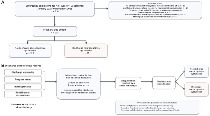
Figure 1: STROBE-aligned cohort derivation and adjudication framework for discharge neurocognitive dysfunction. (A) Flow diagram of patient screening and cohort construction. Among 318 consecutive emergency admissions for acute ischemic stroke, intracerebral hemorrhage, or transient ischemic attack evaluated with neuroimaging, 91 patients were excluded according to prespecified criteria, yielding a final analytic cohort of 227 patients. Of these, 129 were classified as having no discharge neurocognitive dysfunction and 98 as having discharge neurocognitive dysfunction. (B) Prespecified adjudication framework for discharge neurocognitive dysfunction. Discharge-phase clinical records within 24–48 h before discharge were independently reviewed by two trained clinical reviewers blinded to admission homocysteine levels. Disagreements were resolved by a senior neurologist, and final classification was based on predefined discharge-phase neurocognitive documentation criteria. Please click here to view a larger version of this figure.
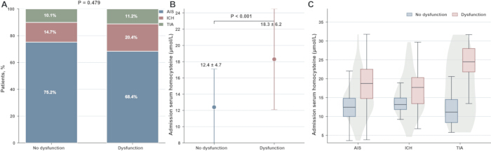
Figure 2: Acute cerebrovascular phenotype composition and admission serum homocysteine profiles according to discharge neurocognitive status. (A) Distribution of acute ischemic stroke, intracerebral hemorrhage, and transient ischemic attack among patients with and without discharge neurocognitive dysfunction. The overall cerebrovascular phenotype distribution did not differ significantly between groups (global Pearson χ2 P = 0.479). (B) Admission serum homocysteine concentrations according to discharge neurocognitive status. Values are presented as mean ± standard deviation. Patients with discharge neurocognitive dysfunction had higher admission homocysteine concentrations than those without dysfunction (18.3 ± 6.2 vs. 12.4 ± 4.7 µmol/L, P < 0.001). (C) Distribution of admission serum homocysteine concentrations across acute ischemic stroke, intracerebral hemorrhage, and transient ischemic attack phenotypes according to discharge neurocognitive status. AIS, acute ischemic stroke; ICH, intracerebral hemorrhage; TIA, transient ischemic attack. Please click here to view a larger version of this figure.
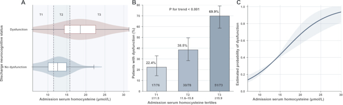
Figure 3: Exposure-response pattern of discharge neurocognitive dysfunction across admission serum homocysteine levels. (A) Distribution of admission serum homocysteine concentrations according to discharge neurocognitive status, with dashed vertical lines indicating the cohort-derived tertile cutoffs at 11.5 and 15.8 µmol/L. T1, ≤11.5 µmol/L; T2, 11.6–15.8 µmol/L; T3, ≥15.9 µmol/L. (B) Prevalence of discharge neurocognitive dysfunction across admission homocysteine tertiles. Dysfunction occurred in 17 of 76 patients in T1 (22.4%), 30 of 78 patients in T2 (38.5%), and 51 of 73 patients in T3 (69.9%), with a significant increasing trend across tertiles (P for trend < 0.001). Error bars indicate 95% confidence intervals for the observed proportions. (C) Unadjusted estimated probability of discharge neurocognitive dysfunction across the continuous range of admission serum homocysteine. The solid line represents the fitted probability from the unadjusted logistic regression model and the shaded band represents the 95% confidence interval. Please click here to view a larger version of this figure.
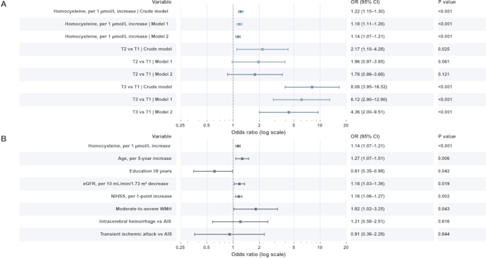
Figure 4: Multivariable logistic regression analyses of the association between admission homocysteine and discharge neurocognitive dysfunction. (A) Sequential logistic regression models showing the association of admission homocysteine, analyzed as a continuous variable and by tertiles, with discharge neurocognitive dysfunction. (B) Selected covariate estimates from the primary fully adjusted model; the complete adjustment set comprised age, sex, education level ≥9 years, eGFR, admission NIHSS score, moderate-to-severe WMH burden, and acute cerebrovascular phenotype. Model 1 was adjusted for age, sex, and education level ≥9 years. Model 2 was additionally adjusted for eGFR, NIHSS score on admission, moderate-to-severe WMH burden, and acute cerebrovascular phenotype. Please click here to view a larger version of this figure.
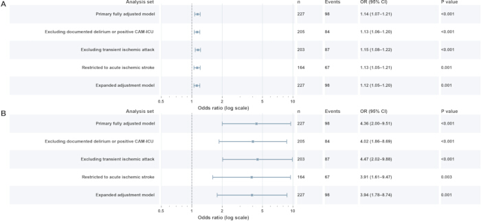
Figure 5: Sensitivity analyses of the association between admission homocysteine and discharge neurocognitive dysfunction. (A) Sensitivity analyses for admission homocysteine modeled as a continuous exposure, expressed as the odds ratio per 1 µmol/L increase. (B) Sensitivity analyses for high homocysteine burden, expressed as the odds ratio for T3 versus T1. Effect estimates were directionally similar across the primary fully adjusted model, the analysis excluding documented delirium or positive CAM-ICU screening, the analysis excluding transient ischemic attack, the acute ischemic stroke-only analysis, and the expanded adjustment model. Please click here to view a larger version of this figure.
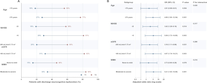
Figure 6: Exploratory subgroup consistency of the association between high homocysteine burden and discharge neurocognitive dysfunction. (A) Absolute prevalence of discharge neurocognitive dysfunction in T1 and T3 homocysteine categories across prespecified clinical subgroups. (B) Adjusted associations for high homocysteine burden, defined as T3 versus T1, across the same subgroups. Adjusted odds ratios, 95% confidence intervals, subgroup-specific P values, and P for interaction are shown. Please click here to view a larger version of this figure.
| Characteristic | Overall (n = 227) | No dysfunction (n = 129) | Dysfunction (n = 98) | P value | |
| Dyslipidemia, n (%) | 103 (45.4) | 59 (45.7) | 44 (44.9) | 0.920 | |
| Previous ischemic stroke, n (%) | 36 (15.9) | 15 (11.6) | 21 (21.4) | 0.076 | |
| Atrial fibrillation, n (%) | 22 (9.7) | 10 (7.8) | 12 (12.2) | 0.325 | |
| Smoking history, n (%) | 98 (43.2) | 53 (41.1) | 45 (45.9) | 0.458 | |
| Admission homocysteine, μmol/L | 14.9 ± 6.0 | 12.4 ± 4.7 | 18.3 ± 6.2 | <0.001 | |
| Fasting plasma glucose, mmol/L | 6.4 ± 1.5 | 6.2 ± 1.4 | 6.6 ± 1.6 | 0.052 | |
| HbA1c, % | 6.5 ± 0.9 | 6.4 ± 0.8 | 6.7 ± 0.9 | 0.031 | |
| Creatinine, μmol/L | 85.6 ± 20.8 | 82.1 ± 19.2 | 90.3 ± 22.0 | 0.004 | |
| eGFR, mL/min/1.73 m² | 80.3 ± 18.0 | 84.3 ± 16.9 | 75.2 ± 18.1 | <0.001 | |
| Acute ischemic stroke, n (%) | 164 (72.2) | 97 (75.2) | 67 (68.4) | 0.479* | |
| Intracerebral hemorrhage, n (%) | 39 (17.2) | 19 (14.7) | 20 (20.4) | ||
| Transient ischemic attack, n (%) | 24 (10.6) | 13 (10.1) | 11 (11.2) | ||
| NIHSS on admission | 5.9 ± 3.1 | 4.8 ± 2.6 | 7.4 ± 3.1 | <0.001 | |
| Cortical involvement, n (%) | 61 (26.9) | 28 (21.7) | 33 (33.7) | 0.049 | |
| Number of lacunes | 1.7 ± 1.3 | 1.4 ± 1.1 | 2.1 ± 1.4 | <0.001 | |
| Moderate-to-severe WMH, n (%) | 121 (53.3) | 55 (42.6) | 66 (67.3) | 0.001 | |
| Cortical atrophy, n (%) | 56 (24.7) | 25 (19.4) | 31 (31.6) | 0.030 | |
| Symptom onset to hospital admission, h, median (IQR) | 5.0 (2.8–9.0) | 4.8 (2.6–8.6) | 5.4 (3.0–9.5) | 0.284 | |
| Length of hospital stay, days, median (IQR) | 8.0 (6.0–10.0) | 7.0 (5.0–9.0) | 9.0 (7.0–11.0) | 0.008 | |
Table 1: Baseline clinical, laboratory, neuroimaging, and temporal characteristics according to discharge neurocognitive status. *P value refers to the global Pearson χ2 comparison of acute cerebrovascular phenotype distribution between groups; recalculated from the counts (97/19/13 vs 67/20/11), P = 0.4785, reported as 0.479. Abbreviations: BMI, body mass index; HbA1c, glycated hemoglobin; eGFR, estimated glomerular filtration rate; NIHSS, National Institutes of Health Stroke Scale; WMH, white matter hyperintensity; IQR, interquartile range.
| Admission homocysteine exposure | Crude OR (95% CI) | P value | Model 1 OR (95% CI) | P value | Model 2 OR (95% CI) | P value |
| Homocysteine, per 1 μmol/L increase | 1.22 (1.15–1.30) | <0.001 | 1.18 (1.11–1.26) | <0.001 | 1.14 (1.07–1.21) | <0.001 |
| Homocysteine tertiles | ||||||
| T1, ≤11.5 μmol/L | Reference | — | Reference | — | Reference | — |
| T2, 11.6–15.8 μmol/L | 2.17 (1.10–4.28) | 0.025 | 1.96 (0.97–3.95) | 0.061 | 1.78 (0.86–3.68) | 0.121 |
| T3, ≥15.9 μmol/L | 8.08 (3.95–16.52) | <0.001 | 6.12 (2.90–12.90) | <0.001 | 4.36 (2.00–9.51) | <0.001 |
| P for trend | <0.001 | <0.001 | <0.001 | |||
Table 2: Association between admission serum homocysteine and discharge neurocognitive dysfunction in logistic regression models. Abbreviations: OR, odds ratio; CI, confidence interval.
Model 1: adjusted for age, sex, and education level ≥9 years. Model 2 (primary fully adjusted model): additionally adjusted for eGFR, NIHSS score on admission, moderate-to-severe WMH burden, and acute cerebrovascular phenotype (AIS, ICH, or TIA).
| Sensitivity analysis | Sample size, n | Events, n | Homocysteine per 1 μmol/L increase OR (95% CI) | P value | T3 vs T1 OR (95% CI) | P value |
| Primary fully adjusted model | 227 | 98 | 1.14 (1.07–1.21) | <0.001 | 4.36 (2.00–9.51) | <0.001 |
| Excluding documented delirium or positive CAM-ICU | 205 | 84 | 1.13 (1.06–1.20) | <0.001 | 4.02 (1.86–8.69) | <0.001 |
| Excluding transient ischemic attack | 203 | 87 | 1.15 (1.08–1.22) | <0.001 | 4.47 (2.02–9.88) | <0.001 |
| Restricted to acute ischemic stroke | 164 | 67 | 1.13 (1.05–1.21) | 0.001 | 3.91 (1.61–9.47) | 0.003 |
| Expanded adjustment model including HbA1c and cortical atrophy | 227 | 98 | 1.12 (1.05–1.20) | 0.001 | 3.94 (1.78–8.74) | 0.001 |
Table 3: Sensitivity analyses of the association between admission serum homocysteine and discharge neurocognitive dysfunction. Abbreviations: OR, odds ratio; CI, confidence interval; TIA, transient ischemic attack; CAM-ICU, Confusion Assessment Method for the Intensive Care Unit; HbA1c, glycated hemoglobin. Primary fully adjusted model: age, sex, education ≥9 years, eGFR, admission NIHSS, moderate-to-severe WMH burden, and acute cerebrovascular phenotype. Expanded adjustment model additionally includes HbA1c and cortical atrophy.
| Subgroup | T3 vs T1 adjusted OR (95% CI) | P value | P for interaction |
| Age | 0.438 | ||
| <70 years | 2.91 (0.98–8.61) | 0.054 | |
| ≥70 years | 4.88 (1.90–12.54) | 0.001 | |
| NIHSS on admission | 0.317 | ||
| ≤5 | 3.42 (1.28–9.16) | 0.014 | |
| >5 | 5.09 (1.73–14.95) | 0.003 | |
| eGFR | 0.286 | ||
| ≥80 mL/min/1.73 m² | 3.16 (1.11–8.96) | 0.031 | |
| <80 mL/min/1.73 m² | 4.97 (1.74–14.21) | 0.003 | |
| WMH burden | 0.214 | ||
| None-to-mild | 2.73 (0.90–8.28) | 0.076 | |
| Moderate-to-severe | 5.21 (1.93–14.06) | 0.001 | |
Table 4: Exploratory subgroup analyses of the association between high admission homocysteine burden and discharge neurocognitive dysfunction. Abbreviations: OR, odds ratio; CI, confidence interval; NIHSS, National Institutes of Health Stroke Scale; eGFR, estimated glomerular filtration rate; WMH, white matter hyperintensity. Adjusted ORs compare T3 with T1. Models include the primary covariates, excluding the stratifying variable from its corresponding subgroup model. P for interaction values are from multiplicative interaction terms. These analyses remain exploratory and are not adjusted for multiple comparisons. All interaction P values are nonsignificant; subgroup differences must not be interpreted as definitive effect modification.
Supplementary File 1: The de-identified analytic dataset underlying the reported tables and figures.Please click here to download this file.
Dyskusja
In this retrospective cohort of 227 patients admitted for acute ischemic stroke, intracerebral hemorrhage, or transient ischemic attack at a high-altitude tertiary care center, higher admission serum homocysteine was associated with clinically adjudicated discharge neurocognitive dysfunction. Patients with dysfunction had substantially higher homocysteine levels at admission than those without dysfunction, and the prevalence of dysfunction increased stepwise from 22.4% in the lowest homocysteine tertile to 69.9% in the highest tertile. The graded association was attenuated but remained evident after adjustment for age, sex, education, renal function, admission NIHSS score, white matter hyperintensity burden, and acute cerebrovascular phenotype. In the fully adjusted model, each 1 µmol/L increase in homocysteine was associated with a 14% higher odds of discharge neurocognitive dysfunction, while patients in the highest tertile had more than fourfold higher odds than those in the lowest tertile. The association was preserved across sensitivity analyses excluding patients with documented delirium or positive CAM-ICU screening, excluding transient ischemic attack, restricting the cohort to acute ischemic stroke, and further adjusting for HbA1c and cortical atrophy. These findings support an association between admission homocysteine and short-term discharge-phase neurocognitive vulnerability but do not establish homocysteine as a causal determinant or validated clinical predictor21.
The present study extends prior work on post-stroke cognitive impairment by focusing on a clinically proximal outcome that is often underrepresented in the literature. Much of the existing evidence has examined cognitive impairment at weeks or months after stroke, or broader functional endpoints, whereas the period immediately preceding discharge has direct implications for bedside care, rehabilitation readiness, medication understanding, and the adequacy of discharge planning22. In our cohort, discharge neurocognitive dysfunction coexisted with older age, lower educational attainment, poorer renal function, higher admission NIHSS scores, heavier white matter hyperintensity burden, more lacunes, and more frequent cortical atrophy. These accompanying clinical and neuroimaging features are compatible with greater neurological and structural brain vulnerability, but they do not establish that the discharge-phase classification represents persistent cognitive impairment rather than a transient acute-phase disturbance. The discharge-phase outcome is therefore best interpreted as a short-term clinically adjudicated neurocognitive phenotype rather than as a substitute for standardized post-stroke cognitive impairment assessed during longer-term follow-up. This interpretation is compatible with prior observations linking stroke severity, structural brain burden, and cognitive outcomes after cerebrovascular events.
The homocysteine findings are also biologically plausible23. Homocysteine has been implicated in endothelial dysfunction, oxidative stress, prothrombotic activity, and microvascular injury, mechanisms that are relevant to cerebrovascular disease and cognitive vulnerability. Prior studies in post-stroke cohorts have reported associations between elevated homocysteine and later cognitive impairment, although the magnitude and consistency of these associations have varied across populations and analytical frameworks. Our results contribute a distinct observation by showing that admission homocysteine is associated with neurocognitive dysfunction documented before discharge, not only with delayed cognitive outcomes. The persistence of the association after adjustment for eGFR is notable, because renal dysfunction is closely related to circulating homocysteine levels. Likewise, the association remained evident after additional adjustment for HbA1c and cortical atrophy, although residual confounding from other metabolic and nutritional determinants cannot be excluded24. These findings do not establish causality, particularly because folate, vitamin B12, dietary pattern, fasting status, and other determinants of homocysteine were not systematically available in this retrospective cohort25.
The high-altitude setting deserves specific consideration. This study was conducted in Xining, Qinghai Province, at approximately 2,275 m above sea level, and cerebrovascular disease in plateau populations may occur within a vascular and metabolic milieu that differs from lower-altitude settings. Previous Chinese data have shown that acute ischemic stroke profiles vary across altitude strata, while studies from Qinghai have highlighted the relevance of early cognitive dysfunction after stroke in high-altitude populations26. In addition, experimental and observational work has suggested that prolonged high-altitude exposure can influence cognition and may relate to circulating homocysteine27, although these data should not be directly extrapolated to acute cerebrovascular disease28. Our study therefore does not claim that altitude caused the observed homocysteine association. Rather, the present findings provide setting-specific evidence from a high-altitude clinical population and require confirmation in multicenter cohorts that include broader geographic and altitude distributions.
The robustness analyses strengthen the internal coherence of the findings. The similar estimates observed after excluding patients with documented delirium or positive CAM-ICU screening reduce, but do not eliminate, concern that acute confusional states contributed to the discharge-phase outcome29. The analyses excluding transient ischemic attack and restricting the cohort to acute ischemic stroke yielded estimates similar in direction and magnitude to the primary analysis, suggesting that the main association was not solely dependent on inclusion of transient ischemic attack or on combining all three cerebrovascular phenotypes30. The exploratory subgroup analyses showed directionally consistent associations across strata defined by age, admission NIHSS, eGFR, and white matter hyperintensity burden. Although point estimates tended to be numerically larger among older patients, those with greater neurological severity, impaired renal function, and moderate-to-severe white matter hyperintensity burden, none of the interaction tests reached statistical significance31. These findings support consistency rather than clear effect modification, and they argue against overinterpreting apparent subgroup differences.
Several limitations should be considered. First, the retrospective single-center design limits causal inference and external generalizability, particularly beyond patients who remained clinically evaluable near hospital discharge. Second, the primary outcome was adjudicated from discharge-phase clinical documentation because standardized instruments such as the MoCA, MMSE, or a formal neuropsychological battery were not systematically available; therefore, the outcome should not be interpreted as a standardized diagnosis of persistent post-stroke cognitive impairment, and residual classification error remains possible. Third, although exclusion of patients with documented delirium or positive CAM-ICU screening yielded similar estimates, unrecognized, resolving, or otherwise undocumented acute confusional states may still have influenced cognitive status near discharge. Fourth, homocysteine was represented by a single measurement obtained within 24 h of admission, fasting status was not uniformly standardized, and acute physiological changes may have contributed to between-patient variation. Fifth, folate, vitamin B12, dietary pattern including vegetarian status, genetic determinants of one-carbon metabolism, inflammatory markers, long-term altitude exposure, and oxygenation-related variables were not systematically available, leaving the possibility of residual confounding. Finally, residual heterogeneity across acute ischemic stroke, intracerebral hemorrhage, and transient ischemic attack remains possible despite phenotype adjustment and sensitivity analyses, and the study did not assess standardized long-term cognitive trajectories or serial homocysteine measurements. In conclusion, among patients admitted for acute cerebrovascular events in a high-altitude tertiary care setting, higher admission serum homocysteine was associated with greater odds of clinically adjudicated discharge neurocognitive dysfunction, with a graded exposure-response pattern and directionally similar estimates across prespecified sensitivity analyses. These findings support further prospective evaluation of admission homocysteine as a potential marker of short-term discharge-phase neurocognitive vulnerability, with future multicenter studies requiring standardized cognitive assessment, serial biomarker measurements, and measurement of relevant nutritional and metabolic determinants.
Oświadczenia
The authors declare that they have no competing interests.
AUTHOR CONTRIBUTIONS:
Conceptualization: N.W., J.S., W.J.; Data curation: N.W., J.S.; Formal analysis: N.W., J.S., W.J.; Investigation: N.W., J.S.; Methodology: N.W., J.S., W.J.; Project administration: W.J.; Resources: N.W., J.S., W.J.; Software: N.W., J.S.; Supervision: W.J.; Validation: N.W., J.S., W.J.; Visualization: N.W., J.S.; Writing - Original Draft: N.W., J.S.; Writing - Review and Editing: W.J.
Materiały
| Nazwa | Firma | Numer katalogowy | Komentarze |
|---|---|---|---|
| Automatic biochemical analyzer | Shenzhen Mindray Bio-Medical Electronics Co., Ltd. | BS-2000M | Routine chemistry testing for homocysteine, fasting glucose, and serum creatinine. |
| Confusion Assessment Method for the Intensive Care Unit (CAM-ICU) | Vanderbilt University Medical Center | N/A (clinical assessment tool) | Delirium screening used in the prespecified sensitivity analysis. |
| Discharge-phase neurocognitive adjudication form | Qinghai Provincial People's Hospital | Institutional prespecified form | Blinded structured review of discharge-phase records within 24–48 h before discharge. |
| Electronic medical record system | Winning Health Technology Group Co., Ltd. | Winning Health EMR | Extraction of demographics, diagnoses, clinical course, and discharge documentation. |
| Fasting plasma glucose assay reagent | Shenzhen Mindray Bio-Medical Electronics Co., Ltd. | GLU reagent kit (BS-series) | Routine fasting plasma glucose measurement. |
| Glycated hemoglobin analyzer | Tosoh Corporation | HLC-723 G8 | HbA1c measurement for the expanded adjustment analysis. |
| IBM SPSS Statistics | IBM Corp. | Version 27.0 | Descriptive statistics, group comparisons, and logistic regression analyses. |
| Laboratory information system | Winning Health Technology Group Co., Ltd. | Winning LIS | Retrieval of laboratory results and verification of sampling times. |
| Magnetic resonance imaging system | Siemens Healthineers | MAGNETOM Skyra 3.0T | Brain MRI assessment of acute lesions and structural imaging features when clinically indicated. |
| National Institutes of Health Stroke Scale (NIHSS) | National Institute of Neurological Disorders and Stroke | N/A (clinical scale) | Neurological severity assessment at hospital admission. |
| Non-contrast computed tomography scanner | GE HealthCare | Revolution CT | Emergency non-contrast brain CT during the index admission. |
| Picture archiving and communication system (PACS) | Neusoft Medical Systems Co., Ltd. | PACS/RIS platform | Storage and review of CT/MRI images and abstraction of neuroimaging features. |
| R software | R Foundation for Statistical Computing | Version 4.3.2 | Statistical analysis and publication-quality figure generation. |
| Serum creatinine assay reagent | Shenzhen Mindray Bio-Medical Electronics Co., Ltd. | CREA reagent kit (BS-series) | Serum creatinine measurement for renal function assessment and eGFR derivation. |
| Serum homocysteine assay reagent | Shenzhen Mindray Bio-Medical Electronics Co., Ltd. | HCY reagent kit (BS-series) | Admission serum homocysteine measurement within 24 h of hospital arrival. |
Bibliografia
- Global, regional, and national burden of stroke and its risk factors, 1990–2021: a systematic analysis for the Global Burden of Disease Study 2021. Lancet Neurol. 2024;23(10):973-1003.
- El Husseini N, et al. Cognitive impairment after ischemic and hemorrhagic stroke: a scientific statement from the American Heart Association/American Stroke Association. Stroke. 2023;54(6):e272-e291.
- Quinn TJ, et al. European Stroke Organisation and European Academy of Neurology joint guidelines on post-stroke cognitive impairment. Eur Stroke J. 2021;28(12):3883-920.
- Zimmerman WD, et al. Transitions of care coordination intervention identifies barriers to discharge in hospitalized stroke patients. Front Neurol. 2021;12:573294.
- Milosevich ET, Moore MJ, Pendlebury ST, Demeyere N. Domain-specific cognitive impairment 6 months after stroke: the value of early cognitive screening. Int J Stroke. 2024;19(3):331-41.
- Abzhandadze T, Buvarp D, Lundgren-Nilsson Å, Sunnerhagen KS. Barriers to cognitive screening in acute stroke units. Sci Rep. 2021;11(1):19621.
- Zhang C, et al. Association between kidney measurements and cognitive performance in patients with ischemic stroke. PLoS One. 2023;18(12):e0292506.
- Lim JS, Lee JJ, Woo CW. Post-stroke cognitive impairment: pathophysiological insights into brain disconnectome from advanced neuroimaging analysis techniques. J Stroke. 2021;23(3):297-311.
- Coenen M, et al. Strategic white matter hyperintensity locations associated with post-stroke cognitive impairment: a multicenter study in 1568 stroke patients. Int J Stroke. 2024;19(8):916-24.
- Luzzi S, et al. Homocysteine, cognitive functions, and degenerative dementias: state of the art. Biomedicines. 2022;10(11):2741.
- Pinzon RT, Wijaya VO, Veronica V. The role of homocysteine levels as a risk factor of ischemic stroke events: a systematic review and meta-analysis. Front Neurol. 2023;14:1144584.
- Ma Y, et al. Blood biomarkers for post-stroke cognitive impairment: a systematic review and meta-analysis. J Stroke Cerebrovasc Dis. 2024;33(8):107632.
- Shi M, et al. Folate, homocysteine, and adverse outcomes after ischemic stroke. J Am Heart Assoc. 2024;13(19):e036527.
- Zhou S, et al. Age-dependent association between elevated homocysteine and cognitive impairment in a post-stroke population: a prospective study. Front Nutr. 2021;8:691837.
- Yan Y, et al. Anterior circulation acute ischemic stroke in the plateau of China: risk factors and clinical characteristics. Front Neurol. 2022;13:859616.
- Liu M, et al. Acute ischemic stroke at high altitudes in China: early onset and severe manifestations. Cells. 2021;10(4):809.
- Yao S, et al. The association between altitude and serum folate levels in Tibetan adults on the Tibetan plateau. Sci Rep. 2022;12(1):17886.
- Joshi SM, Jadavji NM. Deficiencies in one-carbon metabolism led to increased neurological disease risk and worse outcome: homocysteine is a marker of disease state. Front Nutr. 2024;11:1285502.
- Monaghan TF, et al. Blinding in clinical trials: seeing the big picture. Medicina (Kaunas). 2021;57(7):647.
- Vassar M, Holzmann M. The retrospective chart review: important methodological considerations. J Educ Eval Health Prof. 2013;10:12.
- Nam KW, et al. Plasma total homocysteine level is related to unfavorable outcomes in ischemic stroke with atrial fibrillation. J Am Heart Assoc. 2022;11(9):e022138.
- Guo L, et al. The status of readiness for hospital discharge of patients with post-stroke cognitive impairment and its relationship with post-discharge social isolation. BMC Public Health. 2025;25(1):1964.
- Yuan D, et al. Mechanism of homocysteine-mediated endothelial injury and its consequences for atherosclerosis. Front Cardiovasc Med. 2022;9:1109445.
- Lee M, et al. Effects of glycemic gap on post-stroke cognitive impairment in acute ischemic stroke patients. Brain Sci. 2021;11(5):612.
- Lee M, et al. Prediction of post-stroke cognitive impairment after acute ischemic stroke using machine learning. Alzheimers Res Ther. 2023;15(1):147.
- Li X, et al. Early cognitive dysfunction after stroke and related risk factors in the high-altitude and multi-ethnic region of Qinghai, China: a multi-center cross-sectional study. Clin Neurol Neurosurg. 2023;225:107607.
- Sharma VK, et al. Domain specific changes in cognition at high altitude and its correlation with hyperhomocysteinemia. PLoS One. 2014;9(7):e101448.
- Chen X, et al. Mechanism, prevention and treatment of cognitive impairment caused by high altitude exposure. Front Physiol. 2023;14:1191058.
- Breit H, et al. Delirium screening in the neurointensive care unit: comparison of the confusion assessment method for the intensive care unit and intensive care delirium screening checklist. J Neurol Sci. 2025;476:123619.
- Hayes A, et al. Not so transient?: a narrative review on cognitive impairment after transient ischemic attack. Stroke. 2024;55(10):2558-66.
- Wang X, Piantadosi S, Le-Rademacher J, Mandrekar SJ. Statistical considerations for subgroup analyses. J Thorac Oncol. 2021;16(3):375-80.