O uso de porcos em pesquisas tem aumentado nos últimos anos. No entanto, os porcos são caracterizados pela anatomia difícil das vias aéreas. Ao demonstrar como realizar a intubação endotraqueal guiada endoscopicamente, o presente protocolo visa aumentar ainda mais a segurança dos animais de laboratório para evitar o sofrimento dos animais e a morte desnecessária.
É necessária uma assinatura do JoVE para visualizar este conteúdo. Faça login ou inicie seu teste gratuito.
Artigo de método
Intubação endotraqueal usando um endoscópio de intubação flexível como modelo padronizado para o manejo seguro das vias aéreas em suínos
Neste artigo
Aviso de errata
Important: There has been an erratum issued for this article. View Erratum Notice
Resumo
Resumo
A intubação endotraqueal é muitas vezes um requisito básico para a pesquisa translacional em modelos suínos para várias intervenções que requerem uma via aérea segura ou altas pressões de ventilação. A intubação endotraqueal é uma habilidade desafiadora, exigindo um número mínimo de intubações endotraqueais bem-sucedidas para alcançar uma alta taxa de sucesso em condições ideais, o que muitas vezes é inatingível para pesquisadores não anestesiologistas. Devido à anatomia específica das vias aéreas suínas, uma via aérea difícil geralmente pode ser assumida. A impossibilidade de estabelecer uma via aérea segura pode resultar em ferimentos, eventos adversos ou morte do animal de laboratório. Usando uma abordagem de avaliação prospectiva, randomizada e controlada, demonstrou-se que a intubação endotraqueal assistida por fibra óptica leva mais tempo, mas tem uma taxa de sucesso de primeira passagem maior do que a intubação convencional sem causar quedas clinicamente relevantes na saturação de oxigênio. Este modelo apresenta um esquema padronizado para intubação endotraqueal guiada endoscopicamente, proporcionando uma via aérea segura, especialmente para pesquisadores que são inexperientes na técnica de intubação endotraqueal via laringoscopia direta. Espera-se que este procedimento minimize o sofrimento animal e as perdas desnecessárias de animais.
Introdução
A intubação endotraqueal é frequentemente um requisito básico para a pesquisa translacional em modelos suínos para várias intervenções que requerem uma via aérea segura ou altas pressões ventilatórias (como ventilação durante a ressuscitação cardiopulmonar1 ou síndrome do desconforto respiratório agudo2) ou exigem que o fluxo sanguíneo cerebral não seja comprometido por compressão interna por dispositivos de vias aéreas supraglóticas3 , que são ocasionalmente propagadas como alternativas no contexto de uma via aérea difícil prevista em suínos 4,5
Acesso restrito. Inicie sessão ou comece um teste para visualizar este conteúdo.
Protocolo
Os experimentos deste protocolo foram aprovados pelo Comitê Estadual e Institucional de Cuidados com os Animais (Landesuntersuchungsamt Rheinland-Pfalz, Koblenz, Alemanha; aprovação nº G20-1-135). Os experimentos foram conduzidos seguindo as diretrizes do ARRIVE. No geral, 10 suínos machos anestesiados (Sus scrofa domestica) com peso médio de 30 kg ± 2 kg e 12-16 semanas de idade foram utilizados para o presente estudo.
1. Preparação animal
- Mantenha um ambiente normal para os animais para minimizar o estresse. Reter alimentos 6 h antes do experimento programado para diminuir o risco de aspiração, mas perm....
Acesso restrito. Inicie sessão ou comece um teste para visualizar este conteúdo.
Resultados
A intubação endotraqueal foi realizada em 10 suínos machos (idade de 12 a 16 semanas, peso 30 kg ± 3 kg) em um ambiente de estudo prospectivo, randomizado e controlado. Os suínos foram randomizados em dois grupos: um foi convencionalmente intubado laringoscopicamente (grupo IC), e o outro grupo foi intubado assistido por meio de endoscópio de intubação flexível, conforme descrito no protocolo (grupo FIE). A atribuição do grupo foi feita puxando envelopes lacrados. O investigador foi designado aleatoriamente em u.......
Acesso restrito. Inicie sessão ou comece um teste para visualizar este conteúdo.
Discussão
Em estudos anteriores, nosso grupo de pesquisa já descreveu detalhes específicos sobre os benefícios translacionais do modelo suíno 2,27,32,33. Geralmente, a redução do nível de estresse do animal e a dor desnecessária devem ser parte integrante de qualquer protocolo de estudo e é fundamental para gerar dados reprodutíveis de forma confiável. Portanto, a intubação guiada endoscopicamente acorda.......
Acesso restrito. Inicie sessão ou comece um teste para visualizar este conteúdo.
Divulgações
O endoscópio de intubação flexível e seus acessórios foram fornecidos incondicionalmente pelo fabricante apenas para fins de pesquisa. Os autores declaram não haver mais conflitos de interesse financeiros ou outros.
Agradecimentos
Os autores querem agradecer a Dagmar Dirvonskis por seu excelente suporte técnico.
....Acesso restrito. Inicie sessão ou comece um teste para visualizar este conteúdo.
Materiais
| Nome | Empresa | Número de catálogo | Comentários |
|---|---|---|---|
| Ambu aScope Regular | Ambu GmbH, Medizinprodukte, Bad Nauheim, Alemanha | Diâmetro externo de fibra óptica descartável 5 mm | |
| Ambu aView Monitor | Ambu GmbH, Medizinprodukte, Bad Nauheim, Alemanha | monitor | |
| Atracurium Hikma 50 mg/5mL | Hikma Pharma GmbH, Martinsried | atracurium | |
| Azaperone (Stresnil) 40mg/mL | Lilly Deutschland GmbH, Bad Homburg, Alemanha | azaperona | |
| BD Discardit II Spritze 2, 5, 10, 20 mL | Becton Dickinson S.A. Carretera, Mequinenza Fraga, Espanha | seringa | |
| BD Luer Connecta | Becton Dickinson Infusion Therapy, AB Helsingborg, Schweden | 3-way-stopcock | |
| BD Microlance 3 20 G | Becton Dickinson S.A. Carretera, Mequinenza Fraga, Espanha | cânula | |
| Curafix i.v. clássicos | Lohmann & Rauscher International GmbH & Co. KG, Rengsdorf, Alemanha | Curativo de retenção de cânula | |
| Engströ m Carestation | GE Heathcare, Madison USA | ventilador | |
| Fentanyl-Janssen 0,05 mg/mL | Janssen-Cilag GmbH, Neuss | fentanil | |
| Fü hrungsstab, Durchmesser 4.3 | Rü | introdutor de tubo endotraqueal | |
| IBM SPSS Statistics for Windows, Versão 20 | IBM SPSS Statistics for Windows, Versão 20.0. Armonk, NY: IBM Corp.) | Software estatístico | |
| Incetomat-line 150 cm | Fresenius, Kabi Deutschland, GmbH | perfusor line | |
| Intrafix Primeline | B. Braun Melsungen AG, Melsungen, Germany | Linha de infusão | |
| JOZA Einmal Nitril Untersuchungshandschuhe | JOZA, Mü nchen, Alemanha | descartáveis | |
| Laringoscópio, 45.48.50, KL 2000 | Medicon | Cabo de laringoscópio | |
| Littmann Classic III Estetoscópio | 3M Deutschland GmbH, Neuss, Alemanha | estetoscópio | |
| Luer Lock | B.Braun Melsungen AG, Alemanha | ||
| Mutilado Vlieskompresse | Maimed GmbH, Neuenkirchen, Alemanha | Compressa de lã para fixar a língua | |
| Masimo LNCS Adtx SpO2 sensor | Masimo Corporation Irvine, Ca 92618 EUA | clipe de saturação para a cauda | |
| Masimo LNCS TC-I SpO2 sensor de clipe de orelha | Masimo Corporation Irvine, Ca 92618 EUA | Clipe de saturação para a orelha | |
| Masimo Radical 7 | Masimo Corporation Irvine, Ca 92618 EUA | saturação de oxigênio perifêneo | |
| Midazolam 15 mg/3 mL | Hameln Pharma GmbH, Hameln, Alemanha | midazolam | |
| Midmark Máscara Canina Pequeno Plástico com Diafragma FRSCM-0005 | Midmark Corp., Dayton, Ohio, EUA | máscara de ventilação para cães | |
| Octeniderm farblos | Schü lke & Mayr GmbH, Nordenstedt, Alemanha | Desinfetante alcoólico | |
| Seringa Perfusor original 50 mL | B.Braun Melsungen AG, Alemanha | seringa perfusor | |
| Perfusor FM Braun | B.Braun Melsungen AG, Alemanha | bomba de seringa | |
| Propofol 2% 20 mg/mL (frascos de 50 mL) | Fresenius, Kabi Deutschland, GmbH | propofol | |
| RÜ SCH Fü hrungsstab fü r Endotraquealtubus (ID 5,6 mm) | Teleflex Medical Sdn. Bhd, Malásia | Fio guia de tubo revestido de PVC | |
| Rü schelit Super Safety Clear >ID 6/6.5 /7.0 mm | Teleflex Medical Sdn. Bhd, Malásia | tubo endotraqueal | |
| Aço inoxidável Macintosh Grö ß e 4 | Welch Allyn69604 | lâmina para laringoscópio | |
| Sterofundin | B.Braun Melsungen AG, Melsungen, Alemanha | Solução eletrolítica balanceada | |
| Ultrastop Frasco antibeschlagmittel com conta-gotas 25 mL | Sigmapharm Arzneimittel GmbH, Wien, Áustria | Agente antiembaçante | |
| Vasofix Safety 22 G-16 G | B.Braun Melsungen AG, Alemanha | cateter | |
| venosoManômetro de manguito VBM | VBM Medizintechnik GmbH, Sulz a.N., Alemanha | manômetro de manguito | |
| Zelette | Lohmann & Rauscher International GmbH & Co. KG, Rengsdorf, Alemanha | Cotonete de tecido |
Referências
- Kleinman, M. E., Oh, W., Stonestreet, B. S. Comparison of intravenous and endotracheal epinephrine during cardiopulmonary resuscitation in newborn piglets. Critical Care Medicine. 27 (12), 2748-2754 (1999).
- Rissel, R., et al.
Acesso restrito. Inicie sessão ou comece um teste para visualizar este conteúdo.
Reimpressões e permissões
Errata
Formal Correction: Erratum: Endotracheal Intubation Using a Flexible Intubation Endoscope As a Standardized Model for Safe Airway Management in Swine
Posted by JoVE Editors on 4/03/2023. Citeable Link.
An erratum was issued for: Endotracheal Intubation Using a Flexible Intubation Endoscope As a Standardized Model for Safe Airway Management in Swine. The Protocol, Representative Results, and Discussion sections were updated.
In the Protocol, step 1.5 was updated from:
Disinfect the skin with a disinfectant (alcoholic) before inserting a peripheral vein cannula (22 G) into an ear vein. Spray the area, wipe once, then spray again, and allow the disinfectant to dry.
to:
Disinfect the skin with a disinfectant (alcoholic) before inserting a peripheral vein cannula (22 G) into an ear vein. Spray the area, wipe once, then spray again, and allow the disinfectant to dry. Secure the ear cannula with a band-aid (See Table of Materials).
In the Protocol, step 3.7 was updated from:
While maintaining the position of the endoscope, advance the endotracheal tube until it becomes visible in the camera image.
NOTE: If the endotracheal tube cannot be advanced through the glottic plane, there is a possibility that it has become caught on the arytenoid cartilage. In this case, the endotracheal tube must be withdrawn 1 cm and rotated by 90° before gently advancing again. If necessary, this maneuver can be repeated. Similar calibers of flexible intubation endoscope and endotracheal tube can minimize the risk of this issue occurring. If the endotracheal tube cannot be advanced despite this maneuver, it is likely that the subglottic narrowness-the narrowest part of the porcine larynx-cannot be passed. In this case, a smaller endotracheal tube size needs to be selected. Regular commercially available endotracheal tubes in sizes 6.5 cm or 7.0 cm ID should be able to pass the glottis as long as no anatomic abnormalities are present.
to:
While maintaining the position of the endoscope, advance the endotracheal tube until it becomes visible in the camera image.
NOTE: If the endotracheal tube cannot be advanced through the glottic plane, there is a possibility that it has become caught on the arytenoid cartilage. In this case, the endotracheal tube must be withdrawn 1 cm and rotated by 90° before gently advancing again. If necessary, this maneuver can be repeated. Similar calibers of flexible intubation endoscope and endotracheal tube can minimize the risk of this issue occurring. If the endotracheal tube cannot be advanced despite this maneuver, it is likely that the subglottic narrowness-the narrowest part of the porcine larynx-cannot be passed. In this case, a smaller endotracheal tube size needs to be selected. Regular commercially available endotracheal tubes in sizes 6.5 cm or 7.0 cm ID should be able to pass the glottis as long as no anatomic abnormalities are present. Endotracheal tube size requirements vary depending on the piglet size and breed.
In the Representative Results, the sixth paragraph was updated from:
Statistical analyses were performed using commercially available software (see Table of Materials). Normal distribution was examined using the Kolmogorov-Smirnoff test28. If a normal distribution was determined, group differences were analyzed using t-tests of independent samples29 or the Mann-Whitney U test30 for the non-parametric version. Data are presented as mean (± standard deviation). Correlations of ordinal-scale data were examined using Spearman's correlation coefficient31. A significance level of p < 0.05 was assumed.
to:
Statistical analyses were performed using commercially available software (see Table of Materials). Normal distribution was examined using the Kolmogorov-Smirnoff test28. If a normal distribution was determined, group differences were analyzed using t-tests of independent samples29 or the Mann-Whitney U test30 for the non-parametric version. Data are presented as mean (± standard deviation). Correlations of ordinal-scale data were examined using Spearman's correlation coefficient31. A significance level of p < 0.05 was assumed. All tests were performed with exploratory intention; therefore p-values are descriptive. Nevertheless, p < 0.05 was accepted as indicative of statistical significance.
In the Representative Results, the legend for figure 1 was updated from:
Figure 1: Number of intubation attempts in group comparison. For the group that was intubated using a flexible intubation endoscope, every intubation attempt was successful; in the group that was conventionally intubated, it took an average of 1.4 attempts before the endotracheal tube could be placed correctly. Error bars show the standard deviation. Please click here to view a larger version of this figure.
to:
Figure 1: Number of intubation attempts in group comparison. For the group that was intubated using a flexible intubation endoscope, every intubation attempt was successful; in the group that was conventionally intubated, it took an average of 1.4 attempts before the endotracheal tube could be placed correctly. Error bars show the standard deviation. n = 5 (for each group). Please click here to view a larger version of this figure.
In the Representative Results, figure 2 was updated from:
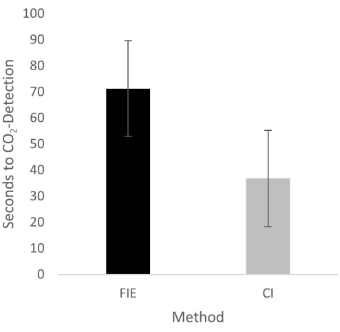
Figure 2: Time until CO2 detection in group comparison. For the group that was intubated using a flexible intubation endoscope, it took significantly longer until end-tidal CO2 could be detected, depicted as mean and standard deviation. Please click here to view a larger version of this figure.
to:
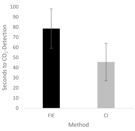
Figure 2: Time until CO2 detection in group comparison. For the group that was intubated using a flexible intubation endoscope, it took significantly longer until end-tidal CO2 could be detected, depicted as mean and standard deviation. n = 5 (for each group). Please click here to view a larger version of this figure.
In the Discussion, the fifth paragraph was updated from:
The increased duration had no clinical significance in this cohort. At no time was the termination criterion-a saturation of less than 93%-reached. This is shown in the results because a procedure change was unnecessary at any time. Prior adequate mask ventilation is a critical step to allow sufficient time for fiberoptic endotracheal tube placement to avoid rapid desaturation34. These results are consistent with previous studies comparing conventional intubation and endoscopically assisted intubations with inexperienced providers35.
to:
The increased duration had no clinical significance in this cohort. At no time was the termination criterion-a saturation of less than 93%-reached. This is shown in the results because a procedure change was unnecessary at any time. Prior adequate mask ventilation is a critical step to allow sufficient time for fiberoptic endotracheal tube placement to avoid rapid desaturation34. These results are consistent with previous studies comparing conventional intubation and endoscopically assisted intubations with inexperienced providers35. We attribute the prolonged duration of fiberoptic intubation to the fact that one must first reorient again after insertion, whereas with conventional intubation, one retains a view of the glottis. It is also important to avoid contact with the mucosa with the flexible intubation endoscope during advancement. This requires occasional corrective maneuvers. Last but not least, after successful placement, retraction of the relatively long endoscope is required, which increases the time to CO2 detection slightly.
