This case report describes a 37-year-old man with von Hippel-Lindau disease and a solitary kidney who underwent robotic-assisted partial nephrectomy for multifocal renal cell carcinoma after disease progression during belzutifan treatment. Three progressive lesions were identified preoperatively, and a fourth lesion was detected intraoperatively.
Relato de caso
Robotic-Assisted Partial Nephrectomy in a Solitary Kidney in a von Hippel-Lindau Renal Cell Carcinoma
Neste artigo
Resumo
Resumo
Von Hippel-Lindau (VHL) disease is a rare autosomal dominant tumor syndrome caused by germline mutations in the VHL gene. Patients may develop multiple, bilateral, and recurrent renal cell carcinomas (RCCs), posing a major therapeutic challenge, particularly in those with a solitary kidney, where preservation of renal function is critical to prevent end-stage renal disease and dialysis dependence. This case demonstrates the surgical management of multiple renal tumors in a solitary kidney. A 37-year-old man with VHL disease (c.257C > G, p.P86R mutation) and a solitary right kidney following previous left radical nephrectomy presented with three progressive clear cell RCC (ccRCC) lesions, the largest measuring 4.8 cm, despite ongoing treatment with belzutifan, a HIF-2α inhibitor. A transperitoneal robotic-assisted partial nephrectomy (RAPN) was performed. A fourth tumor was identified intraoperatively. Complete excision of all tumors with negative surgical margins was achieved. Postoperatively, severe acute kidney injury (AKI) developed, requiring continuous renal replacement therapy (CRRT), which was complicated by forearm necrosis resulting from calcium substitution paravasation. An additional complication included a right pneumothorax. Following intensive care management, including pleural drainage, renal function gradually recovered, and CRRT was discontinued on postoperative day 8. This case demonstrates that RAPN can provide tumor control while preserving renal tissue in patients with VHL-associated multifocal RCC and a solitary kidney. However, the procedure carries a substantial risk of severe postoperative AKI requiring temporary renal replacement therapy.
Introdução
The management of Von Hippel-Lindau (VHL) disease is complex because of its autosomal dominant inheritance and propensity for the development of multiple, recurrent tumors across various organ systems1. A germline mutation in the VHL tumor suppressor gene leads to constitutive activation of hypoxia-inducible factors (HIFs), promoting angiogenesis and tumorigenesis2.
Clear cell renal cell carcinoma (ccRCC) is a leading cause of mortality in patients with VHL, with a lifetime risk of up to 70%3. A key principle in managing VHL-associated renal tumors is nephron-sparing surgery (NSS), which aims to preserve renal function and delay or prevent the need for dialysis4, particularly given the high likelihood of tumor recurrence and the potential need for future interventions.
The therapeutic landscape for VHL-associated ccRCC has recently expanded with the introduction of belzutifan, a HIF-2α inhibitor5. However, not all patients achieve a complete or sustained response, and disease progression during therapy may occur, necessitating surgical intervention5.
In relatively young patients with a solitary kidney, preservation of functional renal parenchyma is particularly important. In such cases, robotic-assisted partial nephrectomy (RAPN) offers the benefits of minimally invasive surgery and enhanced surgical precision, which may facilitate complex tumor resection and renorrhaphy6. A series of RAPN procedures in solitary kidneys demonstrated high rates of dialysis avoidance and successful preservation of estimated glomerular filtration rate (eGFR)7. However, these patients are inherently at increased risk of postoperative acute kidney injury (AKI)8. Severe AKI requiring renal replacement therapy (RRT), although less frequent, remains an important potential complication in this high-risk population.
This case report describes the perioperative management and outcomes of a patient with VHL disease and a solitary kidney who underwent RAPN for multifocal ccRCC that progressed despite belzutifan treatment. The case highlights the technical feasibility of nephron-sparing surgery and the significant challenge of postoperative AKI requiring intensive care and continuous RRT, underscoring the delicate balance between oncologic control and renal function preservation in this complex patient population.
Case presentation
A 37-year-old man with a confirmed diagnosis of Von Hippel-Lindau (VHL) disease was evaluated for multifocal renal tumors. Genetic testing performed in August 2024 identified a germline VHL mutation, c.257C > G (p.P86R). The patient was asymptomatic, and the tumors were suspected on surveillance imaging. The family history was positive for VHL.
The medical history was notable for multifocal clear cell renal cell carcinoma (ccRCC) of the left kidney. An open partial nephrectomy of the left kidney was initially performed at another hospital on November 13, 2024. Histopathological examination confirmed multifocal clear-cell RCC with a positive surgical margin at the lower pole (R1). Because of a postoperative urinoma associated with severe complications, radical left nephrectomy was subsequently performed on December 27, 2024.
On May 1, 2025, treatment with belzutifan at 120 mg daily was initiated as systemic therapy for VHL-associated tumors. Despite treatment, surveillance imaging in October 2025 demonstrated progression of three pre-existing ccRCC lesions in the solitary right kidney (Table 1). The largest lesion, located in the mid-kidney, measured approximately 4.8 cm, compared with 3.1 cm in October 2024, and was in close contact with the renal collecting system. Two additional lesions, measuring approximately 2–2.5 cm, were identified at the upper pole (Figure 1 and Figure 2). Preoperative laboratory values were stable, with a serum creatinine level of 1.2 mg/dL and an estimated glomerular filtration rate (eGFR) of 83 mL/min/1.73 m2 (Table 1). The patient strongly preferred a minimally invasive, nephron-sparing procedure and declined open or radical surgical approaches.
Postoperative laboratory course
Following surgery, acute kidney injury with anuria developed. Serial laboratory assessments demonstrated progressive renal impairment, with serum creatinine reaching a peak of 4.8 mg/dL on postoperative day (POD) 3.
Diagnosis, assessment, and plan
Progressive multifocal ccRCC in a solitary kidney was diagnosed based on imaging findings and the established history of VHL disease. Extensive preoperative planning was undertaken. High-resolution abdominal MRI was performed to delineate the anatomical relationships among the tumors, renal vasculature, and collecting system (Figure 1 and Figure 2). This facilitated detailed preoperative mapping and planning of a sequential tumor resection strategy.
The primary surgical objective was complete tumor excision with negative surgical margins while minimizing warm ischemia time and preserving maximal renal function in the solitary kidney. Given the complex anatomy and preference for a minimally invasive nephron-sparing approach, transperitoneal robotic-assisted partial nephrectomy (RAPN) was selected. The surgical strategy involved sequential tumor resection, beginning with the most complex mid-kidney lesion.
Protocolo
The patient consented in writing to all treatment steps and anonymous publication. The study complies with the Declaration of Helsinki and was approved by the ethics committees of the Westfalen-Lippe Medical Association and the University of Muenster (2023–500-f-S for retrospective data management.
1. Preoperative preparation and planning
- A high-resolution, contrast-enhanced magnetic resonance imaging (MRI scan of the abdomen was performed (Figure 1 and Figure 2).
- The patient was thoroughly counseled regarding the risks of the procedure, including acute kidney injury (AKI, conversion to radical nephrectomy, and the potential need for temporary or permanent renal replacement therapy (RRT).
- The patient was maintained on a daily dose of belzutifan (120 mg until the day of surgery).
- A prophylactic double-J (DJ ureteral stent was inserted in preparation for potential injury to the renal pelvis during tumor resection).
- A central venous catheter was inserted preoperatively.
2. Robotic-assisted partial nephrectomy
- Patient positioning and operating room setup
- The patient was placed in a modified flank position with the right side elevated to 60° using a vacuum beanbag. All pressure points were carefully padded.
- Port placement and robotic docking
- A transperitoneal approach was used. Pneumoperitoneum was established using a Veress needle, and an 8 mm camera trocar was placed at the lateral border of the rectus muscle, 2 cm lateral to the umbilicus.
- Under direct visualization, three 8 mm robotic trocars were placed along the lateral border of the rectus muscle. One trocar was positioned 8 cm cephalad to the camera port, a second trocar was positioned 8 cm caudal to the camera port, and a third 8 mm trocar for the fourth robotic arm was placed 8 cm caudal to the second robotic trocar.
- A 12 mm assistant trocar was placed 2 cm medial to the robotic camera trocar.
- The da Vinci Xi Surgical System was docked with the robotic column aligned with the target anatomy.
- Renal exposure and hilar dissection
- The right colon was mobilized medially along the line of Toldt using medial visceral rotation to expose the retroperitoneum and the solitary right kidney.
- The gonadal vein was identified and followed cephalad toward the renal hilum.
- The renal hilum was dissected, and the main renal artery and vein were isolated using a combination of blunt and sharp dissection.
- Preoperative imaging was used to guide the dissection of the renal artery.
- Intraoperative tumor localization
- A robotic laparoscopic ultrasound probe was used to confirm the location, depth, and margins of the renal tumors relative to the renal surface and adjacent structures.
- The three tumors identified on preoperative MRI were localized: a 4.8 cm dorsal tumor between the middle and lower thirds of the kidney, an approximately 2 cm dorsolateral tumor between the upper and middle thirds of the kidney, and an approximately 2 cm upper-pole tumor. A fourth 2 cm tumor was identified at the lower pole; in retrospect, the lesion was visible on the preoperative MRI. Intraoperative ultrasonography confirmed the presence of the lesion (Figure 1 and Figure 2; Table 2).
- Sequential tumor resection and renorrhaphy
- After intraoperative ultrasonography confirmed the depth and extent of all tumors, the renal artery was clamped using a bulldog clamp. All resections and reconstructions were performed under warm ischemia without intermittent clamp release.
- Resection was initiated with the largest tumor. The tumor was excised with a narrow rim of healthy renal parenchyma using cold scissors. Because of the close relationship of the tumor to the collecting system, the renal pelvis was entered during resection.
- Resection of the upper-pole lesions was subsequently performed under warm ischemia. The tumors were excised using cold scissors while preserving a margin of healthy renal tissue. The renal pelvis was entered during resection of tumor 2, the predominantly endophytic lesion. Finally, the lower-pole tumor was resected using the same technique.
- Defects in the collecting system were closed using 4-0 monofilament sutures. Renorrhaphy of all tumor resection sites was performed using 3-0 monofilament sutures.
- The bulldog clamp was removed after completion of the initial renorrhaphy. The total warm ischemia time was 30 min.
- Following clamp removal, small bleeding vessels were controlled by selective suturing using 4-0 monofilament sutures. An additional layer of renorrhaphy was required for the more endophytic upper-pole tumor.
- Hemostatic agents were applied to the resection sites, and Gerota's fascia was closed with clips.
- Completion of the procedure
- No surgical drain was placed.
- The resected specimens were placed in an endoscopic retrieval bag and removed through a mini-laparotomy incision.
3. Intraoperative monitoring
- Standard intraoperative monitoring included continuous electrocardiography, pulse oximetry, and capnography.
- Invasive arterial blood pressure was monitored through a radial arterial catheter.
- Central venous access was obtained through the right internal jugular vein for fluid management and vasopressor administration.
- Urine output was monitored hourly through a transurethral Foley catheter.
4. Postoperative management and intensive care
- The patient was transferred to the intensive care unit (ICU for close postoperative monitoring).
- Continuous venovenous hemodiafiltration (CVVHDF was initiated on postoperative day (POD 1 because of anuria).
- A chest X-ray revealed a right pneumothorax, which was managed by placement of a pleural drainage system.
- Radiological assessment of the double-J stent was performed on the same day to confirm appropriate positioning and drainage.
- Pneumonia was clinically suspected; therefore, empirical treatment with meropenem (1 g intravenously every 8 h was initiated). Treatment was subsequently expanded to include linezolid (600 mg intravenously every 12 h and azithromycin (500 mg intravenously once daily).
- A trial of furosemide was initiated on POD 8 to assess renal recovery. CVVHDF was subsequently discontinued after adequate diuresis was achieved.
- Necrosis of the left forearm developed during peripheral intravenous calcium replacement associated with continuous renal replacement therapy. The affected area required surgical debridement and vacuum-assisted closure (VAC therapy).
Resultados
The robotic-assisted partial nephrectomy was completed successfully without conversion to radical nephrectomy or open surgery. The total operative time was 120 min, with an estimated blood loss of 300 mL and a warm ischemia time of 30 min (Table 3).
The postoperative course was complex and was primarily characterized by severe acute kidney injury (AKI) requiring continuous renal replacement therapy. Continuous venovenous hemodiafiltration (CVVHDF) was required for 8 days. Renal function subsequently showed signs of recovery, and long-term dialysis was not required.
The primary postoperative complications included severe AKI with anuria requiring CVVHDF for 8 days. A right pneumothorax developed on postoperative day (POD) 2 and was managed with drainage. Additional pleural drainage was required on the right side on POD 11 and the left side on POD 12. Rising inflammatory markers raised concern for infection and were managed with stepwise escalation of antibiotic therapy. In addition, necrosis of the left forearm developed during peripheral intravenous calcium replacement associated with continuous renal replacement therapy (CRRT), requiring surgical debridement and vacuum-assisted closure (VAC) therapy (Table 3).
Despite the complex postoperative course, the primary oncologic and functional objectives were achieved. Complete tumor excision was accomplished, and the solitary kidney was preserved. The patient was discharged from the ICU without requiring permanent dialysis. At the 6-month follow-up, the patient had no complaints and required no dialysis; renal function was preserved. The imaging surveillance indicated no radiographic recurrence while the patient continued Belzutifan.
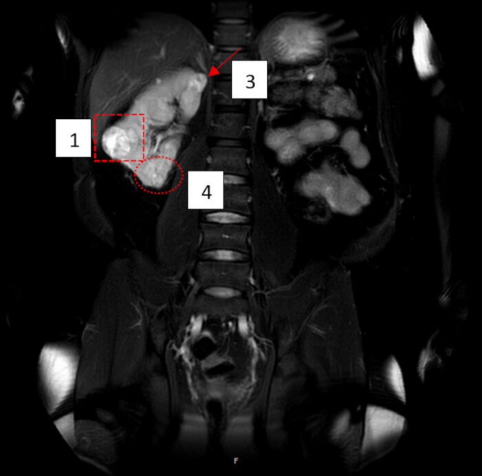
Figure 1: MRI showing the fourth tumor in retrospect. Coronal magnetic resonance imaging (MRI) of the solitary right kidney. (1) Largest tumor (4.8 cm), located in the middle-to-lower portion of the kidney, with a predominantly solid appearance and areas of cystic degeneration. (3) Upper-pole tumor (approximately 2 cm) with a partially endophytic growth pattern. (4) Lower-pole tumor that was not prospectively identified on preoperative MRI but was recognized retrospectively after its intraoperative detection. Please click here to view a larger version of this figure.
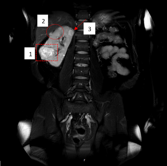
Figure 2: MRI showing the second endophytic tumor. Magnetic resonance imaging (MRI) of the solitary right kidney showing the three tumors identified preoperatively. (1) Largest tumor (4.8 cm), located dorsally between the middle and lower thirds of the kidney. (2) Lateral upper-pole tumor (approximately 2 cm) with a predominantly endophytic growth pattern. (3) Upper-pole tumor (approximately 2 cm). Please click here to view a larger version of this figure.
| Preoperative parameters | |
| latest serum creatinine | 1.2 mg/dL |
| latest eGFR | 83 mL/min/1.732 |
| duration of follow-up | 8 months |
| dialysis-free survival | Not nessecary in a functioning solitary kidney situataion |
| proteinuria | Negative |
| blood pressure | Normal, no antihypertensive medication |
| need for further intervention | Prophylactic insertion of DJC |
| Belzutifan Therapy | |
| duration of treatment | 9 months |
| Dose | 120 mg daily |
| radiological response | Progressive disease |
| RECIST progression | Yes – more than 20% increase in tumor diameter in the largest lesion |
| (progressed from 3.1 cm to 4.8 cm.) | |
| New lesions were suspected. | |
| multidisciplinary decision-making | Tumor progression on belzutifan; patient was young (37 years old), had a solitary kidney, and strongly preferred a minimally invasive, nephron-sparing procedure, declining open or radical surgery. Active surveillance combined with systemic therapy was an option but was declined by the patient. |
| why surgery was preferred over continued systemic therapy or surveillance | |
Table 1: Baseline parameters and systemic therapy.
| First tumor | Second tumor | Third tumor | Fourth tumor | |
| Location | Underpol | Oberpol | Oberpol | Underpol |
| exophytic/endophytic nature | combined | combined | endophytic | exophtic |
| collecting system involvement | Involved | No involvemnt | involved | No involvment |
| pathological stage | pT1a | pT1a | pT1a | pT1a |
| tumour grade | G2 low grade | G2 low grade | G2 low grade | G2 low grade |
| margin status | R0 | R0 | R0 | R0 |
| Size | 4 cm | 1.7 cm | 2.8 cm | 1.3 cm |
Table 2: Tumor characteristics.
| operative outcomes | ||||
| console time | 120 minutes | |||
| docking time | 135 minutes | |||
| estimated preserved parenchyma | 67% | |||
| blood transfusion | 2 reserves (postoperatively) | |||
| exact discharge creatinine | 1.6 mg/dL | |||
| renal function at 3 months | 58 mL/min/1.732 | |||
| First tumor | Second tumor | Third tumor | Fourth tumor | |
| Location | Underpol lateral ventral | Underpol medial caudal | Oberpol ventral | Oberpol lateral dorsal |
| exophytic/endophytic nature | combined | combined | exophtic | endophytic |
| collecting system involvement | involved | No involvment | No involvemnt | involved |
| pathological stage | pT1a | pT1a | pT1a | pT1a |
| tumour grade | G2 low grade | G2 low grade | G2 low grade | G2 low grade |
| margin status | R0 | R0 | R0 | R0 |
| Longest Diameter | 4 cm | 1.3 cm | 1.7 cm | 2.8 cm |
| clamp time for each tumour | 8 minutes | 7 minutes | 7 minutes | 8 minutes |
| selective versus main artery clamp duration | Main artery | Main artery | Main artery | Main artery |
| use of ICG fluorescence | Yes | Yes | Yes | Yes |
| Complications | ||||
| Clavien-Dindo | Cause/indication | |||
| AKI nessicating CRRT | 4a | Acute renal failure postoperativly with anuria | ||
| pneumothorax | 3b | Probably through the prophylactic insertion of a central venous catheter, which may be needed postoperatively for hemodialysis in cases of radical nephrectomy or AKI | ||
| Pneumonia | 2 | Ideopathischer elevation of infect parameters. | ||
| forearm necrosis | 3b | Through paravasation in the calcium substituion therapy, needed in patients undergiong CRRT. | ||
Table 3: Operative outcomes and complications.
Discussão
This report's significance lies in its detailed description of the perioperative management of a complex, high-risk patient, highlighting both the technical feasibility of RAPN and the potential for severe yet reversible postoperative complications.
The primary challenge in this scenario was the dual objective of achieving negative surgical margins for oncologic control while preserving sufficient functional renal parenchyma to avoid long-term dialysis. RAPN, with its enhanced three-dimensional visualization, instrument dexterity, and tremor filtration, is well-suited for such complex resections, particularly in the setting of a solitary kidney.
Despite meticulous surgical techniques and a warm ischemia time of 30 min for all four tumors, severe acute kidney injury (AKI) developed, requiring continuous renal replacement therapy (RRT). Although clinically significant, this outcome is not unexpected in this high-risk population. Reported rates of AKI following partial nephrectomy in solitary kidneys range from 60–80%, with a smaller but substantial subset (5–10%) requiring temporary RRT. Predictors of severe AKI include prolonged warm ischemia time (>25 minutes), lower baseline renal function, and greater parenchymal volume loss9. In this case, the complexity of resecting four tumors, particularly the large, centrally located mid-kidney lesion, likely contributed to the functional impairment despite the reduced warm ischemia time. Renal function recovered sufficiently to discontinue RRT after 8 days, consistent with the literature suggesting that AKI in this setting is often reversible, particularly when the total ischemic insult is limited and optimal supportive care is provided10.
The postoperative course was further complicated by a right pneumothorax and elevated inflammatory markers. This sequence of adverse events underscores the potential for substantial systemic stress following major surgery in a patient with a solitary kidney. It also highlights the importance of a multidisciplinary approach involving urologic surgeons, intensivists, and nephrologists. Immediate, high-level intensive care, including continuous RRT and advanced respiratory support, was critical to achieving a favorable outcome.
RAPN offers several advantages over open surgery in this setting, including smaller incisions, potentially reduced blood loss, and faster wound healing. Enhanced visualization and articulation of the robotic instruments facilitate precise dissection and complex renorrhaphy, which are particularly important in managing multifocal tumors.
The principles and techniques described in this report may also be applicable beyond VHL-associated RCC. They are relevant to patients with a solitary kidney undergoing partial nephrectomy for localized renal masses, whether the solitary kidney results from previous nephrectomy, congenital absence, or a nonfunctioning contralateral kidney.
This case adds to the growing body of literature supporting the safety and efficacy of RAPN in patients with solitary kidneys. However, it also demonstrates that significant postoperative AKI can occur despite meticulous surgical planning and technique. This observation reinforces findings from larger series showing that although dialysis avoidance rates are high, temporary RRT remains a clinically relevant and manageable risk11,12. The use of belzutifan before surgery, followed by surgical intervention because of tumor progression, also reflects the evolving interplay between systemic therapy and surgery in the management of VHL-associated RCC.
Several limitations should be noted. RAPN in such complex cases requires specialized equipment and an experienced team, and does not eliminate the risk of serious complications. Reliance on intraoperative ultrasonography and the inability to palpate the kidney can pose challenges, though this was sufficient in this case. As a single-case report, the findings are subject to potential bias and have limited generalizability. The 6-month follow-up does not allow evaluation of long-term results. Additionally, the choice of surgery timing was influenced by tumor progression, despite a 9-month period of belzutifan treatment and the patient's preference. Although active surveillance combined with systemic therapy might be an option, the patient declined this approach. More extensive studies with longer follow-up are necessary to better understand RAPN’s effectiveness and associated risks.
In conclusion, this case demonstrates that RAPN can achieve both oncologic control and preservation of renal function in a high-risk patient with VHL disease, a solitary kidney, and multiple renal tumors progressing during systemic therapy, even when the postoperative course is complicated by severe AKI requiring temporary RRT. The case underscores the importance of meticulous preoperative planning, advanced surgical techniques to minimize renal ischemia, and access to comprehensive intensive care for managing significant postoperative complications. Long-term surveillance for recurrent RCC and other VHL-associated manifestations remains essential.
Divulgações
The authors have nothing to disclose. The authors used a language model (Gemini) exclusively for language polishing and grammatical refinement. All scientific content was controlled, verified, and approved by the authors, who assume full responsibility for the final manuscript.
Agradecimentos
The authors thank the intensive care unit staff for their dedicated care of this patient. No specific funding was received for this work.
Materiais
| Nome | Empresa | Número de catálogo | Comentários |
|---|---|---|---|
| Azithromycin | Pfizer Inc. | N/A | Macrolide antibiotic used for treatment of susceptible bacterial infections. |
| Belzutifan | Merck & Co., Inc. | N/A | Hypoxia-inducible factor-2 alpha (HIF-2α) inhibitor used as a targeted therapeutic agent. |
| Bülau drainage system | B. Braun Melsungen AG | N/A | Closed thoracic drainage system used for drainage of air or fluid from the pleural cavity. |
| da Vinci Xi Surgical System | Intuitive Surgical, Inc. | N/A | Robotic-assisted surgical platform used to perform minimally invasive surgical procedures. |
| Double-J ureteral stent | Boston Scientific Corporation | N/A | Ureteral stent used to maintain urinary drainage between the kidney and bladder. |
| Furosemide | Fresenius Kabi USA | N/A | Loop diuretic used to promote diuresis and reduce excess fluid accumulation. |
| Linezolid | Pfizer Inc. | N/A | Oxazolidinone antibiotic used for treatment of susceptible Gram-positive bacterial infections. |
| Meropenem | Fresenius Kabi USA | N/A | Broad-spectrum carbapenem antibiotic used for treatment of susceptible bacterial infections. |
Referências
- Schmid S, et al. Management of von Hippel-Lindau disease: an interdisciplinary review. Oncol Res Treat. 2014;37(12):761-771.
- Chappell JC, Payne LB, Rathmell WK. Hypoxia, angiogenesis, and metabolism in the hereditary kidney cancers. J Clin Invest. 2019;129(2):442-451.
- Waeckel T, et al. The Hippo pathway in clear cell renal cell carcinoma (ccRCC): a nexus with the VHL disruption? Transl Oncol. 2026;66:102721.
- Jilg CA, et al. Repeated renal interventions for renal cell carcinoma in von Hippel-Lindau disease: long-term outcomes. Eur Urol Focus. 2026. doi:10.1016/j.euf.2026.07.002.
- Chen AH, Grana AK. Belzutifan's role in the treatment landscape of clear cell renal cell carcinoma. Pharmacotherapy. 2025;45(6):356-366.
- Franz T, Dietel A, Trebst D, Horn LC, Stolzenburg JU. Robotic-assisted laparoscopic partial nephrectomy for multiple synchronous renal tumors in a solitary kidney: a case report. Cureus. 2025;17(10):e94560.
- Grosso AA, et al. Functional and oncologic outcomes in single-kidney patients treated with robot-assisted partial nephrectomy for renal tumors: results from a prospectively maintained dataset of a single tertiary referral center. Cancers (Basel). 2025;17(12):1978.
- Rod X, et al. Impact of ischaemia time on renal function after partial nephrectomy: a systematic review. BJU Int. 2016;118(5):692-705.
- Zhu K, et al. Acute kidney injury in solitary kidney patients after partial nephrectomy: incidence, risk factors and prediction. Transl Androl Urol. 2020;9(3):1232-1243.
- Bravi CA, et al. Impact of acute kidney injury and its duration on long-term renal function after partial nephrectomy. Eur Urol. 2019;76(3):398-403.
- Volpe A, et al. Renal ischemia and function after partial nephrectomy: a collaborative review of the literature. Eur Urol. 2015;68(1):61-74.
- Gul Z, et al. A multi-institutional report of peri-operative and functional outcomes after robot-assisted partial nephrectomy in patients with a solitary kidney. J Robot Surg. 2019;13(3):423-428.