This protocol describes a three‑dimensional laparoscopic artery‑first pancreaticoduodenectomy for malignant pancreatic‑head tumors, aiming to standardize this arterial‑first operative approach and improve surgical safety and oncological outcomes.
Artigo de método
Clinical Application of Three-dimensional Laparoscopic Artery-first Pancreaticoduodenectomy in Treating Pancreatic Head Malignant Tumors
Neste artigo
Resumo
Resumo
Pancreaticoduodenectomy is a technically demanding procedure associated with substantial surgical risk. Conventional laparoscopic approaches have several limitations, including restricted visualization, which can make exposure of the superior mesenteric artery and identification of vessels and lymph nodes challenging. In patients with vascular adhesion or tumor invasion, surgical dissection is particularly difficult and may increase the risk of intraoperative vascular injury and hemorrhage. In addition, poor delineation of anatomical planes may result in incomplete tumor dissection, inadequate lymphadenectomy, and difficult mobilization of the pancreatic head and duodenum, potentially compromising surgical safety and oncological outcomes. To optimize the surgical workflow, this protocol describes a three-dimensional laparoscopic artery-first pancreaticoduodenectomy with the infracolic artery-first approach as the principal technical modification. By combining early arterial exposure and clear delineation of anatomical planes with the stereoscopic visualization of three-dimensional laparoscopy, this technique facilitates identification of vessel–tissue relationships, vascular control, lymphadenectomy, tumor dissection, and tissue mobilization. These features may help reduce inadvertent vascular injury and intraoperative blood loss while enabling complete tumor resection. The protocol provides a standardized surgical approach, particularly applicable to pancreatic head lesions adjacent to or involving major vessels, and is intended to improve the safety and reproducibility of laparoscopic pancreaticoduodenectomy.
Introdução
Laparoscopic surgery, characterized by small incisions, enhanced visualization, reduced patient discomfort, and rapid postoperative recovery, has become a cornerstone of modern surgical practice. Laparoscopic pancreaticoduodenectomy (LPD) has emerged as a key area of development in pancreatic surgery in recent years1,2,3,4. However, LPD is technically demanding and has a long learning curve, and its early adoption in Europe was limited by high complication rates5.
The first case of LPD was reported in 19975, and Chinese surgeons first performed the operation in 20026. After more than 20 years of development, the feasibility, safety, and short- and long-term outcomes of LPD have gradually been established. In the past five years, LPD has been increasingly adopted in China, with numerous hospitals performing or preparing to perform the procedure. However, owing to the pancreas's unique anatomical location, the inherent complexity of the procedure, and the long learning curve and high risks associated with LPD, the implementation of LPD in China remains poorly standardized.
With an improved understanding of pancreatic anatomy and advances in laparoscopic techniques, an increasing number of patients with pancreatic-head cancer, including those with portal vein or superior mesenteric vein (SMV) involvement, can undergo laparoscopic treatment. During resection, selection of an appropriate surgical approach is critical for controlling intraoperative bleeding and achieving oncologically adequate tumor resection. Therefore, the surgical approach should be tailored to the tumor location and the extent of vascular involvement.
The venous-first approach is conventionally used by most surgeons. It enables early assessment of superior mesenteric vein (SMV) and portal vein (PV) involvement but does not permit early evaluation of superior mesenteric artery (SMA) invasion, potentially leading to extensive dissection before SMA invasion is recognized as unresectable. This approach is particularly challenging in patients with venous invasion, extension of the uncinate process to the left posterior aspect of the SMA, or a hypertrophic uncinate process, in whom dissection is more difficult and the risk of intraoperative hemorrhage is increased.
This protocol describes an LPD technique for pancreatic-head lesions, particularly tumors adjacent to the SMV, using an infracolic artery-first approach. The method prioritizes dissection of the SMA and its branches, allowing early control of the arterial blood supply to the pancreatic head and identification of variant arterial anatomy. These features facilitate vascular control and reduce the risk of intraoperative bleeding. The primary goal of this approach is to improve surgical safety while achieving oncologically adequate resection.
The patient was a 63-year-old woman admitted with upper abdominal pain and discomfort accompanied by fever for 10 days. Computed tomography (CT) demonstrated swelling of the pancreatic head with heterogeneous density and patchy, heterogeneous mild-to-moderate enhancement on contrast-enhanced images, measuring approximately 3.3 × 2.3 cm. Mild dilation of the intrahepatic and extrahepatic bile ducts was also observed. The patient had a 10-year history of diabetes mellitus treated with long-term oral metformin and acarbose, with poor glycemic control. Preoperative carcinoembryonic antigen (CEA) and carbohydrate antigen 19-9 (CA19-9) levels were 9.77 ng/mL and 616 U/mL, respectively. Preoperative endoscopic ultrasonography revealed a hypoechoic lesion in the pancreatic head with ill-defined borders and abundant internal vascularity. Pathological examination of the preoperative biopsy specimen demonstrated mild-to-moderate cellular atypia with focal severe atypia, consistent with intraductal papillary mucinous neoplasm (IPMN) of the pancreas. On January 15, 2026, the patient underwent three-dimensional laparoscopic pancreaticoduodenectomy using the infracolic artery-first approach. Postoperative pathological examination revealed a moderately differentiated invasive ductal adenocarcinoma of the pancreatic head, measuring 3.5 × 3 × 2.4 cm, with perineural invasion and invasion of the surrounding fibroadipose tissue. No definite intravascular tumor thrombus was identified, and the surgical margins were negative. Chronic cholecystitis was also observed, and no metastatic carcinoma was identified in the 31 lymph nodes examined (0/31). At the 3-month postoperative follow-up, the patient's CEA and CA19-9 levels had returned to within normal ranges, and the patient had recovered well.
Protocolo
This study was approved by the Institutional Ethics Committee of Dong'e County People's Hospital, Shandong Province (Ethics Approval No. K2026‑024‑01). Written informed consent was obtained from the patient. The research content and methods comply with medical ethical standards and requirements.
1. Patient selection
- Patient-selection criteria
- Select patients with a preoperatively diagnosed resectable or borderline-resectable malignant neoplasm of the pancreatic head or uncinate process that requires radical pancreaticoduodenectomy.
- Confirm that the patient is fit for major laparoscopic abdominal surgery under general anesthesia.
- Confirm adequate hepatic, renal, and hematologic function for major surgery.
- Confirm the absence of distant metastasis on preoperative imaging.
- Ensure that neoadjuvant therapy, if applicable, was completed at least 4–6 weeks before surgery and that the disease is stable or responding.
- Exclusion criteria
- Exclude patients with confirmed distant metastatic disease.
- Exclude patients with unresectable tumors showing >180° circumferential encasement of the superior mesenteric artery (SMA) or celiac trunk.
- Exclude patients with severe cardiopulmonary or systemic comorbidities that preclude major pancreatic resection.
- Preoperative assessment
- Obtain contrast-enhanced thin-section abdominal computed tomography (CT) with multiplanar vascular reconstruction.
- Assess the tumor location and size. Evaluate the liver, peritoneum, and other abdominal organs for distant metastases.
- Assess tumor contact with the SMA, celiac trunk, portal vein (PV), and superior mesenteric vein (SMV).
- Identify vascular variants, including a replaced or accessory right hepatic artery arising from the SMA.
- Identify the dorsal pancreatic artery (DPA), inferior pancreaticoduodenal artery (IPDA), and any common trunk between the first jejunal artery (JA1) and IPDA (Figure 1).
- Assess venous drainage patterns, including the confluence of the left gastric vein (LGV) and inferior mesenteric vein (IMV).
- Evaluate the SMV-PV caliber, vessel-wall integrity, and signs of venous tumor invasion.
- Classify the tumor as resectable, borderline-resectable, or unresectable according to the NCCN pancreatic adenocarcinoma guidelines.
- Consider the infracolic artery-first laparoscopic pancreaticoduodenectomy for resectable and borderline-resectable tumors.
- Discuss borderline-resectable tumors with suspected SMV invasion in a multidisciplinary team meeting. Anticipate possible segmental venous resection.
- Obtain magnetic resonance cholangiopancreatography (MRCP) when additional evaluation of the biliary and pancreatic duct anatomy is required.
2. Preoperative preparation and patient positioning
- Keep the patient nil by mouth for 8 h before the procedure. Allow the patient to consume 250 mL of a clear carbohydrate drink 2 h before surgery.
- Perform lower-extremity venous duplex ultrasound 48 h before surgery. Apply intermittent pneumatic compression devices before beginning the surgical procedure.
- Place the patient in a supine split-leg position with both lower extremities abducted. Elevate the head, lower the feet, and slightly elevate the patient's right side.
- Administer prophylactic intravenous antibiotics 0.5–1 h before skin incision.
3. Surgical procedure
- Surgical team and equipment positioning
- Position the lead surgeon on the patient's right side and the first assistant on the patient's left side.
- Position the camera assistant between the patient's legs. Position the scrub nurse on the patient's right side.
- Position the monitor at the patient's head. Use a three-dimensional (3D) laparoscopic system throughout the procedure.
- Trocar placement and pneumoperitoneum
- Insert a 12 mm trocar for the observation port 2–3 cm below the umbilicus.
- Insert a 12 mm trocar for the main operating port 1–2 cm above the umbilicus at the lateral border of the rectus abdominis.
- Insert a 5 mm trocar 2 cm below the right costal margin along the midclavicular line. Insert another 5 mm trocar at the corresponding position on the left side (Figure 2).
- Establish pneumoperitoneum. Maintain the pneumoperitoneum pressure at 12 mmHg.
- Initial exposure and suspension
- Insert a purse-string suture through the abdominal wall below the xiphoid process. Pass the suture under the liver and fix it to the lesser omentum on the right side of the cardia using a vascular clip.
- Bring the suture out through the abdominal wall. Tie it extracorporeally to retract the liver cephalad.
- Open the gastrocolic ligament to enter the lesser sac.
- Mobilize the inferior border of the pancreas. Expose the anterior wall of the superior mesenteric vein (SMV).
- Retract the transverse colon cephalad.
- Fix the transverse mesocolon to the liver retraction purse-string suture using polymer locking clips.
- Abdominal exploration and SMA exposure
- Carefully inspect the abdominal organs and peritoneum. Confirm the absence of metastatic nodules.
- Adopt the infracolic artery-first approach. Skeletonize the left side of the superior mesenteric artery (SMA) using an ultrasonic scalpel.
- Perform a reverse Kocher incision. Completely mobilize the pancreatic head and duodenum.
- Critical infracolic artery-first dissection
- Identify the ileocolic vein, SMV trunk, third portion of the duodenum, inferior border of the pancreatic neck, and middle colic artery as anatomical landmarks.
- Dissect the jejunal mesentery horizontally toward the left side at the level of the third portion of the duodenum. Follow the jejunal arterial branches using combined dorsal and ventral dissection.
- Divide the proximal jejunum at the predetermined site. Continue the dissection cephalad along the middle colic artery.
- Dissect the lymphatic and adipose tissues surrounding the SMV and SMA using the ultrasonic scalpel. Skeletonize the vessels.
- Vascular variants: identification and safe management
- Review preoperative multiplanar CT angiographic reconstruction for vascular variants. Confirm the vascular anatomy intraoperatively.
- Expose the main trunk of the SMA. Dissect cephalad along its right lateral wall using low-power ultrasonic dissection.
- Identify any replaced right hepatic artery (rRHA) arising from the SMA. Trace its course posterior to the pancreatic head and portal vein toward the hepatoduodenal ligament.
- Identify the dorsal pancreatic artery (DPA). Trace the vessel proximally and distally to confirm its origin and course before division.
- Preserve the rRHA throughout the dissection. Leave a thin layer of perivascular tissue around the vessel to minimize vasospasm and thermal injury.
- Retract the rRHA gently with a vessel loop when it obstructs access to the SMA or retropancreatic plane. Do not divide the rRHA.
- Confirm the origin and distal course of a DPA arising from the rRHA. Ligate the DPA with vascular clips at the inferior border of the pancreas.
- Trace the conventional right hepatic artery from the proper hepatic artery when no rRHA is identified. Preserve the artery during lymph node dissection at station 12.
- Trace an atypically originating DPA along its course before ligation. Exclude an accessory right hepatic artery or other critical vessel before clipping.
- Perform temporary vessel-loop occlusion for 3–5 min when vessel identity remains uncertain. Assess rRHA pulsation, when present, and the color of the right liver lobe before permanent ligation.
- Confirm hepatic arterial flow using intraoperative Doppler ultrasonography when available and required.
- Identify the first jejunal artery (JA1). Divide the JA1 when required to complete SMA skeletonization and perivascular lymph node dissection.
- Identify any common trunk between the JA1 and the inferior pancreaticoduodenal artery (IPDA). Dissect the common trunk proximally to its SMA origin.
- Divide the JA1 and IPDA individually at their origins. Preserve jejunal perfusion while completing IPDA division.
- Gastric and pancreatic dissection
- Dissect the tissues along the greater and lesser curvatures of the stomach. Divide the stomach using a linear stapler.
- Dissect the lymph nodes at stations 8a and 8p along the common hepatic artery.
- Identify the DPA as described in Section 3.6. Confirm its origin and distal course before ligating it with vascular clips at the inferior border of the pancreas.
- Fully expose the pancreatic neck. Divide the pancreatic parenchyma using the ultrasonic scalpel and achieve hemostasis.
- Identify the main pancreatic duct. Divide the duct using scissors and prepare it for pancreaticojejunostomy.
- Middle colic and jejunal artery dissection
- Assess the right branch of the middle colic artery for dense tumor adhesion or suspected invasion. Ligate and divide the branch when required for oncological resection.
- Consider division of the main trunk of the middle colic artery in selected patients with a large pancreatic-head mass when required for adequate exposure and complete oncological resection.
- Carefully expose the JA1 between the SMA and SMV. Ligate and divide the vessel.
- Gastroduodenal artery dissection
- Assess the gastroduodenal artery (GDA) for dense adhesion to adjacent tissues. Apply temporary atraumatic bulldog vascular clamps to the common hepatic artery and proper hepatic artery when vascular control is required.
- Incise the arterial sheath with scissors. Carefully mobilize and divide the GDA.
- Bile duct division and specimen resection
- Fully dissect the bile duct. Divide the bile duct at the predetermined level.
- Confirm close tumor-SMV contact on preoperative CT before applying prophylactic vascular control. Apply bilateral temporary SMV occlusion using vascular clamps in complex cases with dense tumor-vein adhesion.
- Carefully dissect the SMV from the tumor tissue. Release the temporary vascular clamps after completing safe tumor-vein dissection.
- Perform en bloc resection of the tumor and pancreaticoduodenal specimen. Confirm complete macroscopic tumor resection without visible residual disease (Figure 4).
- Pancreaticojejunostomy
- Anastomosis preparation
- Bring the Roux jejunal limb through the retrocolic mesenteric window to the pancreatic stump.
- Create a 2 mm enterotomy on the antimesenteric side of the jejunum adjacent to the closed jejunal end. Match the enterotomy to the main pancreatic duct diameter.
- Posterior U-shaped suture with round ligament bolster
- Place U-shaped sutures through the full thickness of the pancreatic parenchyma at the superior and inferior margins of the pancreatic stump.
- Pass the sutures through the jejunal seromuscular layer using 3-0 non-absorbable polypropylene vascular suture with a 36-mm needle.
- Interpose the ligamentum teres hepatis behind the pancreaticojejunostomy. Secure it with the U-shaped sutures.
- Duct-to-mucosa anastomosis with stent placement
- Place 6–8 interrupted sutures between the main pancreatic duct and jejunal mucosa using 4-0 non-absorbable polypropylene vascular suture with a 26-mm needle.
- Achieve precise mucosa-to-mucosa apposition. Insert a short pancreatic duct stent across the anastomosis.
- Advance the distal end of the stent completely into the jejunal lumen.
- Anterior wall reinforcement
- Place interrupted sutures between the anterior pancreatic parenchyma and jejunal seromuscular layer using 4-0 vascular suture.
- Complete the imbricating modified Blumgart anastomosis.
- Completion
- Maintain uniform suture tension and avoid pancreatic parenchymal laceration.
- Ensure that the anastomosis remains tension-free with the round ligament bolster (Figure 5).
- Anastomosis preparation
- Hepaticojejunostomy
- Perform an end-to-side mucosa-to-mucosa anastomosis between the common hepatic duct and jejunum approximately 10 cm distal to the pancreaticojejunostomy.
- Create a jejunal enterotomy matching the diameter of the transected common hepatic duct. Preserve the periductal blood supply and avoid thermal injury.
- Suture the posterior wall from lateral to medial with a running full-thickness 4-0 absorbable barbed suture. Achieve precise mucosal apposition.
- Suture the anterior wall separately with another running 4-0 absorbable barbed suture. Secure each suture at the end.
- Inspect the completed hepaticojejunostomy for integrity and hemostasis.
- Avoid excessive periductal dissection. Maintain a tension-free anastomosis and omit routine biliary stenting.
- Gastrojejunostomy and completion of the procedure
- Gastrojejunostomy
- Perform a side-to-side gastrojejunostomy on the same jejunal limb 50 cm distal to the hepaticojejunostomy.
- Select the anastomotic site on the posterior gastric wall 2–3 cm proximal to the gastric transection margin.
- Create small enterotomies on the posterior gastric wall and antimesenteric border of the jejunum.
- Close the common opening with a running 3-0 barbed suture.
- Inspect the anastomosis visually for patency. Confirm the absence of suture-line bleeding.
- Drainage placement
- Place drainage tubes behind the pancreaticojejunostomy and hepaticojejunostomy.
- Place an additional drainage tube below the pancreaticojejunostomy.
- Gastrojejunostomy
4. Troubleshooting
- Mobilize the transverse mesocolon sufficiently when SMA/SMV exposure is difficult. Transect the middle colic artery trunk when required to improve exposure.
- Perform gentle blunt dissection along the avascular plane anterior to the SMA root.
- Perform repeated careful, sharp dissection under magnified vision when dense tumor-vessel adhesion is encountered. Avoid blind dissection and excessive traction.
- Control minor intraoperative bleeding with compression and bipolar coagulation.
- Apply temporary vascular occlusion for major venous or arterial laceration. Repair the vascular injury with a running suture.
- Modify or discontinue the approach when SMA encasement exceeds 180°, anatomical planes remain obscured, bleeding is intractable, or unfavorable arterial variants prevent safe dissection.
- Convert to a supracolic or posterior approach when required.
5. Postoperative care and monitoring
- Postoperative management
- Monitor vital signs and abdominal physical findings after surgery. Perform serial laboratory examinations.
- Obtain contrast-enhanced abdominal CT on postoperative day 3, selectively in high-risk patients.
- Evaluate intra-abdominal fluid collections, drain position, intestinal obstruction, pseudoaneurysm, vascular thrombosis, and other major intra-abdominal complications on CT.
- Measure drainage-fluid amylase on postoperative days 1, 3, and 5.
- Monitor conventional blood tests, serum amylase, and hepatic and renal function during postoperative recovery.
- Abdominal drainage management
- Record the volume, color, and consistency of drainage fluid daily.
- Confirm stable vital signs and the absence of abdominal distension or tenderness before drain removal.
- Confirm a daily drainage volume of <100 mL and a drainage-fluid amylase level of <300 U/L before removing the abdominal drain.
- Remove the abdominal drainage tube when all specified removal criteria are satisfied.
- Diet advancement
- Confirm stable postoperative conditions and normal bowel peristalsis before initiating oral intake.
- Begin with a clear liquid diet. Progress gradually to a semi-liquid diet, then to a regular soft diet.
- Suspend oral intake if abdominal distension, nausea, or vomiting occurs.
- Evaluate the patient for delayed gastric emptying, pancreatic fistula, intestinal obstruction, or other complications when feeding intolerance occurs.
- Postoperative complication assessment
- Assess clinically relevant postoperative pancreatic fistula (CR-POPF) using drainage-fluid amylase levels, clinical manifestations, and imaging findings.
- Assess persistent abdominal pain, fever, and other clinical manifestations associated with postoperative complications.
- Evaluate for intra-abdominal hemorrhage, infection, intestinal obstruction, and vascular thrombosis using physical examination, laboratory testing, and abdominal imaging.
- Initiate targeted intervention promptly when a major postoperative complication is confirmed.
Resultados
Preoperative cross-sectional imaging demonstrated a pancreatic-head mass highly suspicious for malignancy. Preoperative percutaneous core-needle biopsy showed intraductal papillary mucinous neoplasm (IPMN). This discordance was most likely due to sampling error of the biopsy procedure: the specimen obtained represented the premalignant IPMN component, whereas the focal invasive carcinomatous area was not sampled. Final postoperative histopathology confirmed invasive ductal adenocarcinoma arising from the malignant transformation of IPMN.
The 3D laparoscopic artery-first pancreaticoduodenectomy was completed successfully. Key vascular anatomy of the infracolic window, including the superior mesenteric artery (SMA) and first jejunal artery (JA1), is shown in Figure 1. Histopathological examination of the resected specimen confirmed moderately differentiated invasive ductal adenocarcinoma of the pancreatic head.
The trocar configuration used for the procedure provided access for 3D visualization and laparoscopic manipulation, with the operative ports positioned as shown in Figure 2. During the initial infracolic-plane dissection, the SMA, superior mesenteric vein (SMV), and middle colic vessels were exposed as shown in Figure 3. The right branch of the middle colic artery was divided intraoperatively due to dense adhesion and suspected tumor involvement.
The common hepatic artery and proper hepatic artery were temporarily occluded during dissection of the gastroduodenal artery (GDA), and the SMV was temporarily occluded during separation of the tumor-associated adhesions from the vessel. Following en bloc specimen removal, inspection of the surgical bed confirmed completion of the resection (Figure 4). Reconstruction was subsequently performed, including a pancreaticojejunostomy using the Blumgart anastomosis technique; the completed pancreaticojejunostomy is shown in Figure 5.
The total operative time was 400 min, with an estimated blood loss of 150 mL and no requirement for blood transfusion. No surgical complications were observed, and the patient had an uneventful postoperative recovery. Drainage fluid amylase decreased from 1,582 U/L on postoperative day (POD) 1 to 124 U/L on POD 3. The postoperative hospital stay was 13 days, and the total hospital stay was 20 days. The relatively prolonged postoperative hospital stay was mainly attributed to the patient's underlying type 2 diabetes mellitus; continuous glycemic optimization was required after oral feeding was tolerated to facilitate wound healing and reduce the risk of surgical site infection. These perioperative and postoperative outcomes are summarized in Table 1. Preoperative and postoperative imaging demonstrated the corresponding changes following surgical treatment (Figure 6).
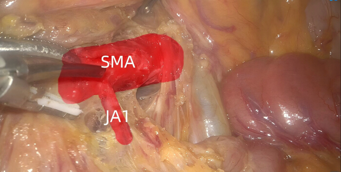
Figure 1: Intraoperative identification of the superior mesenteric artery and first jejunal artery during infracolic artery-first dissection. Intraoperative view showing the exposed superior mesenteric artery (SMA) and first jejunal artery (JA1) during the infracolic artery-first approach. Please click here to view a larger version of this figure.
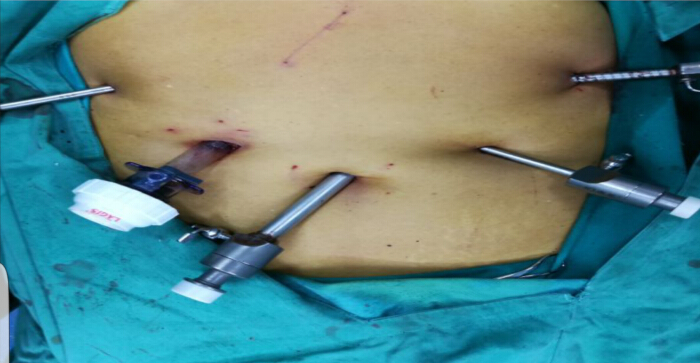
Figure 2: Trocar placement for three-dimensional laparoscopic pancreaticoduodenectomy. Intraoperative view showing the trocar configuration used for the laparoscopic procedure. Please click here to view a larger version of this figure.
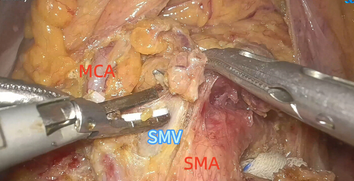
Figure 3: Intraoperative view of the infracolic artery-first approach. Key anatomical structures, including the superior mesenteric artery (SMA), middle colic vessels, and superior mesenteric vein (SMV), are exposed through the infracolic plane during the initial dissection phase. Please click here to view a larger version of this figure.
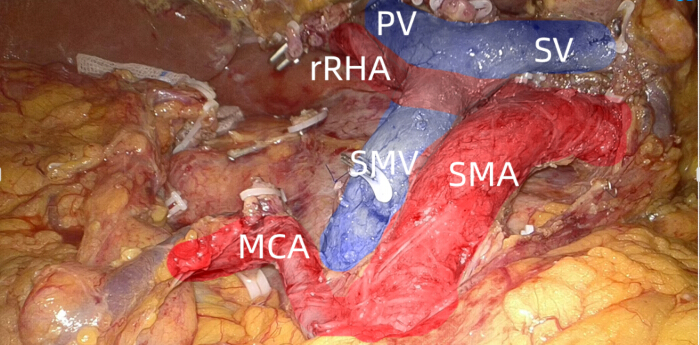
Figure 4: Surgical field following specimen resection. Intraoperative view of the surgical bed after pancreaticoduodenal specimen removal, showing the superior mesenteric artery (SMA), superior mesenteric vein (SMV), portal vein (PV), splenic vein (SV), middle colic artery (MCA), and variant right hepatic artery (rRHA). Please click here to view a larger version of this figure.
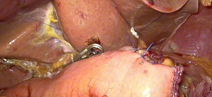
Figure 5: Pancreaticojejunostomy following reconstruction. Intraoperative view of the completed pancreaticojejunostomy. Please click here to view a larger version of this figure.
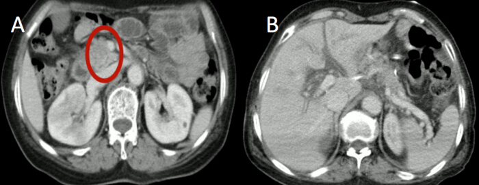
Figure 6: Preoperative and postoperative contrast-enhanced computed tomography (CT) images. (A) Preoperative contrast-enhanced CT showing the pancreatic-head lesion. (B) Postoperative contrast-enhanced CT demonstrating the postoperative appearance after pancreaticoduodenectomy. Please click here to view a larger version of this figure.
| Items | Results |
| Operative time (min) | 400 |
| Blood loss (mL) | 150 |
| Blood transfusion (mL) | 0 |
| Postoperative complications | None |
| Amylase on POD 1 (U/L) | 1582 |
| Amylase on POD 3 (U/L) | 124 |
| Postoperative hospital stay (days) | 13 |
| Total hospital stay (days) | 20 |
Table 1: Perioperative and postoperative outcomes following three-dimensional laparoscopic artery-first pancreaticoduodenectomy. The table summarizes operative time, estimated blood loss, blood transfusion, postoperative complications, drainage-fluid amylase levels on postoperative days (PODs) 1 and 3, postoperative hospital stay, and total hospital stay.
Discussão
Laparoscopic pancreaticoduodenectomy (LPD) has evolved considerably over the past two decades, with increasing recognition of its minimally invasive advantages. However, because of the pancreas's deep anatomical location, its proximity to major vessels, and the complex anatomy of the uncinate process, LPD remains technically demanding. The traditional superior mesenteric vein (SMV)-first approach remains widely used for LPD and enables early assessment of portal vein and SMV involvement7,8. A major limitation is that involvement of the superior mesenteric artery (SMA) may not be recognized until pancreatic neck transection and uncinate process dissection have progressed. This is particularly relevant for pancreatic head tumors associated with venous involvement or a hypertrophic uncinate process extending to the left posterior aspect of the SMA. A meta-analysis of 41 clinical studies evaluated the potential advantages of an SMA-first strategy over the conventional approach in this setting9. The critical step in the present protocol is therefore early identification and skeletonization of the SMA through the infracolic space. Use of the middle colic artery and adjacent vascular structures as anatomical landmarks facilitates orientation, while early identification and control of pancreaticoduodenal vascular branches follow the principles of an artery-first and no-touch oncological approach10,11.
Several technical features are important for the successful implementation of the method. Early SMA exposure allows assessment of tumor-artery relationships before extensive irreversible dissection and may help avoid unnecessary dissection when extensive SMA invasion is identified12. A meta-analysis by Negoi et al. reported a reduction in futile dissection in unresectable cases using an SMA-first approach9. Early arterial control may also provide a clearer operative field during pancreatic transection, uncinate-process dissection, and lymphadenectomy, and meta-analytical evidence has reported lower intraoperative blood loss with artery-first pancreaticoduodenectomy than with conventional approaches13. Another critical step is recognition of vascular variants, particularly an aberrant right hepatic artery arising from the SMA. Early exposure of the SMA facilitates identification and preservation of these vessels and may reduce hepatic arterial injury and subsequent ischemic complications14. The approach may additionally facilitate mesopancreatic dissection along the SMA adventitial plane. A retrospective clinical study has demonstrated superior R0 resection rates with artery-first pancreaticoduodenectomy compared with conventional approaches15; however, these comparative benefits are derived from the published literature and should not be attributed directly to the single patient described in the present protocol.
Modifications of the standard dissection sequence may be required in technically complex cases. In patients with dense venous adhesions or suspected invasion, avoid excessive traction on the pancreatic head and uncinate process, as this may increase the risk of SMV or portal vein injury. Use the SMA as an anatomical reference plane to facilitate controlled dissection between the tumor and the portal venous system and to permit vascular repair, resection, or reconstruction when required10. Temporary vascular occlusion can be used selectively when safe dissection or hemostasis cannot be achieved without vascular control. For dense adhesions or tumor infiltration around the gastroduodenal artery root, temporarily occlude the common hepatic artery and proper hepatic artery with an atraumatic bulldog vascular clamp and release the clamp immediately after completing hemostasis, vessel repair, or perivascular dissection. Limit a single period of warm arterial ischemia under normothermia to no more than 20 min; when prolonged control is required, release the clamp for 5 min before re-occlusion. After release, inspect the vessel wall for integrity and residual bleeding and confirm restoration of distal pulsation. Avoid prolonged arterial occlusion in patients with severe cirrhosis or impaired liver function, and avoid occlusion of the main trunk of a replaced right hepatic artery unless absolutely necessary.
Temporary SMV occlusion should likewise be reserved for selected circumstances rather than performed routinely. Apply temporary venous occlusion for active SMV side-wall bleeding or when dense tumor adherence or inflammatory adhesions prevent safe dissection from the SMV wall. Use an appropriately sized, low-pressure atraumatic bulldog vascular clamp and avoid excessive compression to minimize intimal injury. Keep the occlusion period as short as possible, preferably within 15 min under normothermia, to reduce intestinal congestion. If prolonged control is required, release the clamp for 5 min to restore perfusion before re-occlusion. Perform dissection along the venous adventitia during temporary occlusion and release the clamp immediately after controlling bleeding or completing pancreatic-head dissection from the SMV wall. After clamp release, inspect the venous wall for integrity and residual bleeding and confirm unobstructed venous flow. In the present case, the close relationship between the tumor and the SMV was successfully managed using this dissection strategy.
Several limitations should be considered when applying this protocol. The anatomical sequence differs from that of the conventional venous-first approach and requires familiarity with SMA-centered anatomy, the infracolic plane, the middle colic vessels, SMV, and hepatic arterial variants. Three-dimensional laparoscopic visualization may improve recognition of tissue planes and vascular relationships, but does not eliminate the need for advanced laparoscopic pancreatic surgery experience. The approach is therefore most applicable in centers with established expertise in LPD. Furthermore, the clinical evidence presented here represents a single-center experience involving a single patient. Although the operation was completed without major intraoperative hemorrhage, 31 lymph nodes were harvested, surgical margins were negative, and tumor marker levels returned to within normal ranges at the 3-month follow-up; these observations demonstrate feasibility only and do not establish superiority over alternative approaches16. The 3-month follow-up is also insufficient to evaluate tumor recurrence or long-term survival. Larger prospective studies with comparator groups and longer follow-up are required to assess reproducibility, perioperative outcomes, R0 resection rates, recurrence, and long-term oncological outcomes.
Overall, this protocol provides a standardized approach to three-dimensional laparoscopic pancreaticoduodenectomy using an infracolic SMA-first strategy for pancreatic head lesions, particularly those adjacent to the SMV or portal venous system. Its principal features include early assessment and exposure of the SMA, identification of vascular variants, early control of pancreaticoduodenal arterial branches, and systematic mesopancreatic dissection. These characteristics may facilitate early determination of resectability, controlled vascular dissection, and safe management of anatomically complex tumors while retaining the minimally invasive advantages of LPD. Nevertheless, the method should be considered an alternative technical strategy for appropriately selected patients rather than a proven replacement for the conventional venous-first approach. Future prospective studies should further define patient-selection criteria, technical modifications, perioperative safety, reproducibility, and long-term oncological outcomes associated with the infracolic SMA-first approach.
Divulgações
The authors have no conflicts of interest or financial relationships to disclose.
Agradecimentos
The authors declare no funding support for this work.
Materiais
| Nome | Empresa | Número de catálogo | Comentários |
|---|---|---|---|
| Atraumatic Vascular Bulldog Clamp | Kangji | KJ-101 | Mini atraumatic bulldog clamp for temporary vascular occlusion during SMA/SMV dissection |
| GIA Linear Stapler 60 mm | Ethicon (Johnson & Johnson) | GIA6038S | 60 mm gastrointestinal anastomosis stapler with 3.8 mm staple height |
| Non-absorbable Locking Ligation Clips | Weidu | WD-LC-10 | Polymer locking clips for ligation of medium and large vessels |
| PDS II Suture 4-0 | Ethicon (Johnson & Johnson) | Z209H | Polydioxanone monofilament suture, RB-1 needle, for biliary-enteric anastomosis |
| Prolene Suture 4-0 | Ethicon (Johnson & Johnson) | 8521 | Polypropylene monofilament suture, RB-1 needle, for vascular anastomosis and repair |
| Ultrasonic Scalpel (Harmonic) | Houkai | HK-US-36 | Ultrasonic shears for tissue dissection and vessel sealing up to 5 mm |
| V-Loc 180 Wound Closure Device 4-0 | Covidien (Medtronic) | VLOCL0813 | Absorbable barbed suture, 15mm needle, for running anastomosis (hepaticojejunostomy) |
Referências
- Jin WW, et al. Clinical experience of laparoscopic pancreaticoduodenectomy: a single-center analysis of 233 cases. Chin J Surg. 2017;55(5):354-358.
- Zhou W, et al. Laparoscopic versus open pancreaticoduodenectomy for pancreatic ductal adenocarcinoma: a propensity score matching analysis. Cancer Commun (Lond). 2019;39(1):66.
- Qin RY, Peng F. Progress and directions of clinical research on laparoscopic pancreaticoduodenectomy. Chin J Pract Surg. 2022;42(5):494-497.
- Asbun HJ, et al. The Miami International Evidence-based Guidelines on Minimally Invasive Pancreas Resection. Ann Surg. 2020;271(1):1-14.
- Gagner M, Pomp A. Laparoscopic pancreatic resection: is it worthwhile? J Gastrointest Surg. 1997;1(1):20-25.
- Lu BY, et al. Laparoscopic pancreaticoduodenectomy for ampullary carcinoma: one case report. Chin J Minim Invasive Surg. 2003;3(3):197-198.
- Dai MH, Lin C. Opinions on the development of laparoscopic pancreaticoduodenectomy. J Laparosc Surg. 2016;21(8):573-577.
- Tan CL, et al. Clinical efficacy of pancreaticoduodenectomy with venous resection via inferior mesenteric vein approach for borderline resectable pancreatic head cancer. Chin J Dig Surg. 2018;17(7):602-607.
- Negoi I, et al. Superior mesenteric artery first approach versus standard pancreaticoduodenectomy: a systematic review and meta-analysis. Hepatobiliary Pancreat Dis Int. 2023;22(1):11-22.
- Wu Q, et al. Clinical value of infracolic artery-first approach combined with portal vein resection and allogeneic vascular replacement in radical pancreaticoduodenectomy. Chin J Dig Surg. 2017;16(10):1061-1066.
- Nguyen H, et al. Feasibility of middle colic artery as a landmark for superior mesenteric artery-first approach in laparoscopic pancreatoduodenectomy: a prospective study. World J Surg Oncol. 2024;22(1):141.
- Expert Group on International Consensus of Minimally Invasive Pancreaticoduodenectomy Anatomy. 2023 international consensus on anatomy for minimally invasive pancreaticoduodenectomy. Chin J Dig Surg. 2023;22(5):513-524.
- Negoi I, et al. Perioperative outcomes of artery-first pancreatoduodenectomy versus standard approach: an updated meta-analysis. Langenbecks Arch Surg. 2022;407(7):2647-2660.
- Ke J, et al. Application of the superior mesenteric artery-first approach in laparoscopic pancreatoduodenectomy: a literature review. Asian J Surg. 2024;47(8):3215-3222.
- Li XD. Application value of artery-first approach in radical pancreaticoduodenectomy. Chin J Dig Surg. 2017;16(10):1067-1071.
- Yang N, Wang QD, Sun LZ. Application of infracolic artery-first approach in laparoscopic pancreaticoduodenectomy. Sichuan Med J. 2021;42(9):923-926.
Reimpressões e permissões
Este artigo foi publicado
Vídeo em breve