This protocol describes a porcine abdominal model to quantitatively measure immediate pre-suture incision tension following sequential bilateral subcutaneous dissection.
Методическая статья
A Porcine Abdominal Model for Quantitative Assessment of Incision Tension After Sequential Subcutaneous Dissection
В этой статье
Краткое содержание
Аннотация
This article presents a standardized animal protocol to quantify immediate pre-suture incision tension following varying degrees of bilateral subcutaneous dissection. The model uses healthy domestic pigs, a 10 cm × 4 cm fusiform abdominal incision, sequential dissection at predefined lateral distances, and a sterilizable handheld lateral-force-based incision-tension meter. The protocol enables objective evaluation of how the extent affects local incision-edge recoil under controlled intraoperative conditions. It does not measure post-suture closure tension. Four male domestic pigs (45–60 kg) were used as self-controlled experimental models. The tensions were measured under four conditions: no dissection and bilateral dissection of 2 cm, 6 cm, and 10 cm. In representative experiments, no procedural difficulties were encountered with any of the animals. Immediate pre-suture tension varied significantly among the four measurement nodes, with a non-linear relationship: limited dissection increased tension, but as dissection proceeded, tension decreased between 2 cm and 10 cm. This protocol offers a repeatable method for biomechanical evaluation in this particular model of a 4-cm wide incision in the porcine abdomen. The results cannot be directly applied to other incision sizes, surgical locations, forces applied to close the incisions, or postoperative results.
Введение
The quality of wound healing and scarring is an important consideration in plastic surgery, and one key factor is incision tension. Wound dehiscence, delayed healing, and hypertrophic scars are associated with excessive tension and can affect surgical results1,2,3. Low-tension closure is a central surgical objective4. Available approaches include non-invasive mapping of native skin tension lines using elastic-wave measurements and devices that estimate excisional skin tension; however, these methods primarily characterize skin orientation or excisional biomechanics and are not optimized for repeated sterile measurements during sequential intraoperative dissection5,6.
Subcutaneous undermining is commonly used to improve skin mobility and facilitate wound closure, but the optimal extent remains uncertain, and evidence regarding extensive dissection is mixed2,7. Excessive expansion of the dissection field can increase tissue injury, bleeding, seroma formation, vascular compromise, and impaired healing7. Because incision creation and undermining alter local load paths, the effect on immediate incision-edge recoil may be non-linear and site dependent5,6.
To address this gap, a newly developed, sterilizable, handheld lateral-force-based tension meter was used to quantify local force changes. The device provides a direct local reading, can be sterilized, and can be repeatedly repositioned at a predefined incision site. Prior validation reported an intraclass correlation coefficient (ICC) of 0.999, indicating high measurement reliability8. Porcine abdominal skin was selected because of its similarities to human skin in elasticity and subcutaneous structure6. Immediate pre-suture tension was measured across 4-cm-wide incisions before and after sequential bilateral dissection to 2 cm, 6 cm, and 10 cm. These distances were selected a priori to represent limited, intermediate, and extensive staged dissection. The intended scope is immediate tension in this specific model; application to other incision dimensions, anatomical sites, post-suture forces, or postoperative conditions requires separate validation.
Протокол
All animal procedures were reviewed and approved by the Ethics Committee of Dalian Medical University (approval No. AEE23049). All procedures involving live pigs are conducted in accordance with institutional animal care requirements, JoVE animal experimental guidelines, and the 3Rs principle (Reduction, Replacement, and Refinement)9. All raw data supporting the findings of this study are provided in Supplementary File 1. The reagents and the equipment used are listed in the Table of Materials.
1. Animal selection and preoperative preparation
- Select healthy male domestic pigs weighing 45–60 kg. Confirm that each pig has intact abdominal skin without visible scars, lesions, puncture marks, or previous sutures.
- Use animals enrolled in the same institutionally approved longitudinal experiment. At the end of the planned observation period, perform the scheduled back-tissue collection under general anesthesia, followed by the abdominal incision-tension experiment during the same anesthetic session. Before inclusion, confirm that the abdominal skin is intact and has no visible scars, lesions, puncture marks, or previous sutures.
- Fast each animal according to the local institutional animal anesthesia protocol before surgery. Record the animal identification number and body weight before anesthesia.
- Place the pig in the supine position on the operating table. Remove hair from the abdominal surgical field, disinfect the skin with povidone-iodine solution containing 5 g/L available iodine, and drape the surgical field in a sterile manner.
NOTE: No preanesthetic sedative, preemptive analgesic, neuromuscular blocking agent, or other adjunct injectable anesthetic was administered. - After preparation and sterile draping of the abdominal surgical field, induce general anesthesia with a single intravenous injection of propofol at 4 mg/kg via the auricular vein (following institutionally approved protocols). Following successful induction, perform endotracheal intubation and immediately initiate maintenance anesthesia with 1.5%–2.5% isoflurane in medical oxygen.
- After completion of the scheduled back-tissue collection, proceed directly to the abdominal incision, sequential subcutaneous dissection, and tension measurements while continuously maintaining isoflurane anesthesia until completion of data collection.
- Confirm adequate anesthetic depth before skin incision by assessing the absence of purposeful movement or abnormal response to surgical stimulation. Obtain and record vital signs and responses to surgical stimulation regularly throughout surgery as per institutional protocol. Adjust the isoflurane concentration within the range of 1.5%–2.5% when necessary to maintain stable general anesthesia.
2. Creation of the standardized fusiform incision
- Identify the abdominal midline as the incision site. Using a sterile ruler and skin-marking pen, outline a fusiform incision measuring 10 cm in length and 4 cm in maximum width.
- Align the long axis of the fusiform incision to the abdominal midline. Before cutting, ensure to check the sizes to ensure uniformity across animals.
- Using a sterile scalpel, cut the skin and subcutaneous tissue along the marked line. Continue the incision down to the superficial surface of the muscle fascia.
- Avoid unnecessary thermal injury, excessive traction, or additional tissue separation outside the planned incision area. Verify that the edges of the incision can be seen and aligned, and the plane of tissue is well exposed before dissection is started.
3. Sequential bilateral subcutaneous dissection
- Take note of the baseline measurement node prior to any dissection of the subcutis. Keep the incision in the same field and at the same location for all subsequent measurements.
- Before dissection, note the 2 cm, 6 cm, and 10 cm lateral distances. Perform subcutaneous dissection in the plane immediately superficial to the muscle fascia. Minimise variations and excessive tissue damage by keeping the plane consistent and left and right fields symmetric. Three distances indicate limited, intermediate, and extensive staged release within the available abdominal field.
- Dissect 2 cm laterally from each incision edge on both sides of the incision. Maintain the symmetry of the dissected areas on the left and right sides.
- Extend the same dissection plane 6 cm laterally from each incision edge, adding 4 cm beyond the previous 2 cm dissection range. Avoid creating irregular pockets or damaging visible vessels.
- Extend the dissection plane 10 cm laterally from each incision edge, adding another 4 cm beyond the previous 6 cm dissection range. Avoid perforating the skin flap or creating asymmetric dissection.
- Document the appearance of the dissected plane at each measurement node. Record any bleeding, tissue tearing, skin-flap abnormality, or other procedural event.
4. Calibration and placement of the incision tension meter
- Use a sterilizable handheld lateral-force-based skin incision tension meter with a measurement range of 50–500 g-force (gf) and a scale resolution of 10 gf.
NOTE: This specification was selected from the three measurement-range configurations described in the original device report and covered the expected incision-tension values in the present protocol8. Further details regarding the device design and validation are provided in the original publication8. Inspect the device before each measurement, confirm free pointer movement and return to zero, and perform no additional user calibration beyond this zero check. - At the midpoint of the fusiform incision, place the two hooked test probes into the subcutaneous tissue 0.5 cm outside the incision edges on both sides. Ensure that the hooks penetrate the dermis sufficiently to obtain firm fixation without tearing the skin.
- Align the two probes, overlap them, and lock the device at the zero position. Confirm correct placement by checking that the two incision edges are completely aligned.
- Remove and reposition the probes at the same midpoint if the incision edges do not align after probe locking. Repeat placement until the device is correctly fixed and the incision edges are aligned.
5. Incision-tension measurement
- At each measurement node, leave the operative field undisturbed for 30 s after wound preparation or completion of the planned dissection to allow transient mechanical stress to stabilize. Release the two locked probes and allow them to separate under the lateral recoil force of the skin. Record the reading when no visible pointer movement is observed for at least 3 s.
- Record the incision-tension value in gram-force (gf). Take three separate measurements at each node.
- Use the same mid-point location between repeated measurements for the removal and replacement of probes. Maintain a standardized interval of 20 s between consecutive measurements to allow probe repositioning and tissue recoil stabilization. Use this approach to reduce placement bias.
- Calculate the mean of the three measurements and use this value as the final tension value for that node. Measure the nodes in the following sequence: no dissection, 2 cm bilateral dissection, 6 cm bilateral dissection, 10 cm bilateral dissection.
- Have the same investigator perform all operations and measurements whenever possible. Keep general anesthesia during the entire measurement procedure to reduce the effect of muscle contraction or muscle movement on the tension reading.
NOTE: The approximate time of each procedure recorded in the original experiments was as follows: for the baseline node, 8 min (including 5 min for standardized wound preparation and 3 min for three repeated tension measurements); for the 2 cm bilateral dissection node, 7 min (including 4 min for symmetrical dissection and 3 min for three repeated tension measurements); for the 6 cm bilateral dissection node, 11 min (including 8 min for additional bilateral dissection and 3 min for three repeated tension measurements); and for the 10 cm bilateral dissection node, 11 min (including 8 min for additional bilateral dissection and 3 min for three repeated tension measurements). It took about 37 min per animal to get the full sequence. This time between measurements consisted of 30 s to stabilize the meter before measuring at each node and 20 s between repeated measurements. The same operator and standardized timing were used for all animals to minimize operator-related variability.
6. Post-measurement inspection and euthanasia
- Once all the tensions have been taken, euthanize the animal by intravenous injection of an overdose of pentobarbital sodium (150 mg/kg) to follow the institutionally approved animal euthanasia protocol.
- Do a post-mortem examination of the cut abdomen. Document if tissues are clear and if there is any active bleeding, tearing of skin flaps, necrosis, or excessive tissue damage.
- Use the post-mortem observations to confirm procedural safety and consistency of the dissection process.
7. Data recording and quality control
- For each pig, document the animal identification number, animal body weight, incision size, measurement node, three measurements of tension, mean tension, and any abnormalities in the procedure.
- For all animals, make the same incision size, dissection plane, measurement location, and measurement sequence. Never pool raw readings from multiple measurement nodes prior to computing the mean value at each node.
- If a probe slips, tears tissue, or does not keep tissue fixed during the measurement, discard and repeat the reading. Place the probes back at the same midpoint before taking the measurement again。
8. Statistical analysis
- Express measurement data as mean ± standard deviation (SD). Use repeated measures analysis of variance to compare the different dissection conditions for incision tension. Given that only four animals were present in each condition, no formal normality testing was conducted; the parametric analyses should therefore be considered exploratory.
- Conduct pairwise comparisons using the least significant difference t-test for the full-range analysis. Set the two-sided significance level at α = 0.05.
- For the 2–10 cm bilateral dissection interval, calculate the mean, standard deviation, standard error of the mean, and 95% confidence interval at each node.
- Test for general differences between the 2 cm, 6 cm, and 10 cm dissection range using one-way repeated measures analysis of variance. Use Bonferroni correction for post hoc pairwise comparisons.
- Mark group means at 2 cm, 6 cm, and 10 cm for descriptive visualization and fit a simple LS line. Only three distances are available; treat the fitted equation, the correlation coefficient, the coefficient of determination, and the p-value as exploratory and do not use them as evidence for linearity.
Результаты
General experimental outcomes
All four pigs completed all surgical procedures (Figure 1) and tension measurements (Figure 2) under stable general anesthesia, with no anesthesia-related complications, unexpected movement, or abnormal responses to surgical stimulation. No active bleeding, tissue tearing, or other abnormalities were observed in the incisions or subcutaneous dissected areas during the experiment. Post-mortem anatomical examination confirmed clear tissue layers in the dissected area, with no evidence of necrosis or excessive tissue damage.
Full-range tension comparison
Table 1 presents the individual immediate pre-suture incision tension values for the four pigs at each measurement node. Immediate pre-suture incision tension varied significantly with dissection range. The no-dissection group had the lowest mean tension (170.5 g ± 9.3 g), whereas the 2 cm bilateral dissection group had the highest mean tension (318.5 g ± 9.3 g). As the dissection range expanded to 6 cm and 10 cm bilaterally, tension decreased to 260.5 g ± 9.3 g and 181.5 g ± 9.3 g, respectively, but both values remained higher than the no-dissection baseline.
Repeated-measures ANOVA revealed significant differences in tension among the four groups (F (3, 9) = 724.56, p < 0.001). Pairwise least significant difference t-tests confirmed that all between-group differences were statistically significant (all p < 0.05): 2 cm group vs no-dissection group: t = 36.214, p < 0.001; 2 cm group vs 6 cm group: t = 28.971, p < 0.001; 2 cm group vs 10 cm group: t = 54.321, p < 0.001; 6 cm group vs no-dissection group: t = 28.971, p < 0.001; 6 cm group vs 10 cm group: t = 19.048, p < 0.001; 10 cm group vs no-dissection group: t = 2.589, p = 0.045. The overall trend of immediate incision tension was: no-dissection group, <10 cm bilateral dissection group, <6 cm bilateral dissection group, and <2 cm bilateral dissection group.
Specialized analysis of the 2–10 cm bilateral dissection interval
Descriptive statistics
Formal normality testing was not performed because each node contained data from only four animals. As the bilateral dissection range increased from 2 cm to 10 cm, mean immediate pre-suture tension decreased from 318.5 g to 181.5 g, representing an overall reduction of 43.01%. Detailed descriptive results are shown in Table 2, and the parametric analyses should be interpreted cautiously.
Overall difference and pairwise comparisons
One-way repeated-measures ANOVA demonstrated a significant overall difference in incision tension among the three dissection ranges within the 2–10 cm interval (F (2, 6) = 1234.29, p < 0.001). Bonferroni-adjusted pairwise comparisons indicated significant differences between all three nodes (all p < 0.001): 6 cm group tension was 58.0 g lower than the 2 cm group; 10 cm group tension was 79.0 g lower than the 6 cm group; 10 cm group tension was 137.0 g lower than the 2 cm group.
Exploratory regression analysis
To visually describe the relationship between bilateral dissection range and immediate pre-suture incision tension within the 2–10 cm interval, an exploratory linear regression plot was generated (Figure 3). Immediate pre-suture incision tension decreased as the bilateral dissection range increased. Based on the mean tension values at the 2 cm, 6 cm, and 10 cm dissection nodes, the fitted regression equation was Y = 356.25 − 17.13X, where Y represents immediate pre-suture incision tension (g), and X represents the dissection range on each side (cm). The fitted line showed a strong decreasing linear trend (R2 = 0.992). Because only three dissection levels were included, this regression analysis should be interpreted as exploratory and descriptive.
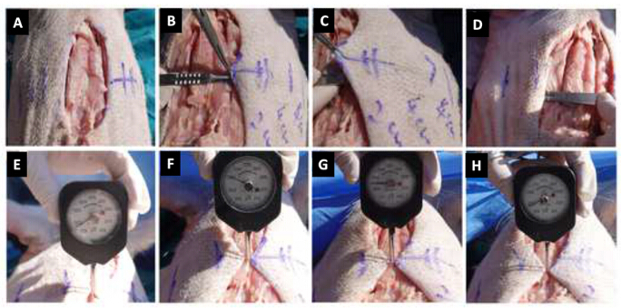
Figure 1: Surgical procedure and incision tension measurements at different stages of subcutaneous dissection. (A) Fusiform midline abdominal incision (10 cm × 4 cm). (B) Bilateral subcutaneous dissection extending 2 cm from the incision margin. (C) Bilateral subcutaneous dissection extending 6 cm from the incision margin. (D) Bilateral subcutaneous dissection extending 10 cm from the incision margin. (E) Measurement of incision tension at the midpoint of the incision before subcutaneous dissection. (F) Measurement of incision tension after 2 cm bilateral subcutaneous dissection. (G) Measurement of incision tension after 6 cm bilateral subcutaneous dissection. (H) Measurement of incision tension after 10 cm bilateral subcutaneous dissection. Please click here to view a larger version of this figure.
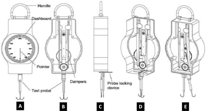
Figure 2: Tensiometer design and incision tension measurement procedure. (A) Front view of the tensiometer. (B) Rear view of the tensiometer. (C) Lateral view of the tensiometer. (D) Schematic illustration of the measurement setup. (E) Procedure for measuring incision tension: (1) Attach the test probes to the subcutaneous tissue adjacent to both sides of the incision. (2) Bring the two test probes together and secure them while ensuring that the dial is set to zero. Proper alignment and closure of the skin incision at this stage indicate correct placement of the tensiometer. (3) Release the probes; the skin tension causes them to separate, and the dial displays the incision tension. Please click here to view a larger version of this figure.

Figure 3: Exploratory regression analysis of the relationship between bilateral subcutaneous dissection distance and immediate pre-suture incision tension. The relationship between bilateral subcutaneous dissection distance (2–10 cm) and immediate pre-suture incision tension is shown. Blue circles and error bars represent the mean ± standard deviation (SD) for four pigs at each dissection distance. The red line represents the exploratory linear regression fitted to the group means. Please click here to view a larger version of this figure.
| Measurement Nodes (Both Sides) | Pig 1(g) | Pig 2 (g) | Pig 3 (g) | Pig 4 (g) |
| No Dissection | 160 | 166 | 175 | 181 |
| 2 cm Dissection | 308 | 314 | 323 | 329 |
| 6 cm Dissection | 250 | 256 | 265 | 271 |
| 10 cm Dissection | 171 | 177 | 186 | 192 |
Table 1: Individual incision tension measurements at each measurement point.
| Bilateral Dissection Range (cm) | Mean ± SD | SEM | 95% CI |
| 2 | 318.5 ± 9.3 | 4.65 | 303.7–333.3 |
| 6 | 260.5 ± 9.3 | 4.65 | 245.7–275.3 |
| 10 | 181.5 ± 9.3 | 4.65 | 166.7–196.3 |
Table 2: Descriptive statistics of incision tension following bilateral subcutaneous dissection (2–10 cm). Data are presented for four pigs (n = 4). Unit: g.
Supplementary File 1: Raw data supporting the findings of this study. Please click here to download this file.
Обсуждение
The principal finding is that subcutaneous dissection did not uniformly reduce immediate pre-suture tension in this porcine abdominal 4-cm-wide fusiform incision model. Limited 2 cm dissection produced an 86.8% increase relative to the no-dissection baseline, whereas extension to 6 cm and 10 cm produced progressive reductions. Even at 10 cm bilateral dissection, the measured pre-suture tension remained slightly above baseline. This finding describes local recoil before suturing and should not be interpreted as evidence that a no-dissection wound would have the lowest closure tension. Incision tension is an important modulator of wound healing and scar morphology1,2,10. Excessive mechanical loading can activate profibrotic signaling, including the TGF-β/SMAD pathway, and promote collagen deposition11,12. Subcutaneous dissection is frequently used to increase tissue mobility13,14, but no standardized dissection range has been established, and the assumption that dissection uniformly reduces local tension lacks direct quantitative support15,16. The present protocol, therefore, focuses on immediate pre-suture force at an open incision rather than on post-closure suture loading. The self-controlled porcine abdominal model offers a broad, relatively flat field for reproducible bilateral dissection, and porcine skin shares several structural and healing characteristics with human skin17,18,19. Nevertheless, porcine skin is not mechanically identical to human skin. Species-related differences in thickness, appendage distribution, subcutaneous attachment, anisotropy, and stiffness remain important; direct testing has shown substantial differences between porcine and human scalp tissue20. In addition, quadrupedal anatomy and the supine experimental position produce gravitational and abdominal-wall loading patterns that do not reproduce those of an upright human. These differences restrict direct clinical extrapolation.
Measurement accuracy and reproducibility depend on several critical steps: maintaining identical incision geometry; keeping the dissection plane immediately superficial to the fascia; creating symmetric left-right dissection ranges; placing both probes at the same midpoint and 0.5 cm from the incision edges; matching edges at the zero position, waiting for a stable pointer, and the same operator, body position, and anesthetic condition. The measured local recoil force will change if any of these steps change. The range of this protocol was 50–500 gf, and the resolution was 10 gf. The device previously demonstrated excellent measurement reliability, with an intraclass correlation coefficient (ICC) of 0.999, further supporting its repeated intraoperative use8. However, reliability in standard experimental conditions does not rule out operator dependence or the accuracy in different incision sizes and anatomical sites.
Common procedural and device issues can be dealt with systematically. Never continue measurement if the pointer fails to return to zero, check the mechanism for debris or deformation, and use another working device that has been previously sterilized. Remove and reposition both probes if the incision edges do not align at the zero position; do not force. If a probe slips or tears tissue, discard that reading and repeat at the same midpoint after secure repositioning. Irregular pockets, asymmetric dissection, active bleeding, or excessive thermal injury should be addressed and documented before the next node; measurements from a technically compromised node should not be pooled with valid readings. The principles of geometric and procedural standardization should be maintained in protocol modification. If the method has been adapted to another anatomical location or incision width, the geometry of the incision, insertion depth of the probe, location of the measurements, dissection intervals, and the order of the measurements should be predetermined and not directly taken from the present 4 cm wide porcine abdominal model. When the force expected is close to the maximum or minimum measurement range of the device, a tension meter with a more suitable measurement range should be used. Within each experiment, the dissection plane, body position, method of probe placement, stabilization time, and operator should be kept constant, regardless of the modification.
In contrast to the non-invasive elastic-wave mapping, which predominantly assesses the orientation of the skin strains rather than directly the force of recoil at an open incision5, and with ex vivo tensile testing, which requires excised tissue, and is incapable of serial intraoperative measurement20, the current handheld meter is able to record the lateral force applied directly at the edge of the cut. Skin-tensiometer methods have also been reported and used to detect excisional skin tension lines and to orient incisions6. The present protocol, however, is based on repeated quantitative measurements during sequential subcutaneous dissection. The device is compact, sterilizable, and free of imaging equipment or complex post-processing, which facilitates its use during surgery. It has the drawbacks of dependence on the operator for probe positioning, measurement of force at a single local point, potential tissue damage from the hooks, and inability to quantify suture forces after closure.
A possible biomechanical explanation is that intact skin and subcutaneous attachments maintain a prestressed, anisotropic equilibrium5,6,13,17,18. Limited separation alters the boundary conditions without releasing sufficient attachment to provide substantial mobility, whereas broader separation permits greater tissue excursion and redistribution of local forces. This interpretation is consistent with the non-linear response of the tissue, but it is a hypothesis, as direct measurements of tissue strain fields, attachment forces, and perfusion were not performed. The response may differ at other anatomical sites. Looser attachment in the face or neck may permit mobility after a smaller dissection, whereas tightly adherent regions, such as the back, may require a broader release. Regional differences in thickness, collagen orientation, subcutaneous architecture, and loading reinforce the need for site-specific validation rather than direct transfer of the present distance thresholds. Immediate pre-suture tension and post-closure suture tension are distinct outcomes. The present protocol does not measure the force borne by sutures after approximation and therefore cannot determine whether a particular dissection range produces a lower-tension closure. Increased mobility after dissection may allow load to be distributed across a broader tissue area, and multilayer tension-relieving closure techniques are designed to transfer load away from the superficial wound edge4,14. However, post-suture load dispersion was not tested in this study. Any connection between the measured pre-suture values and clinically relevant closure tension should therefore be regarded as a hypothesis for future paired pre- and post-closure measurements. From an experimental perspective, objective measurements can identify non-linear local responses that are not apparent from visual inspection alone and may help define candidate dissection ranges for further testing. The device may ultimately support tension-guided surgical planning, but clinical decisions should also consider perfusion, tissue quality, incision geometry, closure technique, and anatomical site. The present data do not justify a universal dissection threshold or direct recommendations for human surgery.
Several restrictions need to be highlighted. First, only four animals and a single 10 cm × 4 cm porcine abdominal incision configuration were evaluated. Second, outcomes were limited to immediate pre-suture tension at a single midpoint, while post-closure suture force, distributed strain, dynamic postoperative loading, wound healing, and scar outcome were not measured. Third, species differences, quadrupedal anatomy, and body-position-related abdominal loading limit transfer to humans. Fourth, skin-flap perfusion was not assessed. Fifth, only three distance levels were used in the exploratory regression; this high R2 does not support a general linear law. More distance nodes and pre- and post-closure measurements are required to establish the true response curve. Future studies should include larger sample sizes, multiple dissection intervals (such as 2 cm, 4 cm, 6 cm, 8 cm, and 10 cm), various incision sizes and locations, before-and-after suture force measurements, perfusion studies, and a longer follow-up period to assess healing and scar outcomes. Reproducibility and clinical relevance would be further verified by validation across breeds, ages, body weights, operators, and centers.
Раскрытие информации
All authors declared that there are no conflicts of interest.
Благодарности
Not applicable
Материалы
| Имя | Компания | Каталожный номер | Комментарии |
|---|---|---|---|
| Drapes/gloves | Shanghai Kebang Medical Latex Materials Co., Ltd. | / | |
| Forceps/scissors | Shanghai Medical Devices Co., Ltd. | JZ CrJ21070 | |
| male domestic pig, 45–60 kg | Dalian Medical University | / | |
| pentobarbital sodium | Mingwang Technology (Zhongshan) Co., Ltd. | 10-0005 | |
| povidone-iodine | Shandong Lierkang Medical Technology Co., Ltd. | / | |
| Prism 9.0 | GraphPad Software (RRID/ID: SCR_002798) | / | |
| propofol | Shanghai Jixing Biotechnology Co., Ltd. | W505102 | |
| Skin incision tension meter | Self-developed | / | |
| Skin-marking pen | Dongguan Tangde Medical Technology Co., Ltd. | TR-03 | |
| SPSS 26.0 | IBM Corp. (RRID/ID: SCR_002865) | / | |
| Sterile ruler | Dongguan Tangde Medical Technology Co., Ltd. | TR-03 | |
| Sterile scalpel | Shanghai Medical Devices Co., Ltd. | JZ CrJ11030 |
Ссылки
- Yang D, Yao L, Zhan Y, Zeng W. Application of remote buried dermal super-tension-reducing sutures for incisional scar prevention. Clin Cosmet Investig Dermatol. 2025;18:2749-56.
- Gong F, Wan B, Qi P, Wei Z. Biomechanical mechanism and clinical management progress of surgical wound tension. Front Surg. 2025;12:1-8.
- Fang X, et al. Hypertrophic scarring and keloids: epidemiology, molecular pathogenesis, and therapeutic interventions. MedComm (2020). 2025;6(10):1-30.
- Min P et al. Using Zhang's supertension-relieving suture technique with slowly absorbable barbed sutures in the management of pathological scars: a multicenter retrospective study. Burns Trauma. 2023;11:tkad021.
- Deroy C, Destrade M, McAlinden A, Ní Annaidh A. Non-invasive evaluation of skin tension lines with elastic waves. Skin Res Technol. 2017;23(3):326-335.
- Paul SP, Matulich J, Charlton N. A new skin tensiometer device: computational analyses to understand biodynamic excisional skin tension lines. Sci Rep. 2016;6:30117.
- Tønseth KA, Hokland BM. Evaluation of microcirculation and wound-closing tension after undermining the skin: a study in a porcine model using laser Doppler perfusion imaging. Eur J Plast Surg. 2004;27(6):295-297.
- Liu M, Chen H, Qu S, Qu W. A technique for the intraoperative assessment of surgical incision tension. Dermatol Surg. 2026;52(4):396-397.
- Louis-Maerten E, Geneviève LD, Hansell L, Blattner CE, Elger BS. Advancing the 3Rs? Researchers’ perspectives on institutional facilitation in Switzerland – Part 1: originative facilitation. Lab Anim. 2026. doi:10.1177/00236772261436225.
- Webster C, Neumayer L, Smout R, Horn S, Daley J, Henderson W, et al. Prognostic models of abdominal wound dehiscence after laparotomy. J Surg Res. 2003;109(2):130-137.
- Mahdavian Delavary B, van der Veer WM, Ferreira JA, Niessen FB. Formation of hypertrophic scars: evolution and susceptibility. J Plast Surg Hand Surg. 2012;46(2):95-101.
- Xu S, Dong W, Shi Y. LncRNA PICSAR binds to miR-485-5p and activates TGF-β1/Smad to promote abnormal proliferation of hypertrophic scar fibroblasts and excessive deposition of extracellular matrix. Med Mol Morphol. 2021;54(4):337-345.
- Feng F, et al. Biomechanical regulatory factors and therapeutic targets in keloid fibrosis. Front Pharmacol. 2022;9(13):906212.
- Ge M, et al. Progress in tension-relieving suturing surgery: revolutionary surgical techniques and patient prognosis evaluation methods. Front Surg. 2025;12:1587582.
- Goldman A, Wollina U, França K, Tchernev G, Lotti T. Chronic encapsulated seroma persisting for three years after abdominoplasty and a successful surgical solution. Open Access Maced J Med Sci. 2018;6(1):82-84.
- Lakatta AC, Steppe C, Teotia SS, Haddock NT. Reduction in seroma rate following deep inferior epigastric perforator flap with umbilectomy utilizing progressive tension sutures. J Reconstr Microsurg. 2024;40(2):118-122.
- Schick S, Leiderer M, Lanzl F, Graw M, Peldschus S. Maximum tensile stress and strain of skin of the domestic pig: differences concerning pigs from organic and non-organic farming. Int J Legal Med. 2020;134(4):1501-1510.
- Purnell CA, et al. Determining the differential effects of stretch and growth in tissue-expanded skin: combining isogeometric analysis and continuum mechanics in a porcine model. Dermatol Surg. 2018;44(1):48-52.
- Sullivan TP, Eaglstein WH, Davis SC, Mertz P. The pig as a model for human wound healing. Wound Repair Regen. 2001;9(2):66-76.
- Trotta A, Ní Annaidh A. Mechanical characterisation of human and porcine scalp tissue at dynamic strain rates. J Mech Behav Biomed Mater. 2019;100:103381.