Diagnosis of hereditary transthyretin (TTR) amyloidosis (ATTRv) relies on identifying TTR amyloid proteins in tissue biopsies. This study presents a sample preparation method for protein extraction from fixed skin tissues, suitable for the identification of TTR amyloid by Western blotting.
Методическая статья
Molecular Identification of Transthyretin Amyloid in Fixed Skin Biopsies: A Simplified Sample Preparation Method
В этой статье
Краткое содержание
Аннотация
Hereditary transthyretin (TTR) amyloidosis (ATTRv) is associated with peripheral neuropathy resulting from the accumulation of fibrillar TTR amyloid deposits in tissues including peripheral nerves and skin. Accurate diagnosis requires molecular identification of amyloid deposits in tissue specimens. Skin punch biopsy is a minimally invasive, well-established diagnostic tool used to confirm small-fiber neuropathy. Congo-red (CR) staining confirms amyloid deposition but does not determine amyloid type. Identification of TTR proteins within deposits commonly employs immunohistochemistry, which is limited by non-specific binding to blood-derived proteins and can lead to misdiagnosis. Liquid chromatography-tandem mass spectrometry (LC-MS/MS) of laser micro-dissected amyloid fibrils precisely identifies deposited proteins but requires specialized equipment available only in dedicated medical centers. This study describes a streamlined protocol for extracting insoluble amyloid proteins from Zamboni-fixed skin biopsies for subsequent Western blotting (WB) and LC-MS/MS. The protocol includes: (a) tissue collection by cryo-sectioning of biopsy samples; (b) removal of soluble background proteins by repeated phosphate buffer rinsing; (c) extraction of insoluble proteins with formic acid; (d) removal of the volatile solvent by vacuum evaporation and lyophilization. In a proof-of-concept cohort of 24 biopsies, 15-kDa and 30-kDa immunoreactive bands consistent with TTR monomer and dimer forms were detected in 10 of 12 CR-positive, genetically confirmed ATTRv samples and in none of 12 CR-negative neurological controls. This corresponded to an estimated sensitivity of 83.3% (95% confidence interval [CI], 55.2%–97.0%) and specificity of 100% (95% CI, 75.8%–100%) within this selected cohort. An in-gel LC-MS/MS analysis of excised 15 kDa and 30 kDa bands confirmed TTR identity in WB-positive samples tested. The findings were also in concordance with an LC-MS/MS analysis of micro-dissected CR-positive fibril deposits in three tested biopsies. This protocol expands the analytical use of routinely collected skin biopsies and provides an accessible proof-of-concept approach for TTR detection.
Введение
Amyloidosis refers to a group of clinically heterogeneous disorders characterized by the extracellular deposition of misfolded fibrillar proteins in various tissues and organs1. Over 40 amyloidogenic precursor proteins have been identified, each associated with distinct disease manifestations and therapeutic requirements2. Transthyretin (TTR)-related amyloidosis (ATTR) is the most common systemic form of the disease, manifesting as either wild-type ATTR (ATTRwt) or hereditary/variant ATTR (ATTRv). ATTRv may be caused by one of more than 130 pathogenic mutations in the TTR gene3. TTR circulates as a tetramer that can dissociate and then misfold, resulting in amyloid fibril formation, which is increased with aging and by the presence of TTR gene variants. Clinically, ATTRv amyloidosis presents with progressive sensory-motor peripheral neuropathy, autonomic dysfunction, and cardiomyopathy. The clinical presentation of ATTRv overlaps substantially with other acquired and hereditary neuropathies; therefore, clinical evaluation alone is insufficient for definitive diagnosis. Amyloidosis diagnostic workflows rely on Congo-red (CR) histochemical staining of tissue biopsies, in which typical apple-green birefringence under polarized light confirms amyloid deposition4. However, CR staining is limited to detecting the amyloid fibril structure and cannot identify the underlying precursor protein. Accurate amyloid typing is essential, particularly when more than one amyloid precursor protein is suspected to underlie the disorder, as occurs in patients with ATTR and paraproteinemia or chronic immunological disorders. In such cases, initiation of disease-modifying interventions, such as TTR stabilizers or gene-silencing therapies, requires molecular identification of TTR as the precursor protein.
Skin punch biopsy is a well-established, minimally invasive diagnostic tool widely used to quantify intraepidermal nerve fiber density, which, when low, supports the diagnosis of small-fiber neuropathy. Specimens collected for neuropathologic evaluation are routinely fixed in Zamboni’s fixative (paraformaldehyde and picric acid) or periodate-lysine-paraformaldehyde to preserve nerve fiber architecture. The fixed tissue is then cryo-protected and snap-frozen to allow preparation of thick sections for analysis5. Standard formalin fixation and paraffin preparation of thin sections is avoided.
Skin biopsies provide diagnostic access to cutaneous amyloid deposits6; however, conventional immunohistochemical techniques have low specificity7, due to cross-reactivity with blood-derived proteins, resulting in false-positive results8,9,10,11that may lead to misdiagnoses12. Advanced techniques, such as immunoelectron microscopy (IEM)13,14and liquid chromatography–tandem mass spectrometry (LC-MS/MS) of laser-microdissected amyloid fibrils15, offer precision and reliability in amyloid typing. Nevertheless, these gold-standard methodologies require specialized equipment, complex sample processing, and high technical expertise, restricting their availability and delaying timely diagnosis and clinical deployment.
The goal was to establish an accessible and reliable diagnostic method for identifying TTR amyloid in routinely collected Zamboni-fixed skin punch biopsies. Building upon previous studies utilizing organic solvents to extract amyloid proteins from formalin-fixed visceral organ biopsies (including gastrointestinal, renal, hepatic, and adipose tissues)16,17,18,19,20,21, a formic acid extraction procedure was adapted to recover amyloid proteins from Zamboni-fixed skin biopsies for their subsequent analysis via Western blotting (WB) and LC-MS/MS. The rationale of the developed protocol relies on the distinct solubility features of fibrillar amyloid proteins. While amyloid proteins remain insoluble during aqueous buffer22 washes that remove background proteins, they readily dissolve when exposed to formic acid.
The developed protocol comprises four core stages: (i) collection of tissue by cryo-sectioning of biopsy samples; (ii) removal of soluble tissue components by repeated aqueous buffer washes of the cryo-sections; (iii) extraction of non-soluble tissue components using formic acid; (iv) removal of formic acid via a centrifugal vacuum concentrator followed by lyophilization. The dried tissue extracts can subsequently be stored at −80 °C until further analysis.
The presented methodology differs significantly from previously reported amyloid extraction protocols that employ detergents and chaotropes, such as SDS, urea, and guanidine salts, which often require a dialysis step23,24,25, typically causing sample loss. In contrast, this proposed method utilizes a volatile organic solvent (formic acid), which can be readily removed by using a centrifugal vacuum concentrator and lyophilization without requiring dialysis. This method may be valuable and applicable for clinical neurology and pathology laboratories evaluating patients with suspected TTR amyloidosis presenting with peripheral neuropathy, for clinical and research assessments.
Протокол
The study was approved by the Sheba Medical Center Institutional Review Board (SMC-1351-24; October 10, 2024, Supplementary File 1), and all participants provided written informed consent. All procedures involving human tissue were performed in accordance with institutional guidelines and the principles of the Declaration of Helsinki. The schematic workflow for skin biopsy processing and molecular analysis is presented in Figure 1.
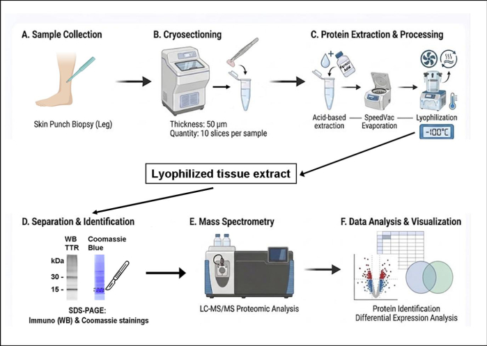
Figure 1: Schematic workflow of skin biopsy processing and molecular analysis. Key features include biopsy sample collection and cryo-sectioning, protein extraction using formic acid, centrifugal vacuum concentrator, and lyophilization. The obtained material can be analyzed by WB and LC-MS/MS. Abbreviations: SDS-PAGE, sodium dodecyl sulfate polyacrylamide gel electrophoresis; LC-MS/MS, liquid chromatography/tandem mass spectrometry; WB, Western blotting. Please click here to view a larger version of this figure.
1. Skin punch biopsy
- Perform skin biopsy (3 mm punch) from the distal leg, 10 cm proximal to the lateral malleolus, under local anesthesia by injecting 3 mL of 1% lidocaine.
NOTE: TTR amyloid deposits are commonly in the deep subcutaneous tissue; thus, a 3–5 mm-deep cylindrical sample should be obtained.
2. Fixation of biopsy specimens
- Prepare Zamboni’s fixative, including 3% paraformaldehyde (PFA) and 15% picric acid; in addition, prepare 15% (w/v) sucrose solution as follows:
- Zamboni’s fixative: Dilute 15 mL of 4% PFA to 20 mL total volume by adding 2 mL of 2 × phosphate-buffered solution (PBS) and 3 mL of picric acid (final PFA concentration: 3%). Store the fixative in a refrigerator at 4 °C for up to 1 month.
- Sucrose solution: Weigh 2.25 g sucrose and dissolve it in 15 mL of 1 × PBS; prepare the solution fresh.
NOTE: PFA is highly toxic and carcinogenic; it can cause severe chemical burns to the skin and eyes. Picric acid is unstable and highly explosive; it is a strong irritant and allergen affecting skin and eyes. Chronic exposure can cause significant damage to the kidneys and liver, potentially leading to renal failure. Preparation of Zamboni’s fixative must be performed in a chemical fume hood.
- Place the collected skin biopsy specimen into a 1.5 mL microcentrifuge tube filled with 1 mL Zamboni's fixative. Incubate for 30 min at room temperature.
- Remove the fixative by aspiration, then wash the fixed biopsy three times with 1 × PBS, 1.5 mL each time. Finally, remove PBS and add 15% sucrose solution for at least 2 h at 4 °C.
3. Tissue embedding and snap freezing
- Prepare embedding medium (10% Tragacanth): weigh 10 g tragacanth and mix it with 100 mL phenol/deionized water solution (1:100). Store at 4–8 °C.
NOTE: Phenol is a highly toxic, corrosive organic compound, particularly upon skin contact, eye contact, or inhalation. Prepare the phenol-containing embedding medium in a chemical fume hood. - Mount the specimen onto a small mound of tragacanth gum inside a specimen mold.
- Pre-cool a container of 2-methylbutane by partially immersing it in liquid nitrogen until properly chilled.
NOTE: 2-methylbutane is a highly flammable liquid and vapor. If swallowed, it can easily be aspirated into the lungs, potentially causing severe chemical pneumonitis, pulmonary edema, or death. All steps of the procedure involving the use of 2-methylbutane must be performed in a chemical fume hood. - Immerse the mold containing the embedded tissue in pre-cooled 2-methylbutane. Freeze for 30 s while stirring with a slow circular motion.
- Transfer the frozen block to a -80 °C freezer for storage until cryo-sectioning.
4. Cryo-sectioning
- Cut 50 µm thick tissue sections, collect 10 sections, and dispense them into a 2 mL microcentrifuge tube. Store at -80 °C until used.
5. Rinsing cryo-sections with PBS
- Place the tube on ice and add 1.5 mL of 1 × PBS.
- Vortex the microcentrifuge tube vigorously for 1–2 min to obtain a homogeneous mixture.
- Centrifuge the tube at 4 °C, 18,000 × g for 15 min using a pre-cooled centrifuge.
- Discard the supernatant using a 1 mL pipette; leave a small amount of PBS (about 20 µL) to ensure the pellet remains intact. Repeat the above-described rinsing and centrifugation procedures twice.
- For the final removal of the supernatant, use a 100 µL pipette with extended-length tips to carefully remove any residual fluid.
NOTE: The procedure can be stopped here; the pellet can be frozen and stored at -20 °C until the extraction step.
6. Amyloid extraction with formic acid
- Add 70 µL of concentrated formic acid (≥98%) to the pellet in the tube. Perform this step in a chemical hood.
NOTE: Formic acid is a flammable liquid. It is harmful if swallowed, toxic if inhaled, and causes severe skin burns and eye damage. - Vortex the microcentrifuge tube vigorously for 1–2 min to obtain a homogeneous mixture.
- Stir gently on a magnetic stirrer overnight at room temperature.
NOTE: Self-made 8 mm length stir bars that fit microcentrifuge tubes may be prepared by clipping 25 G stainless-steel needle shafts. Wear appropriate skin and eye protection during this preparation process. - Centrifuge the tube for 15 min at 18,000 × g, then collect the supernatant.
- Again, add 70 µL of formic acid to the pellet in the tube. Vortex and stir for an additional 2 h at room temperature.
- Centrifuge the mixture (as described in step 6.4), collect the supernatant obtained during this step, and combine it with the supernatant from the first extraction (step 6.4).
- Centrifuge the combined extract (supernatant derived from steps 6.4 and 6.6) to remove any residual insoluble material.
- Measure the total volume of the finally collected supernatant (using a 200 µL pipette).
- Divide the collected supernatant into three microcentrifuge tubes at the ratio of approximately 20%/40%/40% of the total volume. For a typical recovered volume of 125 µL, this corresponds to approximately 25 µL, 50 µL, and 50 µL, respectively.
NOTE: The tube containing 20% of the extracted material (25 µL) is sufficient for LC-MS/MS. The remaining material (40% each) is sufficient for WB analysis and experimental replicates if needed. All the supernatant collection steps must be performed in a chemical fume hood.
7. Centrifugal vacuum concentrator
- Remove formic acid by using a centrifugal vacuum concentrator for 6–12 h or overnight at 18 °C. Ensure that the tube caps remain open.
NOTE: The procedure may be stopped here; the dried material can be frozen and stored at -20 °C until the final lyophilization step.
8. Lyophilization
- Pierce holes in the caps of the microcentrifuge tubes containing the dried tissue extract.
- Add 50 µL of double-distilled water to each tube and close the caps.
- Freeze the tubes at -80 °C for at least 30 min before initiating the lyophilization process.
- Perform lyophilization at -100 °C for 8 h or overnight.
NOTE: Lyophilization is performed to ensure complete removal of residual formic acid.
9. Storage of extracted material
- Replace pierced tube caps with intact, airtight caps. Store the tubes at -20 °C for short-term storage or at -80 °C for long-term storage until the next WB or LC-MS/MS analysis is performed.
10. Downstream analysis
- Western blotting
- Determine the extracted protein concentrations using the Bradford assay26.
- Separate the extracted proteins by sodium dodecyl sulfate polyacrylamide gel electrophoresis (SDS-PAGE) under reducing conditions and immunodetect, as previously described27.
- Load 15–20 µg of protein per well onto a 4–20% Tris-glycine separation gel.
- Following electrophoresis, transfer the separated proteins to a nitrocellulose membrane.
- Probe the membrane with antibodies against TTR (dilution 1:1000), kappa (1:1500) and lambda (1:1000) light chains.
- Mass spectrometry
- For protein-in-gel mass spectrometry, separate the extracted proteins by SDS-PAGE as described above.
- Stain the gel with Coomassie blue and excise the protein band.
- Reduce the in-gel proteins and digest them with trypsin.
- Perform LC-MS/MS according to the previously described methodological details27.
- Analyze the obtained data using LC-MS/MS database search software against the human proteome of the UniProt database (See Table of Materials).
- Filter the peptide- and protein-level false discovery rates (FDRs) to 1% using the target-decoy strategy.
- Quantify the data by label-free analysis using the same software.
- Calculate the relative TTR abundance (TTR%) based on quantitative peptide intensity: divide the intensity of identified TTR peptide sequences by that of total peptide sequences.
- Statistical analysis
- Report the detection proportions with two-sided 95% confidence intervals calculated using the Wilson/Brown method.
NOTE: Because the cohort consisted of deliberately selected positive and negative specimens rather than a prevalence-based clinical population, predictive values were not calculated.
- Report the detection proportions with two-sided 95% confidence intervals calculated using the Wilson/Brown method.
Результаты
The protocol was applied to 24 Zamboni-fixed skin biopsy specimens. Twelve CR-positive biopsy samples were obtained from ATTRv patients, including 10 with the S77Y TTR variant and 2 with the V32A TTR variant. The control group included 12 CR-negative biopsies obtained from individuals undergoing neurological evaluation, who either lacked a family history of ATTRv amyloidosis or tested negative for a pathogenic TTR mutation.
Extracted samples were evaluated by WB. Anti-TTR-immunoreactive bands at approximately 15 kDa and 30 kDa were detected in 10 of 12 CR-positive ATTRv biopsies, consistent with monomeric and dimeric TTR forms, respectively. None of the 12 CR-negative control biopsies showed TTR immunoreactivity. Representative positive and negative immunoblots are presented in Figure 2. Bands at approximately 25 kDa reactive with anti-kappa and anti-lambda antibodies were observed in both groups and were interpreted as blood-derived immunoglobulin light chains. Within this selected cohort, the sensitivity was 83.3% (10/12; 95% CI, 55.2%–97.0%), and the specificity was 100% (12/12; 95% CI, 75.8%–100%).
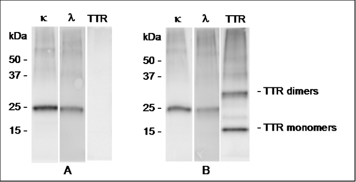
Figure 2: Representative Western blot results from the 24-biopsy cohort. (A) CR-negative skin biopsy from an individual with idiopathic neuropathy. (B) CR-positive biopsy from a patient with ATTRv-polyneuropathy. Skin biopsy extracts were separated by SDS-PAGE using 4–20% gradient polyacrylamide gels under reducing conditions and immunoblotted with antibodies against TTR and kappa and lambda light chains (anti-κ and anti-λ). The CR-negative specimen showed no anti-TTR immunoreactivity, whereas the ATTRv specimen showed anti-TTR-immunoreactive species at approximately 15 kDa and 30 kDa. Bands at approximately 25 kDa reactive with anti-kappa and anti-lambda antibodies were interpreted as blood-derived immunoglobulin light chains. Abbreviations: CR, Congo red; SDS-PAGE, sodium dodecyl sulfate polyacrylamide gel electrophoresis. Please click here to view a larger version of this figure.
Protein-in-gel LC-MS/MS was applied to analyze the excised 15 kDa (n = 5) and 30 kDa (n = 2) bands of WB-positive samples; both 15 kDa and 30 kDa bands showed the presence of TTR peptide sequences. The relative abundance of TTR peptides in the 15 kDa bands was quantified in a subset of biopsies comprising five WB-positive ATTRv samples and five WB-negative controls. TTR% was higher in ATTRv than in controls (p = 0.0001): the TTR% ranged from 0.3% to 17.9% in the WB-positive samples, whereas it was below 0.0001% in all WB-negative controls tested.
Three of the ATTRv biopsy samples were additionally subjected to amyloid typing via laser microdissection of CR-positive deposits, followed by LC-MS/MS analysis. TTR identification by this gold-standard method was in concordance with the WB-positive results and in-gel LC-MS/MS analysis in all three samples.
The possibility of identifying TTR mutations in the obtained mass spectra was also evaluated. Two of the five ATTRv patients tested demonstrated peptide sequences corresponding to the V32A TTR variant, while the S77Y TTR variant was identified in one patient. The obtained results were validated against TTR gene sequencing results. However, no variant-specific peptides were identified in 2 of 5 ATTRv patients tested; thus, MS analysis employing different enzymatic cleavage conditions should be tested to improve this methodological approach.
Supplementary File 1: Ethical approval documentation for the study. The document provides the institutional ethics committee approval for the study protocol, including the relevant approval details.Please click here to download this file.
Обсуждение
This protocol enables recovery of formic acid-soluble proteins from Zamboni-fixed skin punch biopsies for subsequent WB and proteomic analysis. Its principal practical advantages are the use of routinely collected skin biopsy material and minimal transfer of low-input tissue between vessels. Another advantage of the protocol is the easy removal of the extraction solvent by volatilization, avoiding dialysis, which is time-consuming and less suitable for minute amounts of sample material. In a proof-of-concept cohort, TTR immunoreactivity was detected in 10 of 12 CR-positive ATTRv biopsies and in none of 12 CR-negative controls. These findings support the feasibility of this method for assisting with the correct diagnosis of ATTRv, though they do not constitute full clinical validation.
Several steps are critical for reproducible recovery. Biopsy specimens should contain adequate deep dermis and subcutaneous tissue because amyloid deposits may be sparse and unevenly distributed. A consistent tissue input should be used, and sections selected for extraction should be adjacent to those assessed by CR staining whenever possible. After sequential extraction of amyloid proteins from tissue, formic acid should be thoroughly removed by vacuum evaporation and lyophilization. This critical step makes the extracted proteins suitable for long-term storage at -80 °C and prevents low-pH-related adverse effects on electrophoretic run and on tryptic digestion in downstream MS analysis.
TTR immunoreactivity was absent in 2 of 12 CR-positive samples in the cohort. This may result from different reasons, such as limited subcutaneous tissue, low or patchy amyloid burden, pellet loss during washing, or incomplete extraction. Thus, when a CR-positive specimen is WB-negative, the assay should be repeated using adjacent sections when sufficient tissue remains.
This study has important limitations. It was performed at a single center using a small, selected cohort; ATTRv patients mainly carried the S77Y variant, and the confidence intervals for sensitivity and specificity remain wide. The study evaluates the detection of TTR in CR-positive tissue relative to tissue without detectable amyloid and therefore does not establish clinical specificity for distinguishing ATTRv from other systemic amyloid types. Blinded interpretation and prospective consecutive enrollment will also be required for diagnostic validation. The two WB-negative ATTRv biopsies also indicate that a negative result does not rule out ATTR amyloidosis and that low-amyloid-burden specimens should be studied. LC-MS/MS was performed only in a subset of samples, and variant-specific identification was demonstrated for only two TTR variants; thus, additional variants should be studied. Formic acid may lead to formylation of amino acids, mainly lysine, serine, and threonine residues, which may affect sequence retrieval in database searches28, but this was not found to affect TTR identification in the patient samples. The effects of fixation duration, long-term storage, and tissue quantity may also require systematic evaluation. The sensitivity/limit of detection of the method for biopsies with low amyloid burden has yet to be determined.
Compared with immunohistochemistry, WB analysis enables the detection of amyloid deposits based on the characteristic electrophoretic profiles of TTR monomers and dimers and their specific immunoreactivity. This sample preparation is also suitable for proteomic confirmation of the obtained immunochemical data. Compared to laser-microdissection LC-MS/MS, WB requires less specialized tissue-isolation equipment but lacks spatial selection of histologically confirmed deposits.
In conclusion, the presented protocol, including formic acid extraction of proteins from Zamboni-fixed skin biopsies, is compatible with WB and LC-MS/MS analyses. It provides a practical approach for detecting TTR from routinely collected skin specimens. Larger prospective studies that include CR-positive non-TTR amyloidosis controls are needed to precisely establish the method's diagnostic performance and define its appropriate clinical role.
Раскрытие информации
The authors declare that they have no competing financial interests.
Благодарности
The authors thank Pfizer Global Medical Grants (program 65925007). The authors also thank Dr. Tamar Ziv for proteomic data analysis and Dr. Roman Kaplan for his help in the preparation of this manuscript. This work was performed in partial fulfillment of the requirements for a Ph.D. degree of Rivka Goldis, Gray Faculty of Medical & Health Sciences, Tel Aviv University, Tel Aviv, Israel.
Материалы
| Имя | Компания | Каталожный номер | Комментарии |
|---|---|---|---|
| 2-Methylbutane | Thermo Fisher Scientific | 126470010 | Used for rapid freezing of biopsy tissue before cryo-sectioning |
| 4–20% Tris-glycine gels | NuSep (Frenchs Forest, Australia) | NN10-420 | Used for SDS-PAGE separation of extracted proteins |
| Chemical hood | Haroshet | MDF | Used for handling hazardous/volatile chemicals during sample preparation |
| Cryostat | Leica | CM1860 | Used to prepare cryo-sections from fixed skin biopsy samples |
| Formic acid, ≥98% | Sigma-Aldrich | 33015-500 ml | Used for extraction of insoluble tissue components from washed cryo-sections |
| High-resolution Q Exactive plus mass spectrometer | Thermo Fisher Scientific | IQLAAEGAAPFALGMBDK | Used for LC-MS/MS-based identification and analysis of extracted proteins |
| Lyophilizer | Labconco | 4.5 L, -105°C | Used to dry tissue extracts following removal of formic acid; listed specification: 4.5 L, −105 °C |
| Magnetic stirrer | Velp Scientific | F20520262 | Used during preparation/mixing of solutions |
| Paraformaldehyde, 4% | Bio-Lab | 0006450323F | Component of the fixative solution used for skin biopsy fixation |
| Phenol | Sigma-Aldrich | P9346 | Used during preparation/processing of biopsy tissue as described in the protocol |
| Phosphate-buffered solution, 10 x PBS | Gibco | 14200-067 | Used to prepare PBS for washing tissue cryo-sections |
| Picric acid, 98% | Sigma-Aldrich | 197378-500G | Component of the fixative solution used for skin biopsy fixation |
| Polyclonal rabbit anti-human anti-TTR (anti-prealbumin) antibody | Dako (Supplier: Eldan Electronic Instruments) | A0002 (Dako); A0002 (Eldan) | Working dilution 1:Primary antibody used for TTR immunodetection; working dilution 1:1000 |
| Polyclonal rabbit anti-human kappa free light chain antibody | Dako (Supplier: Eldan Electronic Instruments) | A0192 (Dako); A0100 (Eldan) | Primary antibody used for kappa free light chain immunodetection; working dilution 1:1500 |
| Polyclonal rabbit anti-human lambda free light chain antibody | Dako (Supplier: Eldan Electronic Instruments) | A0194 (Dako); A0101 (Eldan) | Primary antibody used for lambda free light chain immunodetection; working dilution 1:1000 |
| Prism | GraphPad Software | 9.5.1 | Used for statistical analysis and/or graphical presentation of study data |
| Proteome Discoverer | Thermo Fisher Scientific | 2.4 | Used for processing and analysis of LC-MS/MS proteomic data |
| Refrigerated microcentrifuge (Eppendorf centrifuge) | Sigma Laborzentrifugen | 1-16K | Used for refrigerated centrifugation during tissue extraction/sample preparation |
| Speed Vac (centrifugal vacuum concentrator) | Labconco | 7310030 | Used for removal of formic acid from tissue extracts before lyophilization |
| Sucrose | Sigma-Aldrich | S7903-250G | Used during preparation/processing of fixed biopsy tissue |
| Tragacath | Sigma-Aldrich | G1128-100G | Used for embedding/support of biopsy tissue for cryo-sectioning |
Ссылки
- Gertz MA, Dispenzieri A. Systemic amyloidosis recognition, prognosis, and therapy: a systematic review. Journal of American Medical Association. 2020;324(1):79–89.
- Buxbaum JN et al. Amyloid nomenclature 2022: update, novel proteins, and recommendations by the International Society of Amyloidosis (ISA) Nomenclature Committee. Amyloid. 2022;29(4):213–219.
- Rowczenio DM et al. Online registry for mutations in hereditary amyloidosis including nomenclature recommendations. Human Mutation. 2014;35(9):E2403–E2412.
- Howie AJ, Brewer DB, Howell D, Jones AP. Physical basis of colors seen in Congo red-stained amyloid in polarized light. Laboratory Investigation. 2008;88(3):232–242.
- Lauria G et al. European Federation of Neurological Societies. EFNS guidelines on the use of skin biopsy in the diagnosis of peripheral neuropathy. European Journal of Neurology. 2005;12(10):747–758.
- Dori A et al. Ser77Tyr transthyretin amyloidosis in Israel: initial manifestations and diagnostic features. Annals of Clinical and Translational Neurology. 2023;10(4):553–567.
- Satoskar AA et al. Strong transthyretin immunostaining: potential pitfall in cardiac amyloid typing. American Journal of Surgical Pathology. 2011;35(11):1685–1690.
- Chee CE, Lacy MQ, Dogan A, Zeldenrust SR, Gertz MA. Pitfalls in the diagnosis of primary amyloidosis. Clinical Lymphoma Myeloma and Leukemia. 2010;10(3):177–180.
- De Larrea CF et al. A practical approach to the diagnosis of systemic amyloidoses. Blood. 2015;125(14):2239–2244.
- Leung N, Nasr SH, Sethi S. How I treat amyloidosis: the importance of accurate diagnosis and amyloid typing. Blood. 2012;120(16):3206–3213.
- Picken MM. Options for amyloid typing in renal pathology: The advantages of frozen section immunofluorescence and a summary of general recommendations regarding immunohistochemistry methods. In: Picken MM, Herrera GA, Dogan A, editors. Amyloid and Related Disorders: Surgical Pathology and Clinical Correlations. Cham: Springer International Publishing; 2015;283–293.
- Lachmann HJ et al. Misdiagnosis of hereditary amyloidosis as AL (primary) amyloidosis. New England Journal of Medicine. 2002;346(23):1786–1791.
- Palstrøm NB et al. Classification of amyloidosis by model-assisted mass spectrometry-based proteomics. International Journal of Molecular Sciences. 2021;23(1):319.
- Pinton S et al. Amyloid detection and typing yield of skin biopsy in systemic amyloidosis and polyneuropathy. Annals of Clinical and Translational Neurology. 2023;10(12):2347–2359.
- Vrana JA et al. Classification of amyloidosis by laser microdissection and mass spectrometry-based proteomic analysis in clinical biopsy specimens. Blood. 2009;114(24):4957–4959.
- Kaplan B, Yakar S, Kumar A, Pras M, Gallo G. Immunochemical characterization of amyloid in diagnostic biopsy tissues. Amyloid. 1997;4(2):80–86.
- Kaplan B et al. Microextraction and purification techniques applicable to the characterization of amyloid proteins in minute amounts of tissue. Methods in Enzymology. 1999;309:67–81.
- Kaplan B, Martin BM, Livneh A, Pras M, Gallo G. Biochemical subtyping of amyloid in formalin-fixed tissue confirms and supplements immunohistological data. American Journal of Clinical Pathology. 2004;121(6):794–800.
- Kaplan B et al. Primary local orbital amyloidosis: biochemical identification of the immunoglobulin light chain kappa III subtype in a small formalin-fixed, paraffin wax embedded tissue sample. Journal of Clinical Pathology. 2005;58(5):539–542.
- Kaplan B et al. Gastrointestinal β2-microglobulin amyloidosis in hemodialysis patients: biochemical analysis of amyloid proteins in small formalin-fixed paraffin-embedded tissue specimens. Modern Pathology. 2005;18(12):1610–1617.
- Goldis R et al. Diagnostic challenges and solutions in systemic amyloidosis. International Journal of Molecular Sciences. 2023;24(5):4655.
- Pras M, Schubert M, Zucker-Franklin D, Rimon A, Franklin EC. The characterization of soluble amyloid prepared in water. Journal of Clinical Investigation. 1968;47(4):924–933.
- Ihse E et al. Amyloid fibrils containing fragmented ATTR may be the standard fibril composition in ATTR amyloidosis. Amyloid. 2013;20(3):142–150.
- Kaplan B, Shtrasburg S, Pras M. Micropurification techniques in the analysis of amyloid proteins. Journal of Clinical Pathology. 2003;56(2):86–90.
- Murphy CL et al. Chemical typing of amyloid protein contained in formalin-fixed paraffin-embedded biopsy specimens. American Journal of Clinical Pathology. 2001;116(1):135–142.
- Bradford MM. A rapid and sensitive method for the quantitation of microgram quantities of protein utilizing the principle of protein-dye binding. Analytical Biochemistry. 1976;72:248–254.
- Kaplan B et al. Amyloid typing in cardiac amyloidosis using Western blotting. Israel Medical Association Journal. 2024;26(3):149–156.
- Zheng S, Doucette AA. Preventing N- and O-formylation of proteins when incubated in concentrated formic acid. Proteomics. 2016;16(7):1059–1068.
Перепечатки и разрешения
Эта статья была опубликована
Видео скоро будет доступно