This narrative review examines contemporary treatment of peri-implant diseases, integrating diagnostic assessment, risk modifiers, nonsurgical and surgical approaches, implant-surface decontamination, and supportive care. Recent evidence is interpreted according to study design, clinical endpoints, and its relevance to therapeutic decisions.
Обзорная статья
Current Therapeutic Approaches to Peri-Implant Diseases: A Narrative Review
В этой статье
Краткое содержание
Аннотация
Dental implants provide durable rehabilitation of partial and complete edentulism, but their retention does not exclude biological complications. Peri-implant mucositis is a biofilm-associated inflammatory condition confined to the soft tissues, whereas peri-implantitis involves inflammation and progressive supporting bone loss. This narrative review examines contemporary therapeutic approaches in the context of diagnosis, pathogenesis, prevalence, and patient-related and local risk modifiers. Literature identified through PubMed, Google Scholar, and Scopus between April and July 2026 was supplemented by targeted searches during revision in October 2026. Clinical guidelines, systematic reviews, and relevant clinical studies inform the synthesis. Mechanical biofilm control and correction of plaque-retentive conditions underpin treatment. Peri-implantitis management proceeds from nonsurgical care to surgical evaluation when inflammatory endpoints are not achieved, with procedure selection guided by defect morphology, implant position, restorative access, and treatment response. Recent trials suggest benefits for selected reconstructive and decontamination approaches, but radiographic gains do not consistently translate into disease resolution. Medication-associated implant failure is distinguished from evidence of peri-implantitis, and quantitative glycemic and smoking findings are interpreted cautiously. Prevention and supportive care are incorporated as components of sustained disease control. Longer comparative studies with standardized outcomes remain necessary to establish durable benefits and appropriate indications for adjunctive therapies.
Введение
Dental implants are widely used to restore oral function in patients with partial or complete edentulism. Long-term studies support their durability, but survival is distinct from freedom from biological or mechanical complications. A 20-year meta-analysis also highlighted the influence of incomplete follow-up on survival estimates, reinforcing the importance of continuing observation1.
The 2017 World Workshop classification distinguishes peri-implant mucositis from peri-implantitis by the presence of progressive supporting bone loss. Both involve biofilm-associated inflammation, but their consequences and therapeutic requirements differ2. Peri-implantitis can threaten implant retention and impose substantial treatment and maintenance burdens. Diagnosis and management therefore require assessment of inflammation, longitudinal tissue changes, and conditions that prevent effective plaque removal3.
This review focuses on current therapeutic approaches, including nonsurgical care, nonreconstructive and reconstructive surgery, implant-surface decontamination, and soft-tissue management. Diagnostic and risk evidence provides the context for treatment selection, while prevention and supportive care are discussed as components of sustained control. Particular attention is given to separating short-term parameter improvements from disease resolution and to distinguishing clinical evidence from preclinical findings4.
Обзор и перспектива
Literature Selection
Literature searches for this narrative review were conducted between April and July 2026 using PubMed, Google Scholar, and Scopus to identify relevant publications. Studies were selected for their relevance to diagnosis, etiopathogenesis, risk factors, prevention, and treatment of peri-implant diseases. The principal evidence comprises clinical practice guidelines, consensus reports, systematic reviews, and clinical studies; preclinical findings are identified separately. Targeted supplementary searches were conducted in October 2026 to address the reviewer comments. This narrative synthesis was not designed as an exhaustive systematic review.
Definition and Diagnosis of Peri-implant Diseases
Peri-implant health is characterized by the absence of clinical inflammation, bleeding or suppuration on gentle probing, and the absence of continuing bone loss. Health can exist with intact or reduced but stable bone support, and no universal probing-depth threshold defines it. Peri-implant mucositis presents with bleeding on probing, sometimes accompanied by swelling, erythema, or suppuration, without bone loss beyond initial remodeling. Increased probing depth may reflect inflammatory enlargement or reduced resistance to probe penetration. Peri-implantitis combines mucosal inflammation with progressive supporting bone loss2,5. When baseline records are available, peri-implantitis diagnosis integrates bleeding and/or suppuration, increased probing depth relative to previous measurements, and radiographic bone loss beyond initial remodeling. Without previous records, bleeding and/or suppuration, probing depth ≥6 mm, and a bone level ≥3 mm apical to the most coronal intraosseous implant portion support the diagnosis. These fallback thresholds should not delay recognition of earlier progression documented longitudinally. Table 1 compares the diagnostic features2. Baseline probing and radiographs after prosthetic rehabilitation provide essential reference points. Follow-up examination should record plaque, bleeding, suppuration, probing changes, recession, and restoration cleansability. Radiographic bone loss alone is insufficient: initial remodeling, malposition, surgical injury, fracture, or other complications must be considered. Retained cement and inaccessible restorative contours can contribute to secondary biofilm-associated inflammation. Serial clinical and radiographic findings should therefore be interpreted together, with particular attention to sites that change despite apparently acceptable plaque scores6,7. Probing depth also depends on tissue dimensions and implant position. A deep but stable site without inflammation is different from a site with increasing depth, bleeding, and new bone loss. Likewise, suppuration is not required in every peri-implantitis case. Consistent examination methods and comparable radiographic projections improve the usefulness of longitudinal records2,5.
Table 1: Diagnostic features of peri-implant health, peri-implant mucositis, and peri-implantitis. Findings should be interpreted using baseline records and evidence of inflammation; criteria for cases without baseline records are shown separately. Suppuration is not required in every peri-implantitis case, and radiographic bone loss alone is insufficient for diagnosis2,5. Please click here to download this file.
Etiopathogenesis and Microbiological Features
Biofilm accumulation at the implant–mucosa interface disrupts microbial–host equilibrium and sustains an inflammatory response. Susceptibility reflects microbial burden together with host regulation and local tissue conditions. Compared with periodontitis lesions, peri-implantitis lesions can contain larger inflammatory infiltrates and extend more directly toward supporting bone; peri-implant connective-tissue organization differs from that around teeth. This biological context helps explain destructive disease behavior without implying that every mucositis lesion progresses8. Mombelli and Décaillet9 described the complex biofilms associated with peri-implant disease, including organisms shared with periodontal infections. Sequencing subsequently demonstrated a pathogen-enriched, commensal-depleted community at diseased sites. Sanz-Martin et al.10 identified increased Porphyromonas and Treponema, alongside less traditional disease-associated taxa. These findings support community-level dysbiosis rather than attribution to a single pathogen. Host inflammatory mediators and proteolytic activity participate in tissue destruction, but microbial or biomarker profiles have not replaced clinical probing and radiography in routine diagnosis11. Figure 1 integrates clinical stages, biofilm, host response, and schematic bone-level changes; its progression arrow indicates a possible pathway rather than an inevitable sequence. Inflammation can itself modify the local ecological conditions that favor a dysbiotic community, creating a reciprocal relationship between host response and microbial composition. Thus, detecting a periodontal pathogen at an implant does not independently establish active peri-implantitis. The biological rationale for treatment remains disruption of the organized biofilm and reduction of the inflammatory burden, accompanied by correction of conditions that favor recolonization11.
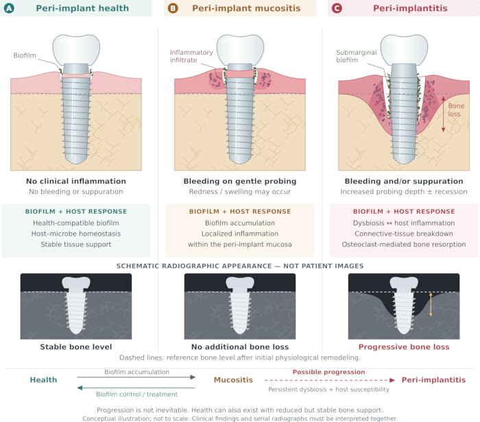
Figure 1. Conceptual model of biofilm–host interactions and tissue changes in peri-implant disease. (A) Peri-implant health, with host–microbe homeostasis and no clinical inflammation. (B) Peri-implant mucositis, with biofilm-associated inflammation confined to the mucosa and no additional supporting bone loss. (C) Peri-implantitis, with dysbiosis, inflammatory tissue breakdown, and osteoclast-mediated bone resorption. Grayscale insets are schematic radiographic representations, not patient images, contrasting stable bone levels with an example of progressive bone loss; clinical defect morphology varies. Dashed horizontal lines indicate the reference bone level after initial physiological remodeling. Lower arrows show mucositis development and potential resolution after biofilm control; the dashed arrow denotes possible, not inevitable, progression to peri-implantitis. Health may also occur with reduced but stable bone support. The illustration is conceptual and not to scale; clinical findings and serial radiographs should be interpreted together2,8,11. Please click here to view a larger version of this figure.
Prevalence and the Influence of Periodontitis History
Prevalence estimates vary with diagnostic definitions, populations, and observation periods. The 2025 AO/AAP systematic review by Galarraga-Vinueza et al. estimated patient-level prevalences of 46% for mucositis and 21% for peri-implantitis. A history of periodontitis was associated with greater odds of mucositis (odds ratio [OR] 1.68; 95% confidence interval [CI] 1.04–2.74) and peri-implantitis (OR 2.92; 95% CI 2.05–4.13). These observational associations identify susceptibility and do not establish causality12. Complementing this evidence, Annunziata et al. synthesized prospective cohorts and reported greater implant-level peri-implantitis risk in patients with a history of periodontitis (risk ratio [RR] 3.24; 95% CI 1.58–6.64), without a significant mucositis difference. The estimates should not be directly equated because study designs, analysis units, and effect measures differ. The evidence supports incorporating periodontal history into counseling and follow-up, while recognizing heterogeneity and uncertainty regarding mucositis13. A periodontal history also differs from active untreated periodontitis at the time of implant assessment. Combining these exposures can obscure whether a study concerns past susceptibility, current inflammatory burden, or both. Smoking, plaque control, and maintenance participation may further influence observed associations. Periodontal history should therefore trigger careful assessment and preventive planning rather than be interpreted as a prediction that an individual implant will develop disease12,13.
Patient-related and Local Risk Modifiers
Previous severe periodontitis, ineffective plaque control, and irregular supportive care are central considerations in risk assessment. Other exposures require distinctions between evidence for peri-implantitis, marginal bone loss, and implant failure. These outcomes are related but not interchangeable; an association with loss of an implant does not establish a medication-specific or metabolic cause of peri-implantitis8,14. Glycemic control provides clinically useful information beyond a diabetes diagnosis alone. In a 2024 systematic review of 35 studies, Bencze et al. reported 1.8 mm greater crestal bone loss in poorly controlled type 2 diabetes (HbA1c >8%) than in metabolically healthy comparators (95% CI 1.0–2.7). Prediabetes, defined as HbA1c 5.7%–6.4%, was associated with a 1.2 mm difference (95% CI 0.4–2.1). Study-level meta-regression associated each one-percentage-point higher HbA1c with approximately 0.24 mm additional bone loss. This is an observational association, not a prediction for an individual implant. The >8% grouping is not a universal implant eligibility threshold; current control, comorbidity, and healing capacity remain relevant15. Smoking exposure should be documented quantitatively. Afshari et al. found approximately 0.58 mm greater marginal bone loss among people smoking >10 cigarettes/day than nonsmokers (95% CI 0.19–0.96). Comparisons involving lighter smoking were less conclusive. These measurements concern marginal bone loss, not a directly estimated peri-implantitis incidence, and do not establish a safe exposure level. Cigarettes/day, duration, and cessation history are useful clinical descriptors; the available synthesis does not justify a universal pack-year cutoff16. Antiresorptive evidence varies by agent, dose, indication, and outcome. Jung et al.’s 2025 meta-analysis did not demonstrate a statistically significant increase in implant failure with bisphosphonates (OR 1.40; 95% CI 0.83–2.34); evidence for denosumab was insufficient for pooled analysis. A nonsignificant result should not be interpreted as proof of equivalence or absence of risk17. Findings dominated by osteoporosis regimens cannot be extrapolated to high-dose oncology treatment. Medication-related osteonecrosis of the jaw is a separate adverse outcome and should not be conflated with biofilm-associated peri-implantitis. Medication history and medical coordination are therefore important, while a consistent independent association with peri-implantitis remains unestablished14. For selective serotonin reuptake inhibitors, Shariff et al. pooled six retrospective studies and found higher implant-failure risk at patient level (RR 2.44; 95% CI 1.68–3.55) and implant level (RR 2.34; 95% CI 1.74–3.15). Confounding by depression, smoking, comedication, and other health characteristics limits causal interpretation. These results do not prove increased peri-implantitis incidence or support automatic discontinuation of antidepressant treatment18. Other proposed systemic or genetic modifiers have less consistent evidence. Interleukin-1 polymorphism findings vary across populations and study methods; they currently offer insufficient support for routine genetic risk testing19. Locally, retained cement and bulky restorative contours can obstruct plaque removal. A systematic review associated convex emergence profiles with peri-implantitis, but the overall marginal-bone-level difference between emergence angles >30° and ≤30° was not statistically significant. Implant connection and platform configuration influenced subgroup findings; 30° should therefore not be treated as an absolute biological threshold20. Limited keratinized mucosa and thin tissues may increase brushing discomfort, inflammation, or recession. Their management should address the patient’s functional difficulties rather than impose a universal width requirement. Exposed rough surfaces complicate cleaning, although surface roughness alone does not establish an independent disease cause7.
Treatment Planning and Management of Mucositis
Treatment begins by defining the diagnosis, implant restorability, and sources of persistent plaque retention. Selection of subsequent treatment depends on defect morphology, implant position, prosthetic access, patient priorities, and response to initial care. Correctable restorative obstacles should be addressed before escalating surgery. Table 2 presents this decision framework without assigning treatment solely to “initial” or “advanced” disease categories21,22. For mucositis, professional mechanical biofilm removal and effective home care are the principal interventions. Instructions should be demonstrated around the actual restoration, including suitable interdental access. Water-based oral irrigation can supplement mechanical cleaning where useful. Reassessment after 2–3 months identifies persistent inflammation and the need for further instrumentation or prosthetic correction. Routine professionally delivered local antibiotics, antiseptics, or similar adjuncts are not supported as replacements for this approach; time-limited patient-administered rinses are considered separately below3,23. The presence of suppuration or persistent extensive bleeding warrants reassessment of the diagnosis and local causes, including comparison with earlier bone levels. A transient improvement in gingival appearance should not be taken as evidence that an inaccessible restoration has become maintainable. Conversely, a single probing measurement should not determine treatment escalation without considering inflammation and longitudinal change5,6. Before treatment, discuss the likelihood of repeated visits, possible recession, limitations of reconstruction, and the continuing need for maintenance. The intended outcome should be explicit: controlling inflammation and arresting further loss may be achievable even when the original bone contour cannot be restored. Such counseling links technical treatment choices to function, esthetics, cost, and the patient’s willingness to undertake further intervention4,21.
Table 2: Treatment selection and supportive care for peri-implant diseases. Treatment selection incorporates response to initial therapy, defect morphology, implant position, restorative access, and maintainability. This table provides a clinical decision framework and is not a validated staging classification; probing measurements alone do not determine the surgical procedure. The European Federation of Periodontology (EFP) clinical endpoints for peri-implantitis treatment include probing depth ≤5 mm, no suppuration, and ≤1 bleeding point per implant, with bone stability assessed longitudinally3,4,21,22. Please click here to download this file.
Nonsurgical Treatment of Peri-implantitis
Initial nonsurgical care reduces biofilm and inflammation while establishing whether the patient and clinician can maintain the site. It includes supra- and submarginal instrumentation, correction of plaque-retentive factors, and treatment of concomitant periodontal disease. Thread geometry, exposed roughness, deep defects, and prosthetic contours limit access, explaining why complete resolution is less predictable than in mucositis. Early reassessment helps distinguish a maintainable residual condition from persistent active disease requiring direct surgical access4,24. Lee et al. randomized 48 patients to debridement with locally delivered 2% minocycline or placebo. At 12 weeks, adjunctive minocycline improved probing-depth and bleeding outcomes. The short follow-up does not establish durable bone stability or sustained disease resolution, and these findings should not be generalized to all local antimicrobial products25. Routine systemic antibiotics are not recommended for nonsurgical peri-implantitis treatment, given uncertain additional benefit and antibiotic stewardship considerations3. Persistent inflammatory pockets or continuing bone loss after initial care prompt surgical evaluation. This transition is a reassessment decision rather than a classification based only on baseline severity. The feasibility of plaque control, access for decontamination, remaining support, and correction of implant or prosthetic problems should be reviewed before selecting a surgical procedure4,22. Where a restoration restricts access, modification or temporary removal may be necessary to permit examination and cleaning. Repeated nonsurgical instrumentation without addressing that obstacle may reduce superficial deposits while leaving the relevant submarginal surface inaccessible. The response to initial therapy therefore provides information about both inflammatory control and the practical limits of access, rather than simply postponing surgery6,21.
Nonreconstructive Surgical Treatment
Access-flap procedures improve visualization, permit removal of inflamed tissue, and facilitate implant-surface cleaning. Resective approaches can reduce poorly maintainable pockets in predominantly horizontal or supracrestal defects. The expected gain in access must be balanced against recession, implant exposure, and esthetic consequences, especially in visible regions. Implant position and restorative contours can limit the benefit of an otherwise technically adequate procedure4,23. Implantoplasty may be considered for exposed supracrestal surfaces when smoothing is expected to improve cleansability. It removes implant material irreversibly, with potential particle release and weakening, particularly in narrow implants. These considerations favor selective application and careful control of instrumentation rather than treating implantoplasty as a mandatory surgical step. Mixed defects may require a combined approach, with resection or surface modification coronally and reconstruction of a suitable contained component4,24. A severely malpositioned implant or an uncorrectable restorative configuration may remain difficult to maintain after surgery. The decision to retain or remove such an implant should incorporate remaining support, structural integrity, expected function, and the feasibility of subsequent rehabilitation. Neither a pocket-depth label nor defect depth alone captures these prognostic considerations21,22.
Reconstructive Surgical Treatment
Reconstruction is most relevant to intrabony defects with favorable containment, particularly defects ≥3 mm deep. Remaining walls, implant position, the ability to stabilize graft material and achieve soft-tissue closure, and long-term cleansability influence suitability. Radiographic defect fill is an important outcome but cannot by itself demonstrate histological re-osseointegration or elimination of inflammation4,23. In a randomized trial involving 52 patients with three- or four-wall defects, Hakkers et al. compared access surgery with and without autogenous bone, xenograft, and a collagen membrane. Reconstruction improved radiographic bone levels and reduced mid-buccal recession at one year, but did not significantly improve probing depth, bleeding, suppuration, or composite disease resolution. These findings support a morphological benefit in selected defects rather than assured superiority across all clinical outcomes26. The distinction matters when discussing grafting: patients may value preservation of the soft-tissue contour, but this benefit should not be described as guaranteed infection control. Results from contained defects cannot automatically be transferred to broad, noncontained lesions or implants outside the bony envelope. Reporting defect configuration alongside clinical endpoints improves interpretation of reconstructive studies4,26. Parma-Benfenati et al.27 reported stable outcomes over ten years after regenerative treatment and maintenance in a single case. The long observation is informative but cannot establish comparative effectiveness. In six beagle dogs with 24 implants, Wang et al. found improved bone formation and bone-to-implant contact with bone morphogenetic protein-2 (BMP-2)-incorporated biomimetic calcium phosphate compared with bovine bone mineral or ungrafted controls. Combined mechanical, chlorhexidine, and photodynamic decontamination also improved early inflammatory parameters. These preclinical findings require controlled human studies and longer safety assessment before clinical translation28.
Implant Surface Decontamination
The objective of decontamination is adequate biofilm disruption with acceptable effects on implant and surrounding tissues. Mechanical, chemical, physical, and combined approaches differ in access, handling, and potential surface alteration. Comparative studies remain heterogeneous, and a favorable short-term parameter does not establish superiority for sustained disease control. Routine systemic antibiotics are not justified as surgical adjuncts3,4. Regidor et al.29 evaluated electrolytic cleaning added to titanium-brush decontamination during reconstructive surgery in 40 patients. At 12 months, composite resolution occurred in 40.0% versus 27.3% of implants, but the difference was nonsignificant; clinical, radiographic, and patient-reported outcomes did not show additional benefit. The numerical difference should therefore not be presented as established efficacy. Kotsakis et al.30 randomized 34 participants to abrasive titanium scalers or nonabrasive polymer microbrushes. Nonabrasive treatment produced 1.50 mm greater radiographic bone gain and 1.93 mm greater probing-depth reduction at 12 months; resolution was 62.5% versus 16.7%. However, the primary titanium-concentration comparison was nonsignificant. The small study supports further evaluation of tissue-preserving instrumentation but does not prove that titanium release caused the observed clinical differences or that all nonabrasive methods are superior. Laser-assisted and photodynamic approaches have also been investigated, but heterogeneous devices, energy settings, cointerventions, and short follow-up limit comparisons. A protocol that performs well in laboratory or animal conditions may behave differently on an implant with a complex clinical defect. Selection of an adjunct should therefore consider demonstrated patient benefit, adverse effects, and added treatment burden, not surface-cleaning performance alone4,24,28.
Soft-tissue Management
Soft-tissue augmentation may improve keratinized mucosal dimensions, tissue thickness, brushing comfort, or recession-related concerns. Free gingival grafts, connective-tissue grafts, and substitute materials address different clinical aims; soft-tissue gain should not be confused with reconstruction of lost implant-supporting bone4. Liu et al.’s systematic review of nine studies found greater keratinized mucosal width and thickness gains with free gingival grafts than collagen matrices, together with lower probing depths. Shrinkage and gingival-index outcomes were comparable. Collagen matrices may avoid a donor site, but their dimensional performance should be discussed when choosing a technique31. Evidence from augmentation studies does not establish an independent improvement in peri-implantitis resolution or implant survival. Augmentation is most defensible when a specific tissue deficiency interferes with comfortable cleaning or the planned surgical result, with the expected benefit weighed against morbidity4,7.
Evaluation of Treatment Outcomes
Outcome assessment should integrate inflammatory control, probing depth, bone stability, and the patient’s ability to maintain the restoration. The European Federation of Periodontology (EFP) guideline uses probing depth ≤5 mm, no suppuration, and no more than one bleeding point at implant level as clinical treatment endpoints, with absence of progressive bone loss assessed longitudinally. Complete absence of bleeding is desirable but is a stricter definition used in some studies, so reported resolution rates require attention to their underlying criteria3. A mean probing-depth reduction or radiographic gain can coexist with residual diseased sites. Clinicians should therefore document site-level findings and recurrence, rather than infer resolution from an average improvement. Patient-reported discomfort, recession, treatment burden, and prosthetic maintainability also affect the value of treatment. Stable reduced bone support may represent an acceptable outcome when inflammation and further destruction are controlled2,4.
Interpretation of the Therapeutic Evidence
The evidence base combines consensus recommendations, systematic reviews, small randomized trials, observational cohorts, case reports, and preclinical experiments. Their roles differ: a long-followed case illustrates feasibility, whereas a comparative trial can estimate an additional treatment effect. Even a statistically significant secondary outcome requires consideration of sample size, follow-up, and consistency with other endpoints. Conversely, a nonsignificant difference in a small study is not proof that treatments are equivalent. The studies discussed here also use different definitions of resolution and different analysis units. Patient-level and implant-level estimates should remain clearly labeled, especially when several implants belong to one patient. These limitations favor cautious interpretation of promising adjuncts and comparison of complete care pathways, including maintenance, rather than isolated technical procedures. This narrative review has no formal pooled analysis or systematic risk-of-bias assessment of its own.
Prevention and Supportive Peri-implant Care
Prevention begins with treatment planning. Retain teeth with a maintainable prognosis, control existing periodontitis, and discuss the continuing care required after implant restoration. Tooth preservation and implant replacement have different indications, and survival percentages alone should not decide between them32. Review medical history, glycemic control, smoking, hygiene capacity, and anticipated maintenance access before treatment. For patients with diabetes, coordinate assessment of current HbA1c and metabolic management with their medical clinician; a single laboratory value should not replace the broader assessment described above15. Daily plaque control requires a restoration that the patient can actually clean. Demonstration, suitable interdental aids, reinforcement at recall visits, and professional deposit removal should be paired with correction of retained cement or obstructive contours. Limited keratinized mucosa warrants attention when it causes pain during brushing or persistent hygiene difficulty; augmentation may then facilitate cleaning, although prevention of future peri-implantitis has not been conclusively demonstrated6,7. Time-limited chlorhexidine or other supported antiseptic rinses may be considered alongside professional treatment of mucositis. Their additional effects are inconsistent, and staining or other adverse effects can limit use. They should not become substitutes for mechanical cleaning or indefinite prophylaxis. Systemic antibiotics have no established routine role in long-term prevention or mucositis management; perioperative prophylaxis for a separate indication should not be confused with prevention of later peri-implantitis3. Immediate placement is not uniformly associated with more failure or bone loss. Patel et al.’s meta-analysis found comparable survival with immediate and delayed placement33. A separate review of randomized trials favored delayed placement for failure, but found no significant marginal-bone-loss difference34. These differences highlight the importance of case selection, socket anatomy, infection control, primary stability, and surgical and prosthetic protocols. Neither timing strategy should be characterized as intrinsically preventive across all sites. Similarly, subcrestal placement is not a general preventive measure. A meta-analysis of ten randomized trials found comparable marginal bone loss and survival with crestal and subcrestal placement35. Outcomes must be interpreted in relation to implant connection and platform design, abutment/restorative configuration, and vertical tissue dimensions. Placement depth should support an appropriate restorative and tissue environment rather than pursue a presumed universal bone-preservation advantage. Supportive visits should update plaque and inflammatory findings, probing measurements, prosthetic access, and relevant medical or smoking changes, with radiographs when clinically indicated. Recall frequency should reflect previous disease and current stability. Long-term observations by French et al. showed increasing peri-implantitis occurrence across later follow-up intervals, despite high overall implant survival; the cohort's maximum follow-up exceeded 22 years, but not every implant was observed that long36. This distinction reinforces that survival alone is insufficient and that monitoring remains necessary after apparent therapeutic success. Recurrent bleeding, suppuration, or increasing probing depth should lead to reassessment rather than automatic continuation of an unchanged recall program. Maintenance also offers an opportunity to reinforce smoking cessation and identify new barriers to self-care, including changes in dexterity or restoration access. A practical plan should make responsibility for follow-up clear and address attendance difficulties so that early changes can be recognized before further supporting tissue is lost6,21.
Выводы
Management of peri-implant diseases should connect accurate longitudinal diagnosis with biofilm control, correction of local obstacles, and reassessment of treatment response. Mucositis warrants prompt professional and patient-performed plaque control. Persistent peri-implantitis requires consideration of surgical access and defect-specific management, with implant position and prosthetic maintainability informing prognosis. Current reconstructive and decontamination studies show that improvement in bone levels or probing depth does not necessarily establish complete disease resolution. Supportive care remains necessary after treatment, including monitoring of periodontal status, glycemic control, smoking, and the ability to clean the restoration. Future trials should use consistent case definitions and composite endpoints, report patient- and implant-level outcomes clearly, and extend follow-up beyond early healing. Priorities include comparisons stratified by defect morphology and restorative configuration, reliable prediction of recurrence, and the long-term safety and clinical value of surface-preserving techniques and biologically based reconstruction. Medication studies need better control of indication-related and behavioral confounding. Research on keratinized mucosal augmentation should measure incident disease and patient comfort, not dimensional gain alone. Patient-reported outcomes, treatment burden, and cost should accompany clinical and radiographic measures. These advances would help identify which patients derive durable benefit from particular interventions and improve the balance between implant retention, function, and continuing care.
Раскрытие информации
Conflict of Interest:
The authors declare no conflicts of interest.
Figure Preparation and AI Assistance:
Figure 1 was newly constructed from custom vector shapes using Python-generated SVG and rendered with Inkscape. ChatGPT (OpenAI) assisted with layout, labels, and drawing code; no image-generation model was used. No published graphical material, patient photographs, or clinical radiographs were reproduced or adapted. ChatGPT also assisted with language editing, literature synthesis, and preparation of the revision materials.
Благодарности
The authors have no acknowledgments.
Ссылки
- Kupka JR, et al. How far can we go? A 20-year meta-analysis of dental implant survival rates. Clin Oral Investig. 2024;28(10):541.
- Berglundh T, et al. Peri-implant diseases and conditions: Consensus report of Workgroup 4 of the 2017 World Workshop on the Classification of Periodontal and Peri-Implant Diseases and Conditions. J Clin Periodontol. 2018;45(Suppl 20):S286-S291.
- Herrera D, et al. Prevention and treatment of peri-implant diseases—The EFP S3 level clinical practice guideline. J Clin Periodontol. 2023;50(Suppl 26):4-76.
- Ramanauskaite A, Schwarz F. Current concepts for the treatment of peri-implant disease. Int J Prosthodont. 2024;37(2):124-134.
- Hirooka H, Renvert S. Diagnosis of periimplant disease. Implant Dent. 2019;28(2):144-149.
- Renvert S, et al. Diagnosis and non-surgical treatment of peri-implant diseases and maintenance care of patients with dental implants: Consensus report of Working Group 3. Int Dent J. 2019;69(Suppl 2):12-17.
- Berglundh T, et al. Etiology, pathogenesis and treatment of peri-implantitis: A European perspective. Periodontol 2000. 2024. Online ahead of print. https://doi.org/10.1111/prd.12549
- Schwarz F, et al. Peri-implantitis. J Clin Periodontol. 2018;45(Suppl 20):S246-S266.
- Mombelli A, Décaillet F. The characteristics of biofilms in peri-implant disease. J Clin Periodontol. 2011;38(Suppl 11):203-213.
- Sanz-Martin I, et al. Exploring the microbiome of healthy and diseased peri-implant sites using Illumina sequencing. J Clin Periodontol. 2017;44(12):1274-1284.
- Belibasakis GN, Manoil D. Microbial community-driven etiopathogenesis of peri-implantitis. J Dent Res. 2021;100(1):21-28.
- Galarraga-Vinueza ME, et al. Prevalence, incidence, systemic, behavioral, and patient-related risk factors and indicators for peri-implant diseases: An AO/AAP systematic review and meta-analysis. J Periodontol. 2025;96(6):587-633.
- Annunziata M, et al. Effectiveness of implant therapy in patients with and without a history of periodontitis: A systematic review with meta-analysis of prospective cohort studies. J Periodontal Res. 2025;60(6):524-543.
- Al-Nawas B, et al. Group 3 ITI Consensus Report: Materials and antiresorptive drug-associated outcomes in implant dentistry. Clin Oral Implants Res. 2023;34(Suppl 26):169-176.
- Bencze B, et al. Prediabetes and poorly controlled type-2 diabetes as risk indicators for peri-implant diseases: A systematic review and meta-analysis. J Dent. 2024;146:105094.
- Afshari Z, Yaghini J, Naseri R. Levels of smoking and peri-implant marginal bone loss: A systematic review and meta-analysis. J Evid Based Dent Pract. 2022;22(3):101721.
- Jung J, et al. Effect of anti-resorptive therapy on implant failure: A systematic review and meta-analysis. J Periodontal Implant Sci. 2025;55(2):87-103.
- Shariff JA, et al. Selective serotonin reuptake inhibitors and dental implant failure: A systematic review and meta-analysis. J Oral Implantol. 2023;49(4):436-443.
- Santostasi N, et al. Relationship between interleukin 1 (IL-1) genetic polymorphism and periimplantitis: Systematic literature review and meta-analysis. Eur Rev Med Pharmacol Sci. 2024;28(10):3566-3582.
- Atieh MA, et al. Influence of implant restorative emergence angle and contour on peri-implant marginal bone loss: A systematic review and meta-analysis. Clin Implant Dent Relat Res. 2023;25(5):840-852.
- Polyzois I. Treatment planning for periimplant mucositis and periimplantitis. Implant Dent. 2019;28(2):150-154.
- Shiba T, et al. Novel flowchart guiding the non-surgical and surgical management of peri-implant complications: A narrative review. Bioengineering. 2024;11(2):118.
- Renvert S, Polyzois IN. Clinical approaches to treat peri-implant mucositis and peri-implantitis. Periodontol 2000. 2015;68(1):369-404.
- Rokaya D, et al. Peri-implantitis update: Risk indicators, diagnosis, and treatment. Eur J Dent. 2020;14(4):672-682.
- Lee JH, et al. Adjunctive use of locally delivered 2% minocycline in the nonsurgical treatment of peri-implantitis: A multicenter randomized placebo-controlled trial. J Dent. 2026;172:106761.
- Hakkers J, et al. Surgical reconstructive peri-implantitis treatment for 3- and 4-wall bone defects: A single-blind randomised controlled trial with a 1-year follow-up. J Clin Periodontol. 2026;53(6):933-946.
- Parma-Benfenati S, et al. Bone stability after regenerative peri-implantitis treatment: A 10-year follow-up case study. Clin Adv Periodontics. 2026;16(3):477-484.
- Wang T, et al. The efficacy of three combinatory decontamination protocols and BMP-2-incorporated BioCaP-based regenerative material in treating implants with peri-implantitis in vivo. Clin Oral Implants Res. 2026;37(7):821-834.
- Regidor E, et al. Adjunctive electrolytic implant surface decontamination in the reconstructive surgical treatment of peri-implantitis: A randomized clinical trial. Clin Oral Implants Res. 2026;37(9):1109-1117.
- Kotsakis GA, et al. Impact of abrasive versus non-abrasive cleaning on peri-implantitis titanium release: A randomized clinical trial. J Periodontol. 2026. Online ahead of print. https://doi.org/10.1002/jper.70158
- Liu Y, Qu J, Zheng J. Clinical efficacy of free gingival graft versus collagen matrix for peri-implant soft tissue augmentation: A systematic review and meta-analysis. Odontology. 2026;114(3):1161-1177.
- Sartoretto SC, et al. Comparing the long-term success rates of tooth preservation and dental implants: A critical review. J Funct Biomater. 2023;14(3):142.
- Patel R, et al. Differences in dental implant survival between immediate vs. delayed placement: A systematic review and meta-analysis. Dent J. 2023;11(9):218.
- Portal-Solera A, Pardal-Peláez B. Survival and marginal bone loss in immediate post-extraction implants versus delayed implants: A systematic review and meta-analysis. Oral. 2024;4(3):325-342.
- Cruz RS, et al. Clinical comparison between crestal and subcrestal dental implants: A systematic review and meta-analysis. J Prosthet Dent. 2022;127(3):408-417.
- French D, Ofec R, Levin L. Long-term clinical performance of 10,871 dental implants with up to 22 years of follow-up: A cohort study in 4,247 patients. Clin Implant Dent Relat Res. 2021;23(3):289-297.