Summary
We will show how to record flash responses from single mouse cones using a suction electrode.
Abstract
Rod and cone photoreceptors in the retina are responsible for light detection. In darkness, cyclic nucleotide-gated (CNG) channels in the outer segment are open and allow cations to flow steadily inwards across the membrane, depolarizing the cell. Light exposure triggers the closure of the CNG channels, blocks the inward cation current flow, and thus results in cell hyperpolarization. Based on the polarity of photoreceptors, a suction recording method was developed in 1970s that, unlike the classic patch-clamp technique, does not require penetrating the plasma membrane 1. Drawing the outer segment into a tightly-fitting glass pipette filled with extracellular solution allows recording the current changes in individual cells upon test-flash exposure. However, this well-established "outer-segment-in (OS-in)" suction recording is not suitable for mouse cone recordings, because of the low percentage of cones in the mouse retina (3%) and the difficulties in identifying the cone outer segments. Recently, an inner-segment-in (IS-in) recording configuration was developed to draw the inner segment/nuclear region of the photoreceptor into the recording pipette 2,3. In this video, we will show how to record from individual mouse cone photoresponses using single-cell suction electrode.Protocol
Making electrodes
- Make the recording electrodes using a micropipette puller and polish the electrodes with heating filament under microscope. For mouse cone single-cell suction recording, the inner diameter on the electrode tip is about 6-7 μm.
- Prepare reference electrodes. Weigh 1% agar in distilled water. Melt the agar solution in hot water bath. Fill the glass pipettes (L = 100 mm, OD/ID = 1/0.75 mm) with 1% agar using syringe. Solidify the agar at room temperature for 10 minutes.
- Cut the pipettes into halves using a diamond knife.
- Soak the reference electrodes in reference solution for at least 24 hours at 4 °C.
Setting up the experiment
- Dark adapt a mouse in a black cage overnight.
- Prepare 100 mL reference solution and 1 L perfusion solution (see below) and filter the solution.
- Dissolve 0.1% BSA and 10 mM Glucose in 20 mL reference solution.
- Calibrate the light intensity of the optical stimulator using photometer.
- Mount the recording chamber on the stage of the inverted microscope.
- Bubble the perfusion solution with 95% O2/5% CO2, incubate it in 40 °C water bath to increase the solubility of CO2. The solution flows from the bottle to the recording chamber passing through a ceramic resistor warming it to 36-38 °C. The heater should be located as close to the recording chamber as possible, ideally at the stage of the microscope.
- Adjust the flow regulator for a flow rate of about 1-1.5 mL/min.
- Fill the recording electrode with reference solution. Insert the electrode in an electrode holder. Make sure there are no bubbles in the electrode. Mount the holder on the headstage of the amplifier. Connect the holder to a suction device, which is built by connecting tubing partially filled with mineral oil to the side-port of the electrode holder. The other end of the tubing is connected to a small reservoir of mineral oil, to which pressure can be applied by mouth.
- Connect the reference electrode to the other pole of the headstage. Bind the thermal couple (TC) to the reference electrode. Make sure the electrode and TC tips are close together so that the temperature reading is taken as close to the cell as possible.
- Turn on the infrared illumination to visualize the electrode. The electrode is viewed by infrared camera connected to a monitor. Center the electrode image on the visual field and of the monitor using a micro-manipulator.
- Adjust the position of the reference electrode tip close (2-4 mm) to the recording electrode tip.
- Adjust the DC voltage for the heater so that the temperature of the solution is about 36-38 °C.
- Run a seal test to check the resistance of recording electrode, normally about 900 kΩ.
Isolating the mouse retina
- Under dim red light, euthanize the dark-adapted mouse with CO2 and cervical dislocation. Mark the eyeballs using the tip of a hot metal torch by cauterizing the most dorsal point of the sclera slightly. Enucleate the eyeballs using scissors. All subsequent procedures are performed under infrared light.
- Hemisect the eyeballs under microscope with infrared illumination and image converters.
- Remove the cornea and lens. Remove as much of the vitreous as possible.
- To record M-cone responses, remove the ventral part of the eyecup 4 with a razor blade judging from the cautery mark.
- Peel the retina from the pigment epithelium layer.
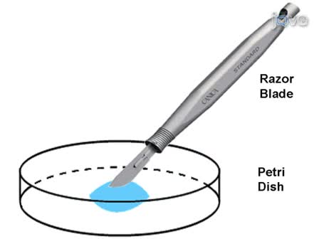
- Flatten the retina and place it on the bottom of the Petri dish filled with 1-2 mL reference solution.
- Slice the retina into small thin pieces using a razor blade and incubate the preparation in a light-tight box saturated with pure oxygen.
Recordings
- Turn off the heater temporarily. Switch the perfusion flow to a bypass tubing to prevent the chamber from drying.
- Drain most of the solution in the chamber using a piece of Kimwipe tissue. Transfer the retina suspension to the recording chamber using a glass pipette.
- Wait for about 2 minutes until the retina slices settle down. Switch the perfusion back on. Turn on the heater.
- Turn on infrared illumination to visualize the preparation. Search for a piece of retina lying on the chamber with photoreceptor outer segments sticking out (see example figure). Draw inner segments gently into the electrode by suction. In this configuration, several nuclei and inner segments should be drawn into the electrode. Record the photoresponse to a bright flash to test whether a cone inner segment is in the electrode and whether the cell is healthy, judging by the response kinetics and amplitude.
- Lower the mineral reservoir to apply slight negative pressure to the tip of the electrode to help hold the inner segment in the electrode.
- Record test-flash responses from dim to bright intensity once a good cell is found. Test flashes are provided from a calibrated optical stimulator 5. Flash intensity and wavelength are controlled by a set of neutral density filters and narrow band interference filters, respectively. Test flash duration (20 ms) is controlled by computer-driven shutters.
Solutions
- Mouse perfusion solution: 112.5 mM NaCl, 3.6 mM KCl, 2.4 mM MgCl2, 1.2 mM CaCl2, 10 mM HEPES (pH 7.4), 20 mM NaHCO3, 3 mM Na succinate, 0.5 mM Na glutamate, 0.02 mM EDTA, and 10 mM glucose.
- Mouse recording electrode solution: 140 mM NaCl, 3.6 mM KCl, 2.4 mM MgCl2, 1.2 mM CaCl2, 3 mM HEPES (pH 7.4), 0.02 mM EDTA.
Subscription Required. Please recommend JoVE to your librarian.
Discussion
Single-cell suction recording from photoreceptor cells was developed 3 decades ago. It enables us to record trans-membrane current change induced by light stimulation without penetrating the cell membrane. Because of high cell-cell adhesion, it is difficult to isolate healthy single rod and cone from mouse retina like amphibian retina and it is hard to find individual cones due to the low percentage (3%) and small size. The inner-segment-in (IS-in) recording method overcomes this difficulty by recording current from the inner segments of several photoreceptors, among which a cone might be included. In wild type mouse, the response should be the combination from rods and cones. A steady background light can be used to suppress rod responses. In this video, we used Tα-/- retina, in which rods can not respond to light stimulation, as they lack the α-subunit of the G-protein transducin 6. Therefore, the photoresponse is only from cones.
Subscription Required. Please recommend JoVE to your librarian.
Acknowledgments
Supported by Career Development Award from Research to Prevent Blindness, NIH grant EY 019312, and unrestricted grant from Research to Prevent Blindness and EY 02687 (Department of Ophthalmology & Visual Sciences at Washington University).
References
- Yau, K. W., Lamb, T. D., Baylor, D. A. Light-induced fluctuations in membrane current of single toad rod outer segments. Nature. 269, 78-80 (1977).
- Nikonov, S. S. Photoreceptors of Nrl -/- mice coexpress functional S- and M-cone opsins having distinct inactivation mechanisms. J Gen Physiol. 125, 287-304 (2005).
- Nikonov, S. S., Kholodenko, R., Lem, J., Pugh, E. N. JR Physiological features of the S- and M-cone photoreceptors of wild-type mice from single-cell recordings. J Gen Physiol. 127, 359-374 (2006).
- Applebury, M. L. The murine cone photoreceptor: a single cone type expresses both S and M opsins with retinal spatial patterning. Neuron. 27, 513-523 (2000).
- Cornwall, M. C., Fein, A., MacNichol, E. F. JR Cellular mechanisms that underlie bleaching and background adaptation. J Gen Physiol. 96, 345-372 (1990).
- Calvert, P. D. Phototransduction in transgenic mice after targeted deletion of the rod transducin alpha -subunit. Proc Natl Acad Sci U S A. 97, 13913-13918 (2000).