A subscription to JoVE is required to view this content. Sign in or start your free trial.

Method Article

Periodic Acid Schiff or PAS Staining: A Method to Assess the Structural Deformities of Glomerulus

7.3K views

April 30th, 2023

In This Article

Abstract

Source: Stevens, M., Oltean, S. Assessment of Kidney Function in Mouse Models of Glomerular Disease. J. Vis. Exp. (2018)

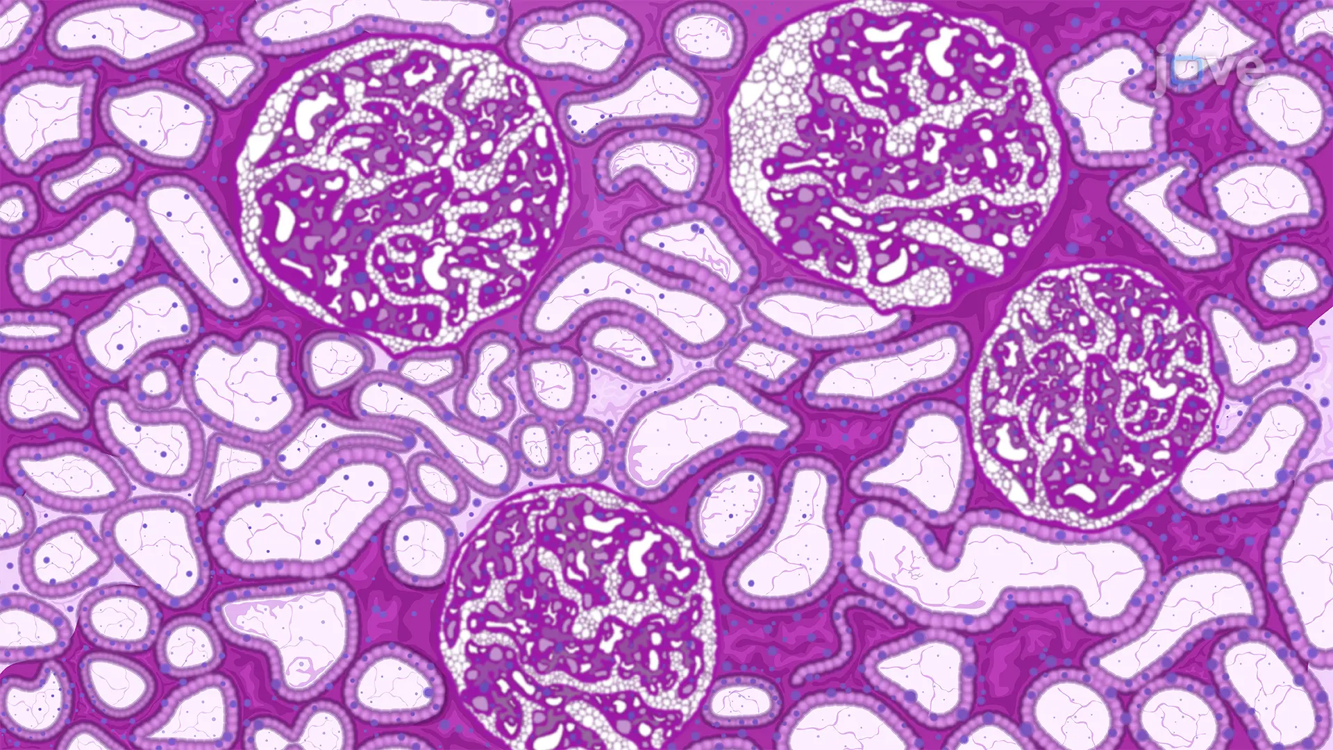
In this video, we describe the protocol for performing Periodic Acid Schiff (PAS) stain to assess the structural anomalies in the kidney glomerulus. The PAS staining method enables appropriate evaluation of the glomerulus pathophysiology.
Protocol

1. Periodic Acid Schiff (PAS) Stain

NOTE: The PAS stain will highlight the basement membranes of glomerular capillary loops and the tubular epithelium. It enables detailed visualization of the glomerular cells, mesangial matrix and potential expansion, and potential changes of the GBM (i.e., thickening and irregularities).

  1. Section the paraffin-embedded, PFA-fixed kidney cortex using a microtome at 5 µm thickness onto poly-L-lysine coated slides. Dry at 37 °C for 1 h. Ensure the section does not contain any folds or holes, which can distort the morphology under the microscope.

Access restricted. Please log in or start a trial to view this content.

Disclosures

No conflicts of interest declared.

Materials

List of materials used in this article
NameCompanyCatalog NumberComments
Periodic Acid-Schiff (PAS) Staining System Sigma-Aldrich 395B-1KT
Hematoxylin Sigma-Aldrich H3136
Xylene MerckMillipore 108298
Poly-Prep Slides Sigma-Aldrich P0425-72EA
Mounting Medium ThermoFisher Scientific 8030
Hydrophobic Pen Abcam ab2601
Fluorescent Light Miscroscope Leica Microsystems
Image J Analysis Software Image J

Tags

Periodic Acid Schiff StainGlomerulus AssessmentKidney Cortex SectionTissue DeparaffinizationAlcohol RehydrationPeriodic Acid TreatmentSchiff Reagent IncubationIron Hematoxylin CounterstainLight Microscopy Examination