A subscription to JoVE is required to view this content. Sign in or start your free trial.

Method Article

Multiplexed Immunofluorescence Imaging to Analyze Immunotherapy-Resistant T Cell Subpopulations

1.1K views

July 8th, 2025

In This Article

Abstract

Source: Granier, C., et al. Multiplexed Immunofluorescence Analysis and Quantification of Intratumoral PD-1+ Tim-3+ CD8+ T Cells. J. Vis. Exp. (2018).

This video demonstrates an in-situ fluorescence multispectral imaging technique to analyze different subpopulations of infiltrating T cells in tumor tissue. A renal cell carcinoma tissue cryosection is treated with antibodies to label CD8+ T cells that express specific inhibitory receptors and exhibit an immunotherapy-resistant phenotype. The labeled section is visualized using a multispectral microscope to identify the distinct CD8+ T cell subpopulations.
Protocol

All procedures involving sample collection have been performed in accordance with the institute's IRB guidelines

1. In Situ Immunofluorescence Staining of tumor-infiltrating lymphocytes (TILs)

  1. Procedural guidelines
    1. Perform all experimental steps at room temperature.
    2. For all steps, perform the dilutions with Tris Buffer Saline (TBS; see Table of Materials).
    3. Perform the wash in TBS for all the steps except after the primary antibody incubation. For the latter, use Tris Buffer Saline Tween20 (TBST, see Table of Materials). For buff....

Access restricted. Please log in or start a trial to view this content.

Results

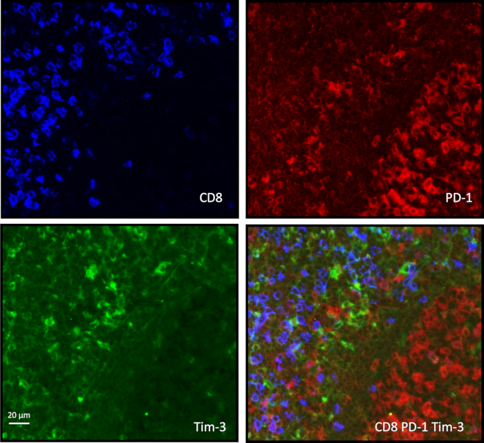
Immunofluorescence markers CD8, PD-1, Tim-3 in tissue sections, microscopy analysis, overlay comparison.
Figure 1: CD8-PD-1-Tim-3 immunostaining on a tonsil paraffin-embedded tissue. Paraffin-embedded tissue sections instead of cryopreserved sections were selected for triple immunostaining. After dewaxing and rehydration, the same protocol of staining .......

Access restricted. Please log in or start a trial to view this content.

Disclosures

No conflicts of interest declared.

Materials

List of materials used in this article
NameCompanyCatalog NumberComments
Vectra 3 Automated Quantitative Pathology ImagingPerkin ElmerCLS142338
inForm cell analysis 2.1.Perkin ElmerCLS135781
R softwarehttps://www.r-project.org
Dakopen delimiting penDakoS2002
Tris Buffer Saline TBS TabletsTakara, Bio Inc.TAKT9141ZpH7.6 100 tablet
Tris Buffer Saline Tween 20 TBS(+Tween20)Takara, Bio Inc.TAKT9142ZpH 7.6 100 tablets
Biotin blocking systemDakoX0590Avidin 0.1% and Biotin 0.01%
Normal donkey serumJackson Immunoresearch017-000-0015% vol./vol. concentration
Fluoroshield with DAPISigma-aldrichF60571.5 µg/mL concentration
Knittel glass coverslipKnittel Gläser,10003924x60 mm 100 cover slips
Rabbit anti-CD8 Clone P17-VnovusNBP1-79055use at 4µg/mL
Mouse anti-PD-1 Clone NATabcamab52587use at 2 µg/mL
Goat anti-Tim-3R&DAF2365use at 3 µg/mL
Rabbit anti-PD-L1 Clone SP142Roche7309457001use at 1 µg/mL
Mouse AF647 labeled pan- Keratin Clone C11Cell Signalling4528use at 0.5 µg/mL
Goat anti-human gal9R&DAF2045use at 0.3 µg/mL
Cyan 5 conjugated donkey anti-rabbitJackson Immunoresearch711-175-152use at 5 µg/mL
Biotinylated F(ab'2) donkey anti-mouse IgGJackson Immunoresearch715-066-150use at 3 µg/mL
Alexa Fluor488 conjugated donkey anti-goat IgGabcamab150133use at 5 µg/mL
Cy3 labeled streptavidinAmershamPA43001use at 3 µg/mL
Negative control mouse IgG1DakoX0931use at 2 µg/mL
IgG from goat serumSigma-aldrichI5256use at 3 µg/mL
IgG from rabbit serumSigma-aldrichI5006use at 4µg/mL

Tags

Immunotherapy Resistant T CellsCD8 T CellsTumor MicroenvironmentMultispectral ImagingPD 1 ExpressionTim 3 ExpressionRenal Cell CarcinomaAntibody LabelingFluorescence Microscopy