All procedures involving sample collection have been performed in accordance with the institute's IRB guidelines.
1. Target Microsphere preparation
- Select a relevant target antigen.
NOTE: In this example, the recombinant protein V1V2-2F5K was used, which was designed to mimic the trimeric apex of native HIV envelope.
- Perform biotinylation using a commercial kit (Table of Materials) according to the manufacturer's protocol.
- Calculate mmol of the biotin reagent to add to the reaction for a 5-fold molar excess using the formula: mmol biotin = mL protein x (mg protein/mL protein) x (mmol biotin/mg protein) x (5 mmol biotin/mmol protein). Then calculate µL of the biotin reagent to add using the formula: µL biotin = mmol biotin x (1,000,000 µL/L) x (L/10 mmol).
- Equilibrate biotin to room temperature before opening. Dissolve protein in 0.5−2.0 mL of phosphate-buffered saline (PBS) according to the calculation made above.
- Prepare a 10 mM solution of biotin reagent in dimethylsulfoxide (DMSO) and add the appropriate volume of 10 mM biotin reagent to the protein solution, and incubate reaction on ice for 2 h or at room temperature for 30 min.
- Remove excess biotin using protein concentrators (polyethersulfone [PES] membranes, 3 kDa molecular weight cut-off [MWCO], 0.5 mL; Table of Materials) according to the manufacturer's instructions.
- Deposit sample into the upper chamber of spin column and add PBS up to 400 µL. Cap, then insert this sample chamber into a collection tube. Centrifuge at 12,000 x g at room temperature for 30 min.
- Discard flow-through and add PBS to 400 µL. Repeat centrifugation. Discard flow-through and add PBS to 100 µL. Measure protein concentration by a spectrophotometer.
NOTE: Protein can be aliquoted and frozen at -80 °C until used.
2. Antibody-Dependent Cellular Phagocytosis (ADCP) Assay Plate Preparation
- Conjugate biotinylated protein to 1 µm microspheres (‘beads'; Table of Materials) according to the manufacturer's instructions.
- Per plate of conjugated beads, incubate 6 µg of protein with 12 µL of stock beads in 200 µL of 0.1% bovine serum albumin (BSA)-PBS at room temperature for 1 h, vortexing gently every 20 min.
- Centrifuge at 13,000 x g for 5 min. Discard supernatant, vortex gently and resuspend in 1200 µL of 0.1% BSA-PBS. Repeat spin and wash step 2x. Resuspend in 1200 µL of 0.1% BSA-PBS.
- Aliquot 10 µL of bead solution per well in a round bottom 96-well plate. Prepare dilutions of antibody or immune sera of interest in 12 µL of 2% HSA HBSS, typically starting at 50 µg/mL of antibody or a 1/100 serum dilution.
NOTE: In the sample data, monoclonal antibody (mAb) 830A was used.
- Add 10 µL of titrated antibody/sera to the bead plate and incubate for 2 h at 37 °C. Add 200 µL of 2% HSA HBSS to each well and centrifuge plate at 2,000 x g for 10 min.
- Carefully remove supernatant by a rapid overturning and decanting of liquid from the plate wells into a sink to avoid disturbing the invisible bead pellet.
3. Breast milk cell isolation
- Obtain human breast milk from healthy lactating women, expressed using double electronic or manual pumps. Isolate cells within ~4 h of expression, keeping milk at room temperature.
- Using 50 mL tubes, centrifuge 50 mL milk at 800 x g for 15 min. Carefully pour off the skim milk and fat while leaving the cell pellet undisturbed. Wipe the inside of the tube with a lint-free wipe to remove all fat from the tube wall.
- Add 10 mL of 2% human serum albumen in Hank's balanced salt solution (2% HSA HBSS [without Ca2+ or Mg+]). Resuspend the pellet by gentle pipetting to avoid cell activation and apoptosis. Transfer to a 15 mL tube and centrifuge at 450 x g for 10 min.
- Pour off supernatant and repeat step 1.3. Then, gently resuspend cells in 1−2 mL of 2% HSA HBSS depending on expected cell number, and count cells by a hemocytometer or an automated cell counter, noting also the cell viability.
4. ADCP assay
NOTE: Methods described here are adapted from Ackerman et al.
- Add 50,000 freshly isolated BM cells to each ADCP assay plate well in 200 µL of 2% HSA-HBSS. Incubate for 2 h at 37 °C.
- For control experiments, pre-incubate cells at 37 °C with 10 µg/mL actin inhibitor (cytochalasin-D), 50 µg/mL Fc receptor blocking agent (FcBlock), or a combination of both prior to their addition to the plates.
- Centrifuge plate at 930 x g for 10 min. Add 200 µL of 2% HSA HBSS and repeat centrifugation. Carefully remove supernatant as in step 2.7 and repeat wash.
- Carefully remove supernatant and stain cells for viability using 0.5 µg/mL (final concentration) fixable viability stain 450 per well in 50 µL of 2% HSA HBSS. Incubate 20 min at room temperature in the dark. Centrifuge plate at 930 x g for 10 min and remove supernatant as in step 2.7. Add 200 µL of 2% HSA HBSS and centrifuge plate again followed by removal of supernatant as in step 2.7.
- After viability staining, stain cells for leukocyte markers of interest, minimally including a CD45-specific stain such as PE-mouse anti-human CD45 (clone HI30) at an optimized concentration (1 µg/µL in 50 µL of 1% BSA HBSS in the example data).
NOTE: Any lineage-specific markers of interest can be included.
- Incubate 20 min at room temperature in the dark. Centrifuge plate at 930 x g for 10 min and remove supernatant as in step 2.7. Add 200 µL of 1% BSA HBSS and repeat centrifugation. Remove supernatant. Fix cells in 200 µL of 0.5% formaldehyde in the dark at room temperature for 30 min or overnight at 4 °C. Refrigerate in the dark until analysis.
5. Analysis by flow cytometry
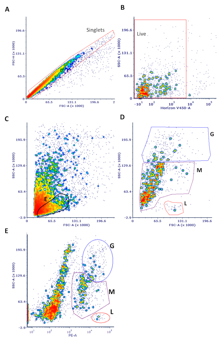
- Perform initial gating to eliminate doublets on a forward scatter (FSC) vs. side scatter (SSC) plot and debris (material smaller than FSC = 5000) (see Figure 1). Use an SSC vs. viability stain (V450 in this case) plot to eliminate dead cells (those that are positive for viability stain).
- Use an SSC vs. CD45 plot to differentiate the major leukocyte classes (granulocytes, monocytes, lymphocytes) as extensively described.
NOTE: This classification is only suggestive and lineage-specific markers are needed to confirm cell type.
- For all CD45+ cells, or for each leukocyte subset of interest, measure ADCP activity by gating with a marker on the bead-positive cells in a histogram of the fluorescein isothiocyanate (FITC) channel, where the fluorescent beads are detected.
NOTE: The negative control wells in which beads were not added will indicate where the bead-positive cells are apparent in the histogram and therefore where to place the gating marker.
- Calculate ADCP scores as (median fluorescence intensity [MFI] of bead-positive cells) x (% of total CD45 + cells in the positive population). Use graphics software to plot scores at each Ab/serum concentration and to perform an area-under-the-curve (AUC) analysis.
NOTE: ADCP is considered positive if the AUC is greater than 3x standard deviation of the ADCP score AUC of a non-specific negative control mAb (in this case, 3865).