| 100 mm Achromatic Lens | THORLABS | AC254-100-B | Broadband, 650 - 1,050 nm, achromatic lens focal length, 100 mm |
|---|
| 20 MHz bandpass filter | Minicircuits | BBP-21.4+ | Lumped LC Band Pass Filter, 19.2 - 23.6 MHz, 50 Ω |
|---|
| 200 mm Achromatic Lens | THORLABS | AC254-200-B | Broadband, 650 - 1,050 nm, achromatic lens focal length, 200 mm |
|---|
| Achromatic Half Waveplate | Union Optic | WPA2210-650-1100-M25.4 | Broadband half waveplate |
|---|
| Achromatic Quarter Waveplate | Union Optic | WPA4210-650-1100-M25.4 | Broadband quarter waveplate |
|---|
| Beam Sampler | THORLABS | BSN11 | 10:90 Plate Beamsplitter |
|---|
| Dichroic Mirror | THORLABS | DMSP1000 | Other dichroics with a center wavelength around 1,000 nm can be used. |
|---|
| DMSO (Dimethyl sulfoxide) | Sigma Aldrich | 472301 | Solvent for calibration of Raman shift. Other solvents with known Raman peaks can be used. |
|---|
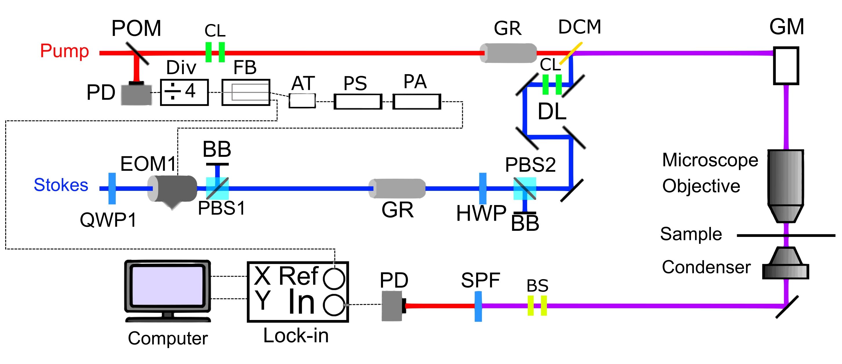
| Electrooptic Amplitude Modulator | THORLABS | EO-AM-NR-C1 | Two EOMs are needed for orthogonal modulation and dual-channel imaging. Resonant version is recommended so lower driving voltage can be used. |
|---|
| False H&E Staining Script | Matlab | | https://github.com/TheFuGroup/HE_Staining |
|---|
| Fanout Buffer | PRL-414B | Pulse Research Lab | 1:4 TTL/CMOS Fanout Buffer and Line Driver, for generating the EOM driving frequency and the reference to the lock-in |
|---|
| Fast Photodiode | THORLABS | DET10A2 | Si Detector, 1 ns Rise Time |
|---|
| Frequency Divider | PRL-220A | Pulse Research Lab | TTL Freq. Divider (f/2, f/4, f/8, f/16), for generating 20MHz from the laser output. |
|---|
| Highly Dispersive Glass Rods | Union Optic | CYLROD01 | High dispersion H-ZF52A Rod lens 120 mm, SF11 Rod lens 100 mm |
|---|
| Insight DS+ | Newport | | Laser system capable of outputting two synchronzied pulsed lasers (one fixed beam at 1, 040 nm and one tunable beam, ranging from 680-1,300 nm) with a repetition rate of 80 MHz. |
|---|
| Lock-in Amplifier | Liquid Instruments | Moku Lab | Lock-in amplifier to extract SRS signal from the photodiode. A Zurich Instrument HF2LI or similar instrument can be used as well. |
|---|
| Mirrors | THORLABS | BB05-E03-10 | Broadband Dielectric Mirror, 750 - 1,100 nm. Silver mirrors can also be used. |
|---|
| Motorized Delay Stage | Zaber | X-DMQ12P-DE52 | Delay stage for fine control of the temporal overlap of the pump and the Stokes lasers. Any other motorized stage should work. |
|---|
| Oil Immersion Condensor | Nikon | CSC1003 | 1.4 NA. Other condensers with NA>1.2 can be used. |
|---|
| Oscilloscope | Tektronix | TDS7054 | Any other oscilloscope with 400 MHz bandwdith or higher should work. |
|---|
| Phase Shifter | SigaTek | SF50A2 | For shifting the phase of the modulation frequency |
|---|
| Photodiode | Hamamatsu Corp | S3994-01 | Silicon PIN diode with large area (10 x 10 cm2). Other diodes with large area and low capacitance can be used. |
|---|
| Polarizing Beam Splitter | Union Optic | PBS9025-620-1000 | Broadband polarizing beamsplitter |
|---|
| Refactive Index Database | | | refractiveindex.info |
|---|
| Retro-reflector | Edmund Optics | 34-408 | BBAR Right Angle Prism. Other prisms or retroreflector can be used. |
|---|
| RF Power Amplifier | Minicircuits | ZHL-1-2W+ | Gain Block, 5 - 500 MHz, 50 Ω |
|---|
| Scan Mirrors | Cambridge Technologies | 6215H | We used a 5mm mirror set with silver coating |
|---|
| ScanImage | Vidrio | ScanImage Basic | Laser scanning microscope control software |
|---|
| Shortpass Filter | THORLABS | FESH1000 | 25.0 mm Premium Shortpass Filter, Cut-Off Wavelength: 1,000 nm. For efficient suppression of the Stokes, two filters may be necessary. |
|---|
| Upright Microscope | Nikon | Eclipse FN1 | Any other microscope frame can be used. If a laser scanning microscope is available, it can be used directly. Otherwise, a galvo scanner and scan lens needed to be added to the microscope. |
|---|
| Water Immersion Objective | Olympus | XLPLN25XWMP2 | The multiphoton 25X Objective has a NA of 1.05. Other similar objectives can be used. |
|---|