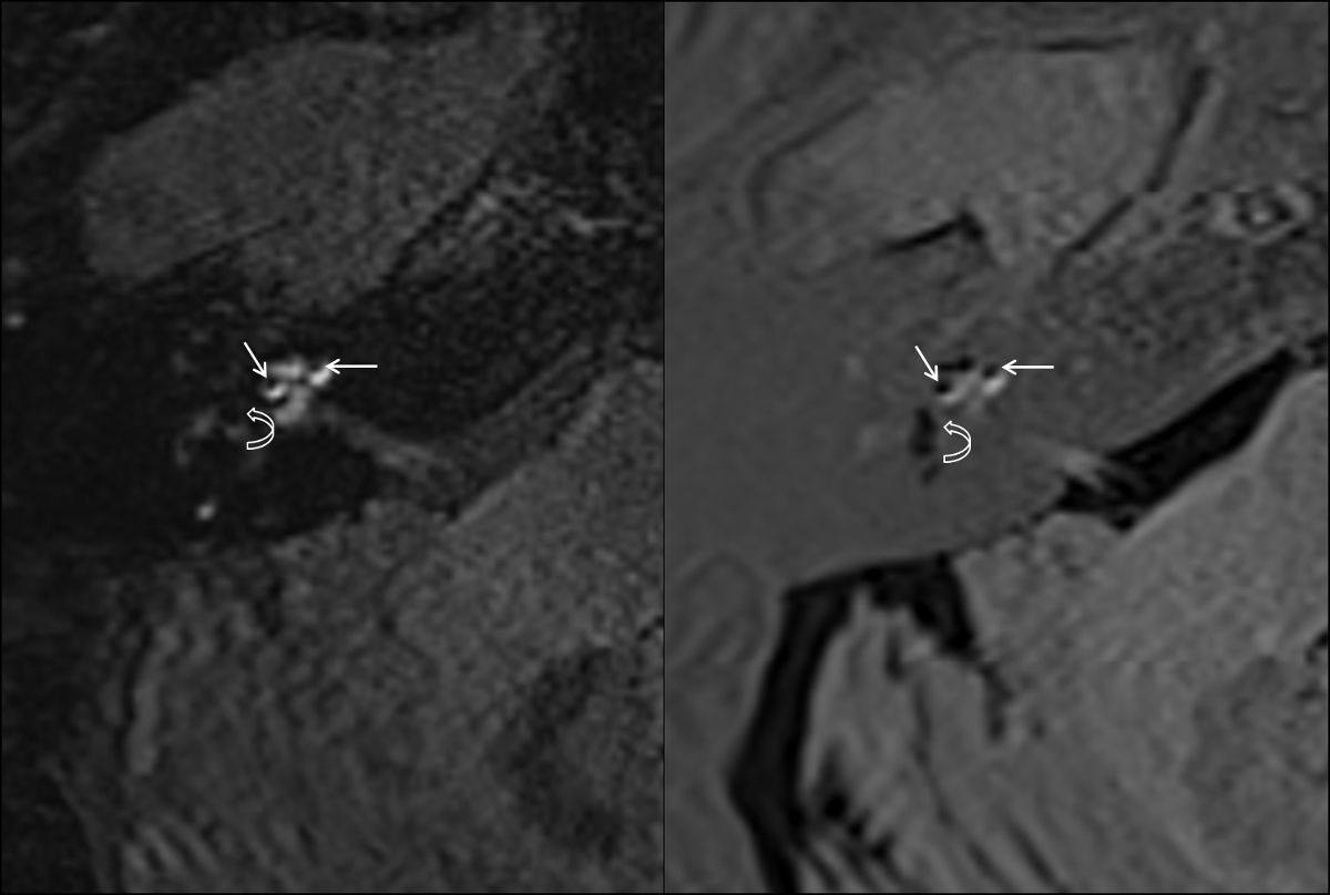
This video demonstrates the use of MRI to analyze cranial nerve morphology in the ears of patients suffering from a chronic inner ear disorder. It outlines the steps for performing a high magnetic field MRI scan on the patient, reconstructing the high-resolution 3D cross-sectional images of the cranial nerves, and measuring nerve dimensions to evaluate the abnormalities.
A subscription to JoVE is required to view this content. Sign in or start your free trial.
Method Article
In Vivo MRI Analysis of Cranial Nerve Morphology in a Patient with Inner Ear Disorder
2.5K views
⸱
May 29th, 2025
In This Article
Abstract
Protocol
All procedures involving human participants have been performed in compliance with the institutional, national, and international guidelines for human welfare and have been reviewed by the local institutional review board.
Access restricted. Please log in or start a trial to view this content.
Results
Access restricted. Please log in or start a trial to view this content.
Materials
| Name | Company | Catalog Number | Comments |
|---|---|---|---|
| MR-scanner, e.g. Siemens Magnetom Verio, or appropriate MR-scans in DICOM format, e.g. 3D-CISS | |||
| Osirix or any other DICOM-Viewer with appropriate evaluation tools | |||
| MedCalc or any other statistical analysis software, e.g. SPSS | |||
| Computer running Windows or MacOSX/macOS |
