This video demonstrates the use of micro-CT imaging to visualize cerebral cavernous malformation (CCM) lesions in a mouse brain. It details the steps for sample preparation, imaging, and 3D image reconstruction to evaluate the density and location of CCM lesions in brain tissue.
A subscription to JoVE is required to view this content. Sign in or start your free trial.
Method Article
Contrast-Enhanced Micro-CT Imaging of Cerebral Cavernous Malformation Lesions in a Mouse Brain
1K views
⸱
May 29th, 2025
In This Article
Abstract
Protocol
All procedures involving animal models have been reviewed by the local institutional animal care committee and the JoVE veterinary review board.
Access restricted. Please log in or start a trial to view this content.
Results
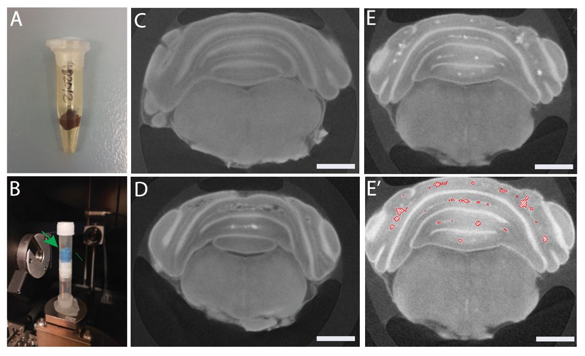
Access restricted. Please log in or start a trial to view this content.
Materials
| Name | Company | Catalog Number | Comments |
|---|---|---|---|
| 4-hydroxy tamoxifen | |||
| Corn oil | |||
| Stereomicroscope | |||
| Lugol's Iodine solution | |||
| Plastic paraffin film | |||
| Micro-CT | |||
| 3D rendering software |
