$$\rightleftharpoonup{xx}$$
$$\longleftharp{xx}$$,
$$\longrightharp{xx}$$,
1. General Reagents and Media Preparation for Membrane Extraction
- Bacterial growth media: Prepare and sterilize 1 L of broth media in a thoroughly cleaned and autoclaved 2 L flask.
- General resuspension buffer (1 M tris(hydroxymethyl)aminomethane buffer (Tris Buffer) pH 7.5; 50 mL): Dissolve 6.05 g of Tris base in 30 mL of H2O. Adjust pH to 7.5 with 5 M HCl. Adjust the final volume to 50 mL with ultrapure H2O.
- Master stock of divalent cation chelation solution (0.5 M ethylenediamine tetraacetic acid, EDTA pH 8; 100 mL): Add 18.6 g of disodium ethylene tetraacetate·2H2O to 80 mL of H2O. Stir vigorously and adjust pH to 8.0 with sodium hydroxide (NaOH). Adjust the final volume to 100 mL with ultrapure H2O.
NOTE: The disodium salt of EDTA will not dissolve until the pH of the solution is adjusted to ~8.0 by the addition of NaOH. - Osmotic buffer A (0.5 M sucrose, 10 mM Tris pH 7.5; 1 L): Weigh 171.15 g of sucrose and transfer it to a 1 L cylinder. Add 10 mL of 1 M Tris pH 7.5. Adjust to a final volume of 1 L with ultrapure H2O. Store at 4 °C.
- Lysozyme (10 mg/mL; 5 mL): Weigh 50 mg of (chicken egg-white) lysozyme and dissolve in 5 mL ultrapure H2O. Store at 4 °C.
- Diluted divalent cation chelation solution (1.5 mM EDTA; 500 mL): Add 1.5 mL of 0.5 M EDTA (Step 1.3) to 497.5 mL ultrapure H2O. Store at 4 °C.
- Osmotic buffer B (0.2 M sucrose, 10 mM Tris pH 7.5; 2 L): Weigh 136.8 g of sucrose and transfer it to a 2 L cylinder. Add 20 mL of 1 M Tris pH 7.5. Adjust final volume to 2 L with ultrapure H2O. Store at 4 °C.
- Nuclease co-factor (1 M magnesium chloride, MgCl2; 10 mL): Dissolve 2.03 g of MgCl2·6H2O in 8 mL of ultrapure H2O. Adjust volume to 10 mL. Store at room temperature.
- Nuclease solution cocktail including RNase and DNase enzymes: See Table of Materials. Store at -20 °C.
- Protease inhibitor cocktail: See Table of Materials. Store at 4 °C.
- Low-density isopycnic sucrose gradient solution (20% w/v sucrose, 1 mM EDTA, 1 mM Tris pH 7.5 Solution; 100 mL): Weigh 20 g of sucrose and transfer to a 200 mL cylinder. Add 100 µL of 1 M Tris Buffer pH 7.5 and 200 µL of 0.5 M EDTA pH 8. Adjust the final volume to 100 mL with ultrapure H2O. Store at room temperature.
- Medium-density isopycnic sucrose gradient solution (53% w/v sucrose, 1 mM EDTA, 1mM Tris pH 7.5 Solution; 100 mL): Weigh 53 g of sucrose and transfer to a 200 mL graduated cylinder. Add 100 µL of 1 M Tris Buffer pH 7.5 and 200 µL of 0.5 M EDTA pH 8. Adjust the final volume to 100 mL with ultrapure H2O. Store at room temperature.
NOTE: Prepare this solution in a graduated cylinder to ensure accuracy due to the high percentage of sucrose. Add a magnetic stir bar and stir until the sucrose is completely dissolved in the solution. This process may take several hours. - High-density isopycnic sucrose gradient solution (73% w/v sucrose, 1 mM EDTA, 1 mM Tris pH 7.5 Solution; 100 mL): Weigh 73 g of sucrose and transfer to a 200 mL graduated cylinder. Add 100 µL of 1 M Tris Buffer pH 7.5 and 200 µL of 0.5 M EDTA pH 8. Adjust the final volume to 100 mL with ultrapure H2O. Store at room temperature.
NOTE: Prepare this solution in a graduated cylinder to ensure accuracy due to the high percentage of sucrose. Add a magnetic stir bar and stir until the sucrose is completely dissolved. This process may take several hours. - Isolated-membrane-storage buffer (10 mM Tris Buffer pH 7.5; 1 L): Add 1 mL of 1 M Tris Buffer pH 7.5 to a 1 L flask and adjust the final volume to 1 L with ultrapure H2O.
2. Preparation of Bacteria for Membrane Extraction
- Streak the bacteria from frozen glycerol stocks onto fresh agar plates. Store the plates at 4 °C once colonies develop. Inoculate a single colony into a 5 mL tube filled with broth media and culture the bacteria as desired overnight.
- Back-dilute the overnight bacterial culture into 1 L of preferred broth media and culture the bacteria until the desired optical density is achieved.
NOTE: Inoculating a single bacterial colony into 1 L of broth media is recommended for mutant genotypes that are prone to suppressing growth phenotypes, but some Gram-negative bacteria simply grow more slowly than others. If it is not possible to achieve a sufficient culture density by single-colony inoculation, back-diluting an overnight culture into 1 L of media is one strategy to synchronize growth. Bacterial-membrane composition varies depending upon the growth phase of the culture (logarithmic vs stationary phase). Growth curves measuring the change in optical density for the bacterial cultures as a function of time should be performed with all strains to correlate culture density with growth phase. - Set the flasks containing the broth cultures on ice. Read the optical density at 600 nm (OD600) and calculate the volume of culture that is equivalent to between 6.0 and 8.0 x 10¹¹ bacterial colony-forming units (CFU). For S. Typhimurium, this corresponds to 1 L of culture at an OD600 of between 0.6 - 0.8, since an OD600 of 1.0 is equal to roughly 1.0x10⁹ CFU/mL. Add this volume to a centrifuge tube and ensure that the remaining cultures stay on ice until they are to be used.
- Pellet the bacteria by centrifugation at 4 °C at 7,000-10,000 x g in a fixed-angle high-speed centrifuge for 10 min.
NOTE: Pre-cool and maintain the centrifuges at a low temperature. Maintain the samples on ice during the entire procedure. - Decant and discard the supernatant carefully.
NOTE: If the membrane fractions are not going to be extracted immediately, the pellet can be flash frozen and/or stored at -80 °C. However, it is recommended to proceed directly with plasmolysis on the same day the cells are harvested, especially for non-enterobacterial species.
3. Dissociation of the Outer Membrane and Plasmolysis
- Thaw the cell pellets on ice if they were previously stored at -80 °C, and retain the samples on ice for the remainder of the procedure. Resuspend each cell pellet within the centrifuge tube in 12.5 mL of buffer A. Add a magnetic stir bar to the suspension of cells.
- Add 180 µL of 10 mg/mL lysozyme (final concentration of 144 μg/mL) to each cell resuspension. Keep the samples on ice while stirring for 2 min.
- Add 12.5 mL of 1.5 mM EDTA solution to each cell resuspension and continue stirring on ice for an additional 7 min.
- Decant the suspension into a 50 mL conical tube and centrifuge at 9,000-11,000 x g for 10 min at 4 °C.
- Discard supernatants into a biohazard waste container and retain the pellets on ice.
- Add 25 mL of buffer B to the cell pellet.
- Add 55 µL of 1 M MgCl2, 1 µL of RNase/DNase nuclease reagent (to avoid viscosity problems associated with bacteria undergoing plasmolysis prior to homogenization), and 1 µL of protease inhibitor cocktail to the volume of buffer B that sits atop the cell pellet
- Resuspend the pellet in the buffer B mixture. Vigorously pipette and vortex until observing a homogenous solution.
NOTE: It is very important to have a homogeneous solution before proceeding to Step 4 of this protocol. The resuspended cells should have a viscous cake-batter-like appearance and consistency. - Vortex each sample for 15 s. Retain suspensions on ice and proceed to Step 4.
4. Pressurized Homogenization and Lysis
NOTE: Several methods can be used for lysis. Sonication is not ideal due to the generation of heat. Osmotic lysis can be achieved, but is often inefficient. Therefore, we recommend high-pressure lysis. High-pressure lysis can be achieved using a variety of instruments. We suggest homogenization machines, such as the French Press or the Emusliflex. We work with many types of Gram-negative bacteria whose response to high osmolar sucrose solutions varies. The high-pressure homogenization step improves efficiency, reproducibility, and yield.
- Prechill the French Press cell at 4 °C or insert the metal coil from the homogenizer machine on ice.
- Pour the sample into the French-pressure cell or the homogenizer-sample cylinder and bring the cell under the desired homogenization pressure (10,000 psi should be adequate when using a French Press or 20,000 psi when using a homogenizer).
- Adjust the outlet flow rate to approximately one drop per second while maintaining the pressure if utilizing a French Press.
- Collect the cell lysate in 50 mL conical tubes while keeping samples on ice.
- Repeat steps 4.2-4.4 three to five times to achieve complete lysis, which is typically indicated by a gradual increase in the sample's transparency.
NOTE: The sample chamber should be washed and equilibrated with Buffer B in between samples. - Keep lysed cells on ice.
5. Total Membrane Fractionation
- Centrifuge the lysed bacterial samples at 6,169 x g for 10 min at 4 °C to pellet the remaining intact cell material. (e.g., unlysed bacterial cells).
- Distribute the remaining portion of the supernatant, which now contains the homogenized membranes, into a polycarbonate bottle for ultracentrifugation.
CAUTION: If needed, cell samples can be balanced by diluting with buffer B. - Ultracentrifuge the cell lysates at 184,500 x g for at least 1 h, at 4 °C. This step can be performed overnight without affecting the quality of the membranes.
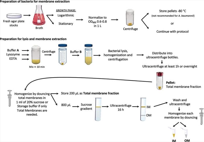
- Discard the remaining supernatant present in the ultracentrifuge tube and retain the membrane pellets on ice (Figure 1).
- Resuspend the membrane pellets in 1 mL of the low-density isopycnic-sucrose gradient solution using a glass-Dounce homogenizer. Transfer the sample homogenate to a 1.5 mL microcentrifuge tube using a glass Pasteur pipette and retain on ice.
NOTE: If only total membrane composition analysis is desired, substitute 1 mL of low-density isopycnic-sucrose gradient solution for 1 mL of isolated membrane storage buffer. Step 5.5 is the endpoint of isolation if only total bacterial membrane samples are desired. Store samples at -20 °C until further downstream analysis is required.
6. Density Gradient Ultracentrifugation to Separate the Dual Membranes
- Gather the appropriate number of 13 mL polypropylene or ultra-clear open-top tubes specified for a swinging bucket rotor and ultracentrifuge.
- Hold the tube in a slightly tilted position and prepare the sucrose gradient by slowly adding sucrose solutions from higher density to lower density in the following order:2 mL of 73% w/v sucrose,1 mM EDTA, 1 mM Tris pH 7.54 mL of 53% w/v sucrose, 1 mM EDTA, 1 mM Tris pH 7.5
- Next, add the total membrane fraction (1 mL), which has been resuspended in the 20% w/v sucrose solution (step 5.5). Avoid mixing the membrane fraction with the sucrose solution that lies beneath it. Divisions should be visible between each of these layers.
- Finally, fill the tube with the low-density isopycnic-sucrose gradient solution (approx. 6 mL). Polypropylene and ultra-clear open-top tubes should be filled as full as possible (2 or 3 mm from the tube top) for tube support.
- Adjustment for Acinetobacter baumannii 17978
- Adapt an adjusted sucrose gradient for use with different bacterial specimens. For A. baumannii, the following sucrose gradient afforded more complete separation of the membranes (Figure 2).2 mL of 73% w/v sucrose, 1 mM EDTA, 1 mM Tris pH 7.54 mL of 45% w/v sucrose, 1 mM EDTA, 1 mM Tris pH 7.5
- Next, add the total membrane fraction (1 mL), which has been resuspended in the 20% w/v sucrose solution (step 5.5). Avoid mixing the membrane fraction with the sucrose solution that lies beneath it. Divisions should be visible between each of these layers.
- Finally, fill the tube with low-density isopycnic-sucrose gradient solution (approx. 6 mL). Polypropylene and ultra-clear open-top tubes should be filled as full as possible (2 or 3 mm from the tube top) for tube support.
- Ultracentrifuge the samples using a swinging-bucket rotor at 288,000 x g and 4 °C overnight.
NOTE: For the volumes used in the previous steps, we recommend centrifugation times between 16 h and 23 h. - Cut the end of a P1000 pipette tip about 5 mm from the point. Using the pipette, remove the upper-brown inner membrane (IM) layer. Transfer the IM fraction into a polycarbonate bottle for ultracentrifugation.
- Leave about 2 mL of the sucrose solution above the 53-73% interface to ensure that the lower white outer membrane (OM) is not cross-contaminated with the IM fraction. Repeat the pipetting procedure from step 6.4 for the OM fraction (Figure 1).
NOTE: The membranes can also be collected by puncturing the centrifuge tubes at the bottom with a needle and collecting them as fractions dropwise. - Fill the remaining void of the ultracentrifuge tube with isolated-membrane storage buffer and mix by inversion or pipetting. Retain the samples on ice.
- Collect the now washed and isolated membranes by ultracentrifugation at 184,500 x g for 1 h at 4 °C.
- Discard the supernatant and resuspend the membranes by Dounce homogenization. Add 500-1000 μL of storage buffer. Collect samples in 2 mL microcentrifuge tubes.
- Store the bacterial membrane samples at -20 °C.