This video demonstrates the use of fluorescent nanoparticles to visualize endo-lysosomal remodeling in human epithelial cells infected by intracellular bacteria. Following bacterial invasion, the pathogens manipulate the host cytoskeleton and endo-lysosomal system, forming vacuoles with tubular projections that alter vesicle transport. Fluorescently labeled nanoparticles are then used to trace these changes, allowing for high-resolution imaging of pathogen-induced intracellular trafficking events.
A subscription to JoVE is required to view this content. Sign in or start your free trial.
Method Article
Nanoparticle-Mediated Visualization of Endo-Lysosomal Remodeling in Bacterially-Infected Cells
575 views
⸱
August 29th, 2025
In This Article
Abstract
Protocol
- Culture of HeLa Cells
- HeLa cells permanently expressing LAMP1-GFP (Lysosome-associated membrane glycoprotein 1-Green fluorescent protein) are cultured in Dulbecco’s Modified Eagle Medium, or DMEM with 10% fetal calf serum (FCS) and grown at 37 °C in an atmosphere containing 5% CO2.
- Seed the cells at a density of 50,000 per well in an 8-well chamber slide (Ibidi) and incubate overnight (O/N).
- Culture of Bacteria
- Use Salmonella enterica serovar Typhimurium NCTC 12023 wild-type (WT) strain. For comparison, use mutant strains ΔssaV defective in the SPI2-T3SS and ΔsifA lacking key effector SifA for SIF. Use strains harboring plasmid pFPV25.1 for constitutive expression of enhanced GFP.
NOTE: Bacterial strains are routinely cultured in Luria-Bertani broth (LB) with the addition of 50 µg/ml carbenicillin (WT) and LB with the addition of 50 µg/ml carbenicillin and/or 50 µg/ml kanamycin (ΔssaV, ΔsifA) of required to maintain plasmids. - Inoculate a single colony of bacteria in 3 ml LB with appropriate antibiotics and grow O/N at 37 ºC under shaking conditions for aeration, then dilute 1:31 in fresh LB and continue growth for 3.5 hr. At this time-point, the cultures reach the late log phase and bacteria are highly invasive. A ‘roller drum’ is convenient to incubate test tube cultures with aeration.
- Use Salmonella enterica serovar Typhimurium NCTC 12023 wild-type (WT) strain. For comparison, use mutant strains ΔssaV defective in the SPI2-T3SS and ΔsifA lacking key effector SifA for SIF. Use strains harboring plasmid pFPV25.1 for constitutive expression of enhanced GFP.
- Infection of HeLa cells by Salmonella and Gold-BSA-Rhodamine Nanoparticles or NPs Pulse Chase-labeling (see Figure 1 for scheme)
- Measure OD600 of the sub-cultured bacteria and dilute to OD600 = 0.2 in 1 ml Phosphate-buffered saline or PBS (~3 × 10⁸ cfu/ml), add appropriate amounts of bacteria to HeLa cells in 8-well chamber slides to achieve a multiplicity of infection (MOI) of 100.
- Incubate for 30 min in the cell incubator, wash 3 times with PBS to remove non-internalized bacteria (this time point was set as 0 hr post-infection, or 0 hr p.i.). Add 300 µl of fresh culture medium containing 100 µg/ml gentamicin and maintain for 1 hr. Then replace the medium with fresh medium containing 10 µg/ml gentamicin for the rest of the incubation time.
- After incubation with culture medium containing 100 µg/ml gentamicin for 1 hr, the medium is replaced by imaging medium (Eagle's MEM without FCS, L-glutamine, phenol red, and sodium bicarbonate, with 30 mM HEPES, pH 7.4) containing 10 µg/ml gentamicin. Gold-BSA-rhodamine NPs are added to HeLa cells to obtain a final concentration of OD520 = 0.1. (Bovine Serum Albumin, or BSA)
NOTE: Gold-BSA-rhodamine NPs may also be added to cells before infection, or at various time-points p.i. - After 1 hr incubation, remove the medium, wash 3 times with PBS, and add 300 µl of fresh imaging medium containing 10% FCS and 10 µg/ml gentamicin for the rest of the incubation time.
NOTE: Duration of incubation may vary depending on concentration of NPs and cell lines used. For RAW264.7 macrophages, we observed that 30 min incubation with NPs at a concentration of OD520 = 0.05 allowed sufficient internalization.
Access restricted. Please log in or start a trial to view this content.
Results
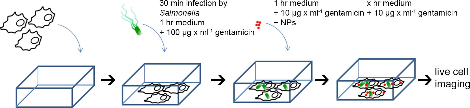
Figure 1. Scheme for the preparation of HeLa cells for live cell imaging. HeLa cells were seed...
Access restricted. Please log in or start a trial to view this content.
Materials
| Name | Company | Catalog Number | Comments |
|---|---|---|---|
| Gold chloride | Sigma-Aldrich | 520918 | |
| Bovine serum albumin | Sigma | A2153 | |
| NHS-Rhodamine | Pierce | 46406 | |
| DMSO | Sigma | D8418 | |
| HEPES | Sigma | H3375 | |
| Gentamicin | Applichem | A1492 | |
| Kanamcyin | Roth | T832 | |
| Carbenicillin | Roth | 6344 | |
| 8-well chamber slides | Ibidi | 80826 | tissue culture treated, sterile |
