1. Prepare Instruments, Culture Media, Solutions, and Petri Dishes
1. Autoclave toothpicks in a 50 mL beaker covered with aluminum foil to keep them sterile.
2. Sterilize all dry goods that will be used (tips, sticks, glassware, etc.) in an autoclave.
NOTE: Caution! Follow all safety directions for the autoclave model used. Autoclaves pose a risk for explosions, burns from steam, and contact with hot surfaces, materials, and media.
3. Prepare the MS agar (Mannitol Soya Flour (SFM)) to grow cultures.
1. Add 5 g of soya flour and 5 g of agar to a 1 L flask.
2. Dissolve 5 g of mannitol in 250 mL of tap water and dispense into the flask containing the soy flour and agar.
3. Add a foam plug and foil to the opening of the flask and autoclave at 121 °C, 15 psi for at least 30 min.
CAUTION: This medium easily boils over. Use a flask that can hold at least four times the actual volume of MS agar being autoclaved. A standard pressure cooker can be used in place of an autoclave with similar safety considerations. Alternatively, a microwave oven may be used to sterilize media and materials if access to an autoclave is not possible. High school teachers may also want to collaborate with local colleges or universities to access autoclaved supplies.
4. Cool media until comfortable enough to handle. Be careful not to cool it for too long because agar solidifies at temperatures below 50 °C.
5. Pour MS agar from the flask into sterile Petri dishes until the bottom of each Petri dish is approximately half full (approximately 25 mL of media per Petri dish). Allow to solidify before using agar plates. Store agar plates in a plastic bag at room temperature until needed.
NOTE: Agar plates may also be stored in a refrigerator, especially if antibiotics are needed in the media.
2. Streak Streptomyces onto Plates for Propagation
1. Streak Streptomyces strains onto MS agar plates to single colonies using a standard method, such as the quadrant streak method.
NOTE: An inoculating loop or sterile toothpicks may be used. Optional: Other media are commonly used for Streptomyces species, such as R2YE and minimal media.
2. Incubate plates at 30 °C.
3. Restreak plates by picking a single colony every 4–6 days. As colonies age, they become prone to random mutation.
3. Compare and Record Visual Appearance
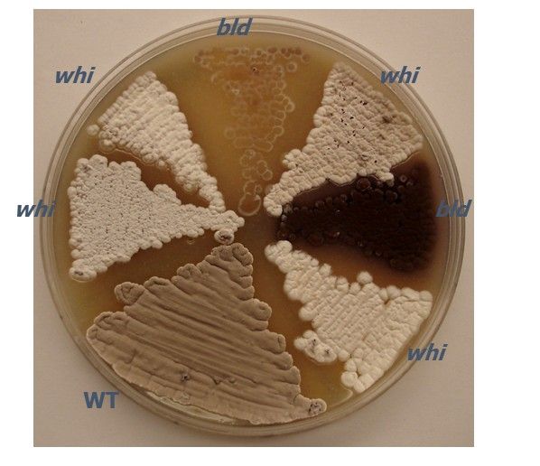
1. Take note of colony morphology for each mutant as compared to the wild-type parent strain after streaking all strains as in step 2. Compare the new isolate to that of a well-studied species, such as S. coelicolor or S. venezuelae, when characterizing new Streptomyces species. Note characteristics (Figure 1) such as basic shape, surface of colony (fuzzy, bald, wrinkled, etc.), opacity, elevation, and pigmentation (distinguish between vegetative mycelium, aerial mycelium, and/or surrounding medium).
2. Label an MS agar plate and streak wild-type and mutant strains in wedge patterns (Figure 1). Be careful that no strain touches another strain to avoid cross-contamination. Note the date and time that strains are streaked onto the plate. Incubate the plates at 30 °C.
3. Place grown Petri dishes on colored or white paper to homogenize the background. Write on the paper the strain name, date, incubation temperature, and time from first plate streaking. Take a digital picture(s) of the plate with the latter information written on the paper background so that confusion is minimized.
NOTE: The writing can be cropped from the photograph at a later time if it is to be used for figure preparation.