This video demonstrates the use of phase-contrast microscopy to examine phenotypic differences between wild-type and mutant Streptomyces coelicolor. It outlines the steps involved in preparing and imaging aerial filaments to detect developmental defects in spore formation.
A subscription to JoVE is required to view this content. Sign in or start your free trial.
Method Article
Phase-Contrast Microscopy for Evaluating Developmental Mutants in Streptomyces
582 views
⸱
September 26th, 2025
In This Article
Abstract
Protocol
1. Perform Phase-Contrast Microscopy
- Sterilize tweezers by washing in 70% ethanol and then passing through a Bunsen burner flame to evaporate the ethanol. Allow to cool.
- Sterilize coverslips by washing them in 70% ethanol and then allowing them to dry in a sterile Petri dish.
- Prepare a coverslip lift of the bacterial growth to be examined.
- Pick up a sterile coverslip with sterile tweezers and place the coverslip on a mannitol soya flour (MS) agar plate where Streptomyces coelicolor bacterial growth is dense. Press on the back of coverslip gently with tweezers to ensure sufficient transfer of bacterial spores and aerial mycelium.
- Pick up the coverslip from the plate and place it so that the side with the cell material is facing toward the surface of a microscope slide, with a 15 µL drop of 50% glycerol prepared in water for mounting. Reduce air bubbles by placing the coverslip at a 45° angle to the slide and letting it fall onto the 50% glycerol.
- (Optional) Seal with clear nail polish for preservation of the slide.
- Perform phase-contrast microscopy at various time intervals during the life cycles.
NOTE: Repeated imaging allows for a more complete analysis and the ability to detect delays in development. Independently repeat the observations to ensure accuracy and reproducibility.- Place the prepared slide on the microscope stage and add a drop of immersion oil to the center of the coverslip. Rotate the 100X phase objective in place and set the condenser turret to the proper "matching" phase setting. Focus the image using only the fine adjustment knob once the objective lens is in contact with the oil.
- Examine several fields of view to discern noticeable and consistent differences between mutant strains and the wild type, such as the ability to form spores, spore size and shape, and overall number of spores (see Figure 1).
NOTE: Alternatives to phase-contrast microscopy include differential interference contrast (DIC) microscopy and simple staining techniques for use with bright field microscopes, such as crystal violet staining.
Access restricted. Please log in or start a trial to view this content.
Results
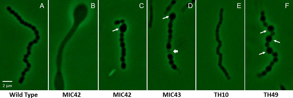
Figure 1: Phase-contrast micrographs showing a representative aerial filament for Strept...
Access restricted. Please log in or start a trial to view this content.
Materials
| Name | Company | Catalog Number | Comments |
|---|---|---|---|
| 50% Glycerol | Sigma-Aldrich | G5516 | |
| Immersion Oil (Type DF) | Cragille | 16482 | |
| Sterile Water | GeneMate | G-3250-1L | |
| 100% Ethanol | Sigma-Aldrich | E7023 | |
| Soy Flour | Bob's Red Mill | 1516C164 | |
| D-Mannitol | Sigma-Aldrich | M4125-1KG | |
| Agar | Sigma-Aldrich | A1296-1KG | |
| Glycerol 100% | VWR amresco life science | 0854-1L | |
| Petri dishes | Sigma-Aldrich | P5856-500EA | |
| Cover slips #1.5 | Thomas Scientific | 64-0721 | |
| Slides | Carolina | 63-2010 | |
| Phase-Contrast Microscope | Olympus | BX40 | |
| Forceps | Carolina Biological | 624504 | |
| Bunsen Burner | Carolina Biological | 706706 | |
| Clear nail polish | OPI | 22001009000 | |
| Camera for Microscope | Olympus | DP72 | |
| Nitrile Examination Gloves (Med) | Bio Excell | 71011002 |
